A 46-year-old man with alcoholic chronic pancreatitis was admitted to our hospital with severe upper abdominal pain and high fever (temperature up to 38.5°C) for 1 day. A week ago, he was scheduled to undergo EUS-guided intervention for a pseudocyst in the pancreatic tail, about 9 cm in size, which was identified on magnetic resonance imaging (
Fig. 1A). Laboratory tests revealed low hemoglobin (9.8 g/dL) and elevated inflammatory markers (white blood cell count 17,500/µL and C-reactive protein 324.56 mg/L). Abdominal computed tomography revealed that the hemorrhagic pancreatic pseudocyst had ruptured, with direct extension to the splenorenal ligament and subcapsular splenic space. However, there was no evidence of active contrast extravasation (
Fig. 1B,
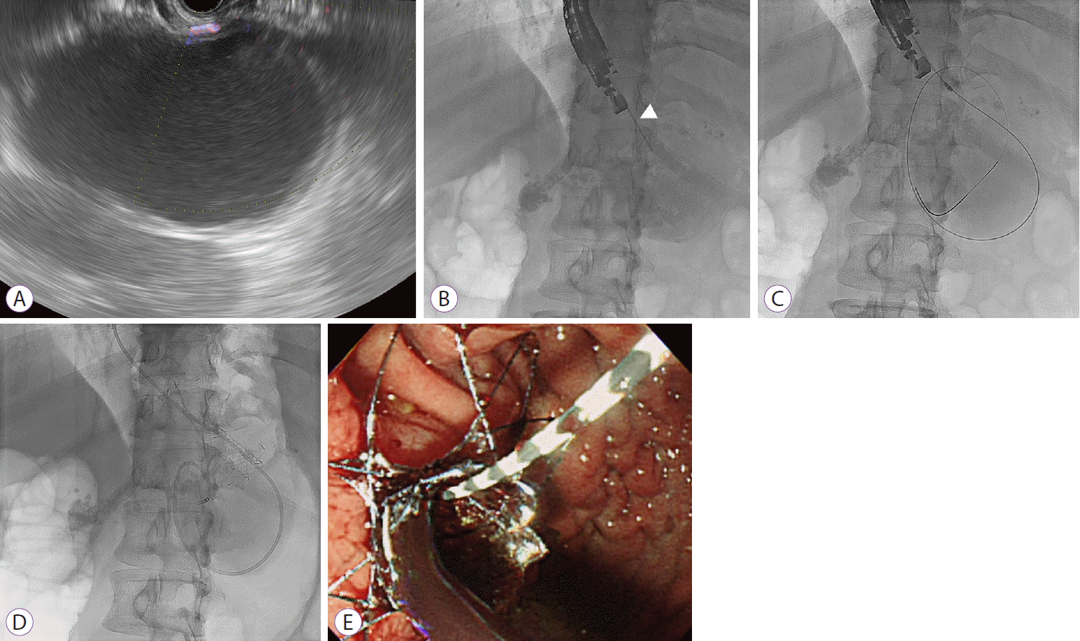
C). EUS showed an anechoic thick-walled cystic lesion, 9 cm in size (
Fig. 2A). Transgastric wall puncture was performed using a 19-gauge needle for fine-needle aspiration (EZ shot 3plus
TM; Olympus, Tokyo, Japan), avoiding intervening vessels (
Fig. 2B). Thick and turbid bloody fluid with debris was aspirated, and a 0.025-inch guidewire (Visiglide
®; Olympus Medical Systems, Tokyo, Japan) was coiled into the pseudocyst cavity under fluoroscopic guidance (
Fig. 2C). Subsequently, tract dilation was performed using a 6-Fr cystotome (Wilson-Cook Medical Inc., Bloomington, IN, USA), and an FCSEMS (BONA-AL
® 10 mm diameter, 5 cm total length; Standard Sci Tech Inc., Seoul, Korea) with a 7-Fr nasocystic drainage tube was inserted for additional lavage and aspiration (
Fig. 2D,
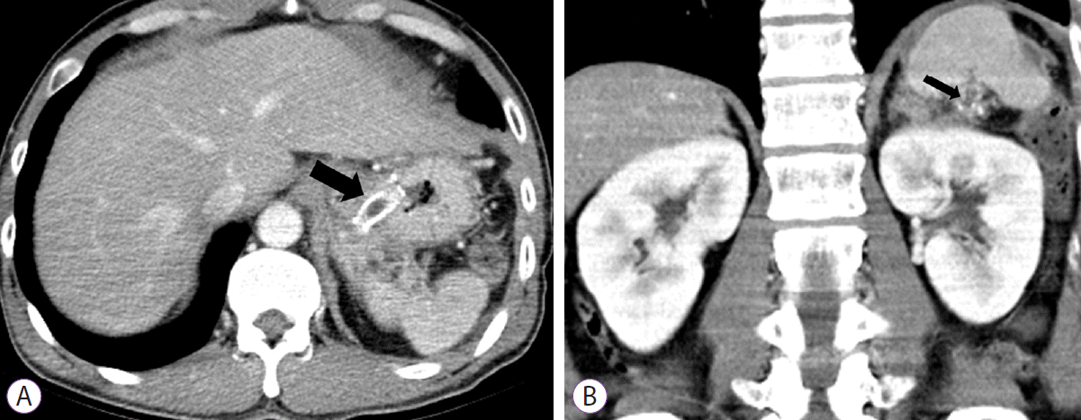
E). The patient inadvertently removed the nasocystic tube 2 days after the procedure, but the FCSEMS was found to be well positioned on the X-ray. The clinical hallmarks improved, and the patient was discharged 12 days later. An abdominal computed tomography scan 4 weeks later showed complete resolution of the ruptured pseudocyst and perisplenic fluid collection (
Fig. 3A,
B). The metal stent that was placed for gastrocystostomy was removed. The patient recovered without any recurrent symptoms.
 | Fig. 1.(A) Axial T2-weighted magnetic resonance imaging showing the main pancreatic duct dilation (open black arrow) with multiple pancreatoliths (black arrowheads) and a pseudocyst (open white arrow), about 9 cm in size, in the pancreatic tail. Abdominal computed tomography scan of a hemorrhagic pancreatic pseudocyst that ruptured spontaneously. (B) Axial and (C) coronal views showing mural discontinuity (black arrows) of the hemorrhagic pancreatic pseudocyst (asterisks), extension of fluid into the perisplenic space (white arrows), and multiple main pancreatic duct stones with pancreatic atrophy. 
|
 | Fig. 2.Endoscopic ultrasound-guided gastrocystostomy. (A) Large anechoic cystic lesion with hyperechoic material on endoscopic ultrasound, (B) The pseudocyst
was punctured with a 19-gauge needle for fine-needle aspiration (white arrowhead). (C) Passage of a 0.025-inch guidewire under fluoroscopic guidance, (D) Placement of a metal stent with a nasocystic drainage tube, and (E) Endoscopic view showing gushing thick and turbid bloody fluid gushing out, and the presence of the metal stent. 
|
 | Fig. 3.Abdominal computed tomography scan showing complete resolution of the ruptured pancreatic pseudocyst with the metal stent (black arrow) on axial (A) and coronal (B) views. 
|


 PDF
PDF Citation
 Print
Print


 XML Download