This article has been
cited by other articles in ScienceCentral.
Abstract
Superficial colonic neoplasms sometimes extend into a diverticulum. Conventional endoscopic mucosal resection of these lesions is considered challenging because colonic diverticula do not have a muscularis propria and are deeply inverted. Even if the solution is carefully injected below the mucosa at the bottom of the diverticulum, the mucosa is rarely elevated from the diverticular orifice, and it is usually just narrowed. Although endoscopic submucosal dissection or full-thickness resection with an over-the-scope clip device enables the complete resection of these lesions, it is still challenging, time consuming and expensive. Underwater endoscopic mucosal resection without submucosal injection (UEMR) is an innovative technique enabling en bloc resection of superficial colon lesions. We report three patients with colon adenomas extending into a diverticulum treated with successful UEMR. UEMR enabled rapid and safe en bloc resection of colon lesions extending into a diverticulum.
Go to :

Keywords: Colonic diverticulum, Colonic neoplasm, Underwater endoscopic mucosal resection
INTRODUCTION
Colorectal cancer was the third most common cancer and the second most common cause of cancer death worldwide in 2018 [
1]. A national polyp study revealed that colonoscopic removal of colorectal adenomas can reduce the incidence and mortality of colorectal cancer [
2,
3]. Since then, the role of endoscopic resection of colorectal adenomas has been greatly enhanced. However, superficial colorectal neoplasms sometimes occur where endoscopic removal is technically difficult. A lesion extending inside a colonic diverticulum is a difficult scenario because of the lack of the muscularis propria. Some of these patients undergo surgical resection [
4] or advanced endoscopic treatment such as endoscopic submucosal dissection (ESD). Binmoeller et al. first reported underwater endoscopic mucosal resection without submucosal injection (UEMR), an innovative technique enabling
en bloc resection of superficial colon lesions [
5,
6]. The first case of UEMR for a colon adenoma extending into a diverticulum was reported in 2020 [
7]. We report successful UEMR of colorectal adenomas extending into a diverticulum.
Go to :

CASE REPORTS
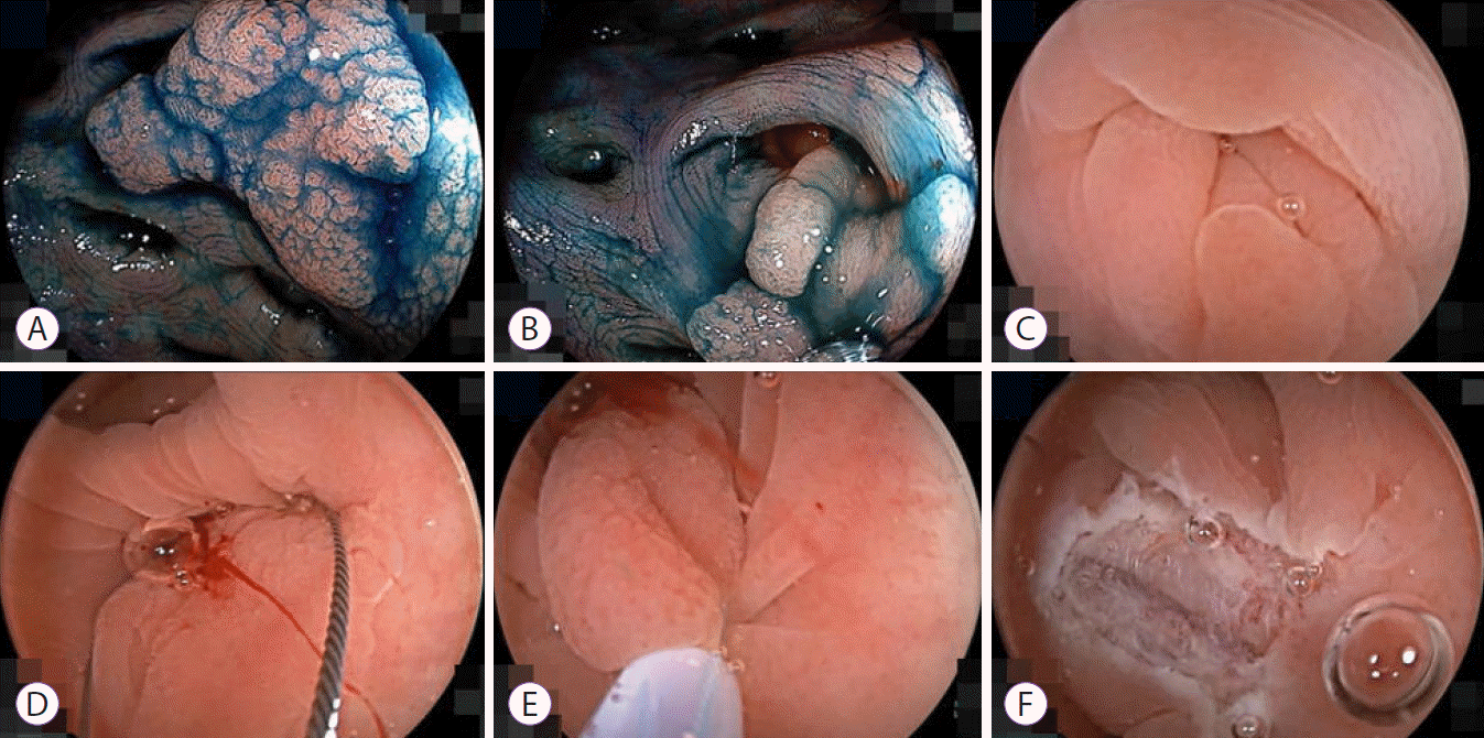
Patient 1 was a 65-year-old man with a positive fecal immunochemical test who underwent colonoscopy showing a flat lesion extending into a diverticulum in the cecum. The lesion was 1.5 cm in diameter and suspicious for an adenoma. After water immersion, the mucosa in the diverticulum was everted through the orifice and the diverticulum-side demarcation of the lesion was easily identified. We snared the entire lesion and safely resected it in an
en bloc fashion (
Fig. 1,
Supplementary Video 1).
 | Fig. 1.Underwater endoscopic mucosal resection without submucosal injection of a lesion in the cecum. (A) A 1.5-cm flat lesion in the cecum. (B) This lesion extended into a diverticulum. (C) The border of the lesion in the diverticulum was identified using water immersion. (D) The snare tip was anchored outside the lesion in the diverticulum. (E) The entire lesion was snared and cut using pure-cut mode diathermy. (F) The mucosal defect without any residual lesion was closed with hemoclips. Histology of the resected specimen showed an adenoma with negative resection margins. 
|
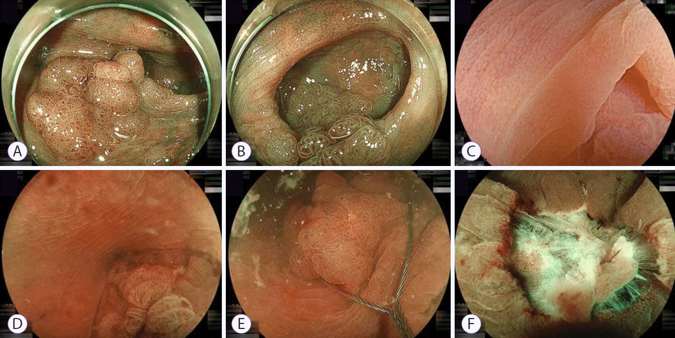
Patient 2 was a 70-year-old woman with a positive fecal immunochemical test who underwent colonoscopy revealing a flat lesion extending into a diverticulum in the ascending colon. The lesion was 1.5 cm in diameter and suspicious for an adenoma. After water immersion, part of the lesion was everted from the diverticulum and further everted using the edge of a distal cap. We snared the entire lesion and safely resected it in an
en bloc fashion (
Fig. 2,
Supplementary Video 1).
 | Fig. 2.Underwater endoscopic mucosal resection without submucosal injection of the lesion in the ascending colon. (A) A 1.5-cm flat lesion in the ascending colon. (B) This lesion extended into a diverticulum. (C) Lesion demarcation in the diverticulum was done after everting the lesion with edge of a distal cap after water immersion. (D) The snare tip was placed outside the lesion in the diverticulum. (E) The entire lesion was snared and cut using pure-cut mode diathermy. (F) The mucosal defect without any residual lesion was closed using a reopenable clip with an 11-mm opening width (SureClip; Micro-Tech, Nanjing, China). Histology of the resected specimen showed an adenoma with negative resection margins. 
|
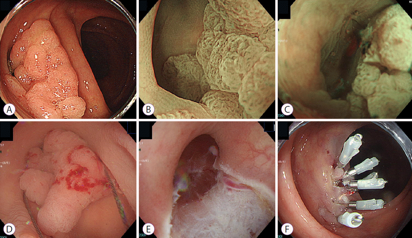
Patient 3 was an 80-year-old man who underwent routine colonoscopy before ESD for gastric cancer, which revealed a flat lesion extending into a diverticulum in the ascending colon. The lesion was 2 cm in diameter and suspicious for an adenoma. After water immersion, the mucosa in the diverticulum was everted through the orifice and the diverticulum-side demarcation of the lesion was easily identified. We snared the entire lesion and safely resected it in an
en bloc fashion (
Fig. 3,
Supplementary Video 1).
 | Fig. 3.Underwater endoscopic mucosal resection without submucosal injection of the lesion in the ascending colon. (A) A 2-cm flat lesion in the ascending colon. (B) This lesion extended into a diverticulum. (C) Lesion demarcation in the diverticulum was done after water immersion. (D) The snare tip was placed outside the lesion in the diverticulum. The entire lesion was snared and cut using pure-cut mode diathermy. (E) The mucosal defect without any residual lesion. (F) The defect was completely closed with hemoclips while maintaining water immersion. Histology of the resected specimen showed an adenoma with negative resection margins. 
|
All procedures were performed on an outpatient basis. Endoscopic treatment utilized a distal attachment (Olympus, Tokyo, Japan), 15-mm RotaSnare (Medi-Globe GmbH, Achenmühle, Germany), distilled-water irrigation and pure cutting mode diathermy. All post-UEMR mucosal defects were closed with hemoclips immediately while maintaining water immersion. There were no adverse events in any patient. Pathologic evaluation of all lesions showed adenomas with negative margins. The three cases in this report are all of the consecutive patients in our experience of UEMR for colonic lesions extending into a diverticulum. Therefore, we have successfully achieved en bloc resection in all cases without failure so far.
Go to :

DISCUSSION
Superficial colonic neoplasms sometimes extend into a diverticulum. Conventional endoscopic mucosal resection (EMR) of these lesions is considered challenging because colonic diverticula lack a muscularis propria and are deeply inverted. Even if the solution is carefully injected below the mucosa at the bottom of the diverticulum during EMR, the mucosa is rarely elevated from the diverticular orifice and is usually just narrowed. It is very difficult to resect neoplasms extending into a diverticulum in an en bloc fashion using conventional EMR.
UEMR is an innovative technique enabling en bloc resection of superficial colon lesions. The colonic mucosa and submucosa appear to float above the circular muscularis propria in the underwater view of endoscopic ultrasound. Therefore, even a flat lesion can be easily snared. The essential point to perform UEMR is to completely aspirate the intestinal gas in order to collapse the intestinal lumen and subsequently fill it with water like when performing endoscopic ultrasound.
UEMR has some advantages compared to conventional EMR; it allows one to reliably snare the entire lesion in a reasonable time without concern for dispersion of the injected solution leading to flattening of the mucosal elevation, as seen in the EMR procedure. Dispersion of the injected solution makes snaring a flat lesion more difficult or impossible [
5]. UEMR also allows one to snare a lesion repeatedly until the entire lesion is captured, sometimes with contraction [
8]. The EMR procedure sequence includes two irreversible steps: elevating the mucosa with an injection and snaring of the elevated mucosa. Each step demands a certain level of skill. If one of the steps fails, it will be difficult to achieve EMR with a complete
en bloc resection of the lesion.
We recommend cutting the captured lesion with pure-cut mode diathermy to avoid thermal injury to the muscularis propria. When cutting a flat lesion with pure-cut mode, there is no need to worry about immediate massive bleeding because these lesions rarely have feeding arteries which would massively bleed. Meanwhile, too much thermal injury in a diverticulum presents a risk of post-polypectomy coagulation syndrome because of the lack of the muscularis propria. After removing the lesion, the postprocedural mucosal defect can be closed with fewer hemoclips because the defect stays smaller with water immersion than with gas insufflation.
If massive bleeding occurs immediately, blood will quickly interfere with endoscopic visibility. We recommend the use of a water-irrigating pump to easily control the water supply and clear the endoscopic view. We also recommend the preparation of hemoclips before cutting, to deal with any immediate bleeding. If possible, a reopenable clip is ideal.
There has been a report of perforation associated with UEMR [
9] probably caused by stretching of the colonic wall with a retroflexed colonoscope. When the lesion was snared after submucosal injection even after water immersion, differing from UEMR, the colonic wall stretched and the procedure resulted in a perforation [
10]. In our opinion, accidental intramuscular injection may also allow snaring of the muscularis propria of the lesion even with water immersion. Although ESD or endoscopic full-thickness resection with an over-the-scope clip device [
11,
12] enables complete resection of superficial colonic lesions extending into a diverticulum, it is still challenging, time consuming and expensive. Some recently introduced traction devices facilitate ESD of the lesions [
13-
15].
This report is the first case series of UEMR of colon adenomas extending into a diverticulum. UEMR enabled rapid and safe en bloc resection of colon adenomas extending into a diverticulum without adverse events. UEMR may be useful for endoscopic removal of colonic neoplasms extending into a diverticulum.
Go to :


 PDF
PDF Citation
 Print
Print


 XML Download