Linked color imaging (LCI) provides bright observation suitable for screening of the entire stomach [1], and its high color contrast to the surrounding mucosa makes it easier to detect early malignant lesions [2,3]. Compared to white light imaging, LCI enhances subtle differences in the color tone by advanced post processing steps [4]. LCI has a higher emission intensity of 410 nm, which can influence the mucosal color of various lesions [1,4]. Clear images of gastric cancer have been reported in the background with a history of Helicobacter pylori infection. Here, we report a semi-pedunculated gastric adenocarcinoma in H. pylori-negative normal mucosa imaged using LCI.
A 61-year-old asymptomatic man underwent screening esophagogastroduodenoscopy and was diagnosed with a red polypoid lesion on the greater curvature in the proximal gastric body, and biopsy revealed adenocarcinoma. He was referred for further examination and treatment. He had not undergone H. pylori eradication therapy and had no family history of malignancies. He consumed about 20 g of alcohol daily and had a smoking history of 20 cigarettes per day from ages 20 to 30. Physical examination revealed no abnormalities. No long-term use of acid blockers was reported when the lesion was detected. Baseline laboratory data were unremarkable. The serum anti-H. pylori IgG antibody (<3 U/mL), H. pylori stool antigen test at our hospital, and the Giemsa stain of the biopsy specimen from the surrounding mucosa of the lesion from the referring clinic were all negative.
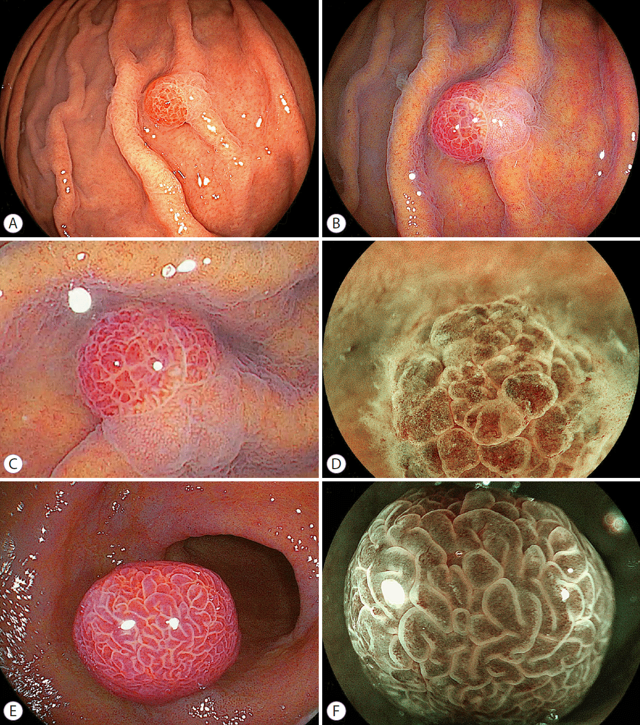
White light images of endoscopy showed a regular arrangement of collecting venules on the lesser curvature from the gastric angle to body without atrophic changes, with no evidence of an H. pylori infection. In addition, a 7-mm red semi-pedunculated polyp was observed in the proximal gastric body, with a color similar to red hyperplastic polyps in H. pylori-positive mucosa (Fig. 1A). There were no polypoid lesions seen in other areas. Using LCI, this polyp appeared purple–red with an irregular surface pattern, that is, a polygonal white zone of varied size (Fig. 1B, C). Magnifying blue laser imaging showed irregular micro-vascular and irregular surface patterns suspicious for gastric adenocarcinoma (Fig. 1D). These findings were different from those of a hyperplastic polyp with elongated white zones in the background of H. pylori-positive mucosa (Fig. 1E, F). In LCI imaging of the current lesion, the surrounding mucosa appeared as light orange (white-apricot) in all of the mucosa with fundic glands (Fig. 1B, C) [5-7] and no intestinal metaplasia in the antrum was observed although a patchy purple distribution is useful with LCI to diagnose intestinal metaplasia, which spreads from the antrum to the body in H. pylori active or previously infected areas [1-3]. These data strongly suggested an area negative for H. pylori infection. A line of demarcation was apparent between the malignant lesions and surrounding mucosa (Fig. 1A-C).
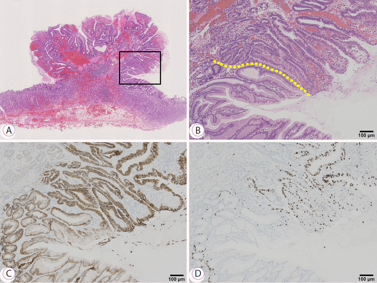
Endoscopic mucosal resection was performed. Histological findings revealed papillary and tubular proliferation of atypical cells, suggesting differentiated adenocarcinoma. Malignant cells were observed in most areas of the polyp (Fig. 2A, B). There was no evidence of atrophic changes, intestinal metaplasia, or inflammatory cell infiltration around the lesion. Immunohistochemical examination revealed malignant cells positive for mucin (MUC) 5AC (Fig. 2C), MIB-1 proliferation (Fig. 2D), and suppressive protein p53, but were negative for MUC6, MUC2, and CD10 (not shown). According to the Japanese classification system, the final diagnosis was adenocarcinoma (tub1), pT1a(M), 7 mm × 5 mm × 5 mm, Type 0–I, UL0, Ly0, V0, pHM0, pVM0. The patient is disease-free two years after resection.
LCI provides clear mucosal visualization with high color contrast between a malignant lesion and the surrounding mucosa without magnification and is used to screen for early gastric cancers [1-4]. In white light imaging, H. pylori-negative gastric cancers display several shapes, including elevated, flat, and depressed types and several colors such as discolored and red mucosa [8]. A recent series reported red and semi-pedunculated gastric adenocarcinomas arising in normal mucosa [9,10]. In this patient, the color and shape of the malignancy seen with white light imaging were similar to those seen in hyperplastic polyps in H. pylori-positive mucosa. However, LCI showed endoscopic findings consistent with a malignant lesion and a background different from hyperplastic polyps. The background mucosa was light orange seen without an active or previous H. pylori infection [1-4].
In addition, an irregular surface pattern was visualized with various sized white zones corresponding to marginal crypt epithelium in the near view, suggesting a malignant lesion spreading over the entire area of the polyp. These findings of surface pattern have been observed in several patients with similar malignant lesions and play an essential role in distinguishing this type of cancer from a benign polyp [9,10].
We reported that early gastric cancers show orange–red, orange–white, and purple colors using LCI [3]. It is noted that red cancers imaged with white light imaging change to purple with LCI [3]. In this lesion, LCI revealed the same color changes from red to purple or purple–red mucosa for a semi-pedunculated gastric adenocarcinoma in H. pylori-negative normal mucosa. The color patterns of gastric lesions could be influenced by the absorption of 410-nm violet light in the shallow layer of the mucosa. Most early gastric cancers have abundant abnormal glandular cells in the shallow layers, which may absorb violet light resulting in mainly orange color mucosa. However, a semi-pedunculated gastric adenocarcinoma has broad interstitial areas with few glandular cells in the shallow layer, which do not absorb violet light leading to a visible purple color on the mucosal surface [4].
LCI provides useful information about semi-pedunculated purple malignant lesions in H. pylori-negative background mucosa although white light imaging showed a shape and color similar to that seen in hyperplastic polyps in H. pylori-positive mucosa.
REFERENCES
1. Osawa H, Miura Y, Takezawa T, et al. Linked color imaging and blue laser imaging for upper gastrointestinal screening. Clin Endosc. 2018; 51:513–526.

2. Kanzaki H, Takenaka R, Kawahara Y, et al. Linked color imaging (LCI), a novel image-enhanced endoscopy technology, emphasizes the color of early gastric cancer. Endosc Int Open. 2017; 5:E1005–E1013.

3. Fukuda H, Miura Y, Osawa H, et al. Linked color imaging can enhance recognition of early gastric cancer by high color contrast to surrounding gastric intestinal metaplasia. J Gastroenterol. 2019; 54:396–406.

4. Shinozaki S, Osawa H, Hayashi Y, Lefor AK, Yamamoto H. Linked color imaging for the detection of early gastrointestinal neoplasms. Therap Adv Gastroenterol. 2019; 12:1756284819885246.

5. Dohi O, Yagi N, Onozawa Y, et al. Linked color imaging improves endoscopic diagnosis of active Helicobacter pylori infection. Endosc Int Open. 2016; 4:E800–E805.

6. Yasuda T, Hiroyasu T, Hiwa S, et al. Potential of automatic diagnosis system with linked color imaging for diagnosis of Helicobacter pylori infection. Dig Endosc. 2020; 32:373–381.
7. Jiang ZX, Nong B, Liang LX, Yan YD, Zhang G. Differential diagnosis of Helicobacter pylori-associated gastritis with the linked-color imaging score. Dig Liver Dis. 2019; 51:1665–1670.

8. Yamamoto Y, Fujisaki J, Omae M, Hirasawa T, Igarashi M. Helicobacter pylori-negative gastric cancer: characteristics and endoscopic findings. Dig Endosc. 2015; 27:551–561.

Fig. 1.
(A) White light imaging shows a 7-mm red semi-pedunculated polyp along the greater curvature in the proximal gastric body. (B) The polyp appears purple–red in an area of light orange normal mucosa using linked color imaging. (C) The near-view using linked color imaging shows an irregular polygonal shaped white zone, suggestive of a malignant lesion. (D) Magnifying blue laser imaging shows an irregular surface pattern with a varied size white zone. (E) The near-view of a hyperplastic polyp using linked color imaging shows elongated white zones distinct from an irregular polygonal shaped white zone, suggestive of a benign lesion. Purple intestinal metaplasia is seen in the background mucosa. (F) Magnifying blue laser imaging of a hyperplastic polyp shows elongated white zones.

Fig. 2.
The resected specimen (A) (loupe, hematoxylin and eosin [H&E] stain) and (B) (H&E stain, ×10). The area in the black box shows papillary proliferation of atypical cells, suggesting well-differentiated adenocarcinoma and hyperplastic foveolar epithelium in the deeper layer. The yellow line shows the border between the malignant portion and the foveolar epithelium. Cancer is found on most of the polyp’s surface and is limited to the mucosa and broad interstitial areas are seen. Immunostaining for mucin 5AC (C) in the malignant area stains in a manner similar to that observed in the background foveolar epithelium and MIB-1 (D) is positive in the entire area of adenocarcinoma.


 PDF
PDF Citation
 Print
Print


 XML Download