Abstract
Vanishing white matter (VWM) disease is an autosomal recessive disorder that affects the central nervous system of a patient, and is caused by the development of pathogenic mutations in any of the EIF2B1-5 genes. Any dysfunction of the EIF2B1-5 gene encoded eIF2B causes stress-provoked episodic rapid neurological deterioration in the patient, followed by a chronic progressive disease course. We present the case of a patient with an infantile-onset VWM with the pre-described specific clinical course, subsequent neurological aggravation induced by each viral infection, and the noted consequent progression into a comatose state. Although the initial brain magnetic resonance imaging did not reveal specific pathognomonic signs of VWM to distinguish it from other types of demyelinating leukodystrophy, the next-generation sequencing studies identified heterozygous missense variants in EIF2B3, including a novel variant in exon 7 (C706G), as well as a 0.008% frequency reported variant in exon 2 (T89C). Hence, the characteristic of unbiased genomic sequencing can clinically affect patient care and decisionmaking, especially in terms of the consideration of genetic disorders such as leukoencephalopathy in pediatric patients.
Leukoencephalopathy with vanishing white matter disease (VWM; OMIM#603896) is an autosomal recessive disorder that affects the central nervous system in a patient; its severity ranges from the mild adult-onset type to the most severe congenital type characteristics. In this sense VWM is clinically manifested as a stress-provoked episodic rapid neurological deterioration, which is usually followed by a chronic progressive disease course. The pathological evidence of VWM has suggested progressive axon demyelination, which is eventually entirely replaced by fluid, and such progressive characteristics are seen to be illustrated from the use of a serial brain magnetic resonance imaging (MRI), revealing symmetric and diffuse abnormality and later disappearance of the cerebral white matter [1].
The occurrence of VWM is attributed to pathogenic mutations in any of the five housekeeping genes (EIF2B1-5), and research has revealed several causative EIF2B1-5 mutations in various locations [2-4]. Although reportedly prevalent among whites, the precise incidence among non-white Asian patients with VWM remains today still unassessed; besides this notation, to date only Chinese and Japanese cases have been reported among Asians and Asian demographic populations. Recently, we experienced a Korean patient with the infantile-onset VWM carrying heterozygous missense variants, including a novel variant.
Informed consent was obtained from her parents.
A 20-months-old baby presented with acute motor developmental delay since she was 15 months old, when she experienced a severe fever higher than 38°C and irritability, which spontaneously regressed after 3 days. The patient was the first and only daughter of nonconsanguineous healthy Korean parents, and upon review of the family history, it was noted as unremarkable for causation factors in this case. She experienced no prenatal history and had not shown any definite abnormal developmental milestones as compared to similarly aged peers at the same developmental stages. Her motor development stopped and regressed after 15 months old, and she could not walk independently more than eight to ten steps, and she showed a bilateral foot drop.
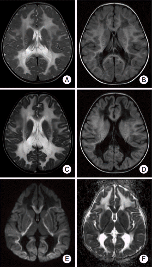
The neurological examination showed increased muscle tone and exaggerated deep tendon reflex in all limbs. The brain MRI showed extensive abnormal signal intensity at the periventricular and deep cerebral white matter relatively sparing subcortical U-fibers (Fig. 1A, 1B), which agreed with the leukoencephalopathy. She was first diagnosed of Krabbe disease, because only her β-galactosylcerebroside enzyme activity level was noted as having been moderately decreased from a quantitative analysis of several enzyme levels.
One month later, she showed rapid motor regression again, and could neither sit alone nor control her head movements after another mild viral upper respiratory infection had occurred. The neurological examination revealed that spasticity of her lower extremities had been noted to have worsened, and seizures were seen to have newly appeared. But at that time, the result of a Sanger sequencing of GALC gene to confirm the diagnosis of Krabbe disease showed no pathologic variants.
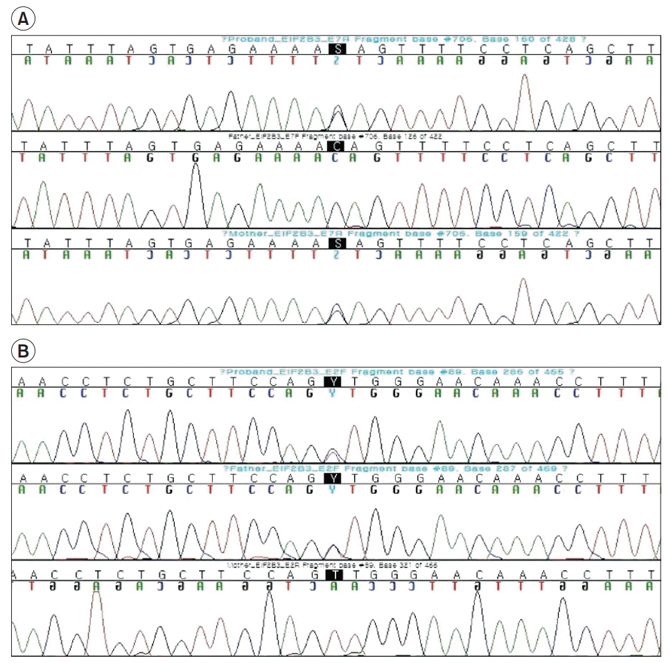
Then, a whole exome sequencing was performed using the Agilent SureSelect Target Enrichment protocol for Illumina paired-end sequencing library (ver. B.3, June 2015), together with the 200 ng input genomic DNA and the HiSeq 2500 platform (Illumina, San Diego, CA, USA) [5]. Finally, there were heterozygous missense variants which were found in the EIF2B3 (Fig. 2): a novel variant in exon 7 (c.706C>G/p.Gln236Glu) from the patient’s mother and 0.008% frequency (gnomAD_exome_ALL) reported variant in exon 2 (c.89T>C/p.Val30Ala) from the father. In addition, the patient’s parents were identified as heterozygous carriers for each variation which occurs in trans. This novel variation in exon 7 (c.706C>G/p.Gln236Glu) has not been reported in control databases, such as the 1000 Genomes Project, Exome Aggregation Consortium, gnomAD and the dbSNP Database. Moreover, both variations in EIF2B3 are located in conserved sequences across species (c.706C>G: GERP 5.93, phyloP20 1.048, c.89T>C: GERP 6.02, phyloP20 1.193) and have been predicted to be deleterious by several in silico analysis tools. Otherwise, we observed no other potential variants in the EIF2B1-5 gene sequences of the patient. Since any compound heterozygous mutation in eIF2B complex-encoding genes is known to independently cause VWM, we considered the two EIF2B3 variants (c.89T>C and c.706C>G) as conditions of pathogenic causes for VWM.
At that time, the patient was diagnosed of VWM and the patient suffered another deterioration at her age of 28 months, and could not even respond to any external stimuli as given during subsequent testing of the patient’s condition. An 8-month follow-up brain MRI finally showed progression of rarefaction and cystic degeneration of affected white matter, showing the same signal intensity of cerebrospinal fluid and a radiating stripe-like pattern suggesting remaining tissue strands (Fig. 1C, 1D). Also, a restricted diffusion was more evident adjacent to, or at the border of white matter than already cystic degenerated lesion (Fig. 1E, 1F). The restricted diffusion was found in an early and active demyelination stage, thus, this diffusion weighted image (DWI) findings suggest that the myelin degeneration is still going on and spreading to whole white matter, which is correlated with VWM [6,7]. In the early stage, however, it was difficult to differentiate from many other genetic disorders similarly affecting white matter because the pathognomonic characteristic of VWM only appeared in the patient’s follow-up MRIs. As a consequence, the radiologic diagnosis was delayed and made later than the genetic diagnosis.
A study reported that among more than the identified 200 different mutations in the EIF2B1-5 genes, there are approximately 120 pathogenic mutations for VWM, which are typically missense types and reside most frequently in EIF2B5 [4]. However, recently, several pathogenic EIF2B3 mutations have been reported to be more frequent in Chinese people than in Caucasians [8]. Although previous studies have demonstrated a correlation between the EIF2B3 mutation in Caucasians and the milder type with adult-onset compared to mutations in other subunits [9], several EIF2B3 mutations are being reported in Asians and Asian populations with early-onset and with more severe clinical courses, which are consistent with the findings seen in our patient [10]. Hence, it is imperative that we discover more pathogenic variants of EIF2B3 mutations and determine their clinical severities, particularly in the Asian populations.
A review of the MRI during VWM reveals a symmetric, diffuse abnormality of the cerebral white matter, which is known to begin as early as in the presymptomatic stage of the disease [1,4]. However, in the early stage, it is challenging to distinguish from other leukodystrophies that similarly affect the white matter by progressive demyelination. Likewise, our patient was misdiagnosed with Krabbe disease at first notice, because it was seen that a deep white matter lesion was primarily involved rather than a diffuse pattern. The pathognomonic cystic characteristics of our patient appeared only in the follow-up MRI scan, thereby delaying the radiological diagnosis, even later than the genetic diagnosis.
In conclusion, this case study reports EIF2B3 missense variants in a Korean patient with infantile-onset VWM, including a novel variant in exon 7. Notably, our patient was accurately diagnosed by the use of next-generation sequencing, much before the brain MRI scan revealed a definite cystic alteration in the affected white matter of the brain. Hence, unbiased genomic sequencing can clinically affect patient care and decision-making, regarding the identification and management of genetic disorders of leukoencephalopathy, especially in pediatric patients.
ACKNOWLEDGMENTS
We thank the family and the patient for their cooperation in this study. This work was supported by the National Research Foundation of Korea (NRF) grant funded by the Korea government (MSIT) (No. 2017R1C1B5014840).
Notes
Conceptualization: Ryu JS, Jang DH. Methodology: Ryu JS, Jang DH, Hyun SE. Formal analysis: Choi BS, Jang JH, Jeon I. Funding acquisition: Jang DH. Visualization: Jeon I, Jang JH, Hyun SE. Writing – original draft: Hyun SE. Writing – review and editing: Jang DH, Choi BS, Ryu JS. Approval of final manuscript: all authors.
REFERENCES
1. van der Knaap MS, Pronk JC, Scheper GC. Vanishing white matter disease. Lancet Neurol. 2006; 5:413–23.

2. van der Knaap MS, Leegwater PA, Konst AA, Visser A, Naidu S, Oudejans CB, et al. Mutations in each of the five subunits of translation initiation factor eIF2B can cause leukoencephalopathy with vanishing white matter. Ann Neurol. 2002; 51:264–70.

3. Fogli A, Schiffmann R, Hugendubler L, Combes P, Bertini E, Rodriguez D, et al. Decreased guanine nucleotide exchange factor activity in eIF2B-mutated patients. Eur J Hum Genet. 2004; 12:561–6.

4. Pronk JC, van Kollenburg B, Scheper GC, van der Knaap MS. Vanishing white matter disease: a review with focus on its genetics. Ment Retard Dev Disabil Res Rev. 2006; 12:123–8.

5. Oh JY, Do HJ, Lee S, Jang JH, Cho EH, Jang DH. Identification of a heterozygous SPG11 mutation by clinical exome sequencing in a patient with hereditary spastic paraplegia: a case report. Ann Rehabil Med. 2016; 40:1129–34.
6. Ding XQ, Bley A, Ohlenbusch A, Kohlschutter A, Fiehler J, Zhu W, et al. Imaging evidence of early brain tissue degeneration in patients with vanishing white matter disease: a multimodal MR study. J Magn Reson Imaging. 2012; 35:926–32.

7. van der Lei HD, Steenweg ME, Bugiani M, Pouwels PJ, Vent IM, Barkhof F, et al. Restricted diffusion in vanishing white matter. Arch Neurol. 2012; 69:723–7.

8. Zhang H, Dai L, Chen N, Zang L, Leng X, Du L, et al. Fifteen novel EIF2B1-5 mutations identified in Chinese children with leukoencephalopathy with vanishing white matter and a long term follow-up. PLoS One. 2015; 10:e0118001.
9. La Piana R, Vanderver A, van der Knaap M, Roux L, Tampieri D, Brais B, et al. Adult-onset vanishing white matter disease due to a novel EIF2B3 mutation. Arch Neurol. 2012; 69:765–68.

10. Gowda VK, Srinivasan VM, Bhat M, Benakappa A. Case of Childhood ataxia with central nervous system hypomyelination with a novel mutation in EIF2B3 gene. J Pediatr Neurosci. 2017; 12:196–8.
Fig. 1.
Magnetic resonance imaging of brain at the patient’s 20 months old (A, B) and 28 months old (C-F). The T2-weighted image (A, C) shows the diffuse hyperintense abnormality of deep cerebral white matter spreading to whole white matter. The FLAIR image (B, D) shows that more parts of the abnormal white matter have a low signal intensity in (D) than (B), similar to cerebrospinal fluid, indicative of progressive cystic degeneration. Within the abnormal white matter lesion, a pattern of radiating stripes is revealed suggestive of remaining tissue strands. The diffusion weighted image (E) and apparent diffusion coefficient map (F) show restricted diffusion around white matter lesion, indicative of ongoing spreading and active demyelinating process within the white matter.


 PDF
PDF Citation
 Print
Print


 XML Download