Journal List > Ann Rehabil Med > v.41(6) > 1150195

Chae, Park, Choi, Jung, Kim, Sohn, and Im: Rapid, Objective and Non-invasive Diagnosis of Sudomotor Dysfunction in Patients With Lower Extremity Dysesthesia: A Cross-Sectional Study

Abstract

Objective

To determine whether patients with lumbosacral (LS) radiculopathy and peripheral polyneuropathy (PPNP) exhibit sudomotor abnormalities and whether SUDOSCAN (Impeto Medical, Paris, France) can complement nerve conduction study (NCS) and electromyography (EMG).

Methods

Outpatients with lower extremity dysesthesia underwent electrophysiologic studies and SUDOSCAN. They were classified as normal (group A), LS radiculopathy (group B), or PPNP (group C). Pain severity was measured by the Michigan Neuropathy Screening Instrument (MNSI) and visual analogue scale (VAS). Demographic features, electrochemical skin conductance (ESC) values on hands and feet, and SUDOSCAN-risk scores were analyzed.

Results

There were no statistical differences in MNSI and VAS among the three groups. Feet-ESC and hands-ESC values in group C were lower than group A and B. SUDOSCAN-risk score in group B and C was higher than group A. With a cut-off at 48 microSiemens of feet-ESC, PPNP was detected with 57.1% sensitivity and 94.2% specificity (area under the curve [AUC]=0.780; 95% confidence interval [CI], 0646–0.915). With a SUDOSCAN-risk score cut-off at 29%, NCS and EMG abnormalities related to LS radiculopathy and PPNP were detected with 64.1% sensitivity and 84.2% specificity (AUC=0.750; 95% CI, 0.674–0.886).

Conclusion

SUDOSCAN can discriminate outpatients with abnormal electrophysiological findings and sudomotor dysfunction. This technology may be a complementary tool to NCS and EMG in outpatients with lower extremity dysesthesia.

INTRODUCTION

Dysesthesia accompanied by pain in the lower extremities is a common complaint in outpatient clinics. It can result from diverse causes that include overuse of back muscles, zygapophyseal joint degeneration, disc herniation, spine instability such as spondylolisthesis, spondylolysis, lumbosacral (LS) radiculopathy, and peripheral polyneuropathy (PPNP) [1]. LS radiculopathy and PPNP are frequently present in patients with lower extremity dysesthesia [23]. Physical examination and pain questionnaires may not be adequate for a diagnosis because of their subjective nature, which underestimates detection compared to other tools [4]. Imaging of only structural lumbar lesions can be insufficiently revealing and provides limited information in cases of LS radiculopathy. Nerve conduction study (NCS) and needle electromyography (EMG) can help diagnose LS radiculopathy and PPNP [5678]. However, these tools can only reveal abnormalities of the large fiber nerves and not those involving the small fibers [9]. Small fiber dysfunction has been linked to pain associated with diabetic neuropathy, chronic kidney disease, autonomic diseases, vasculitis and fibromyalgia [10].
There is a need for tests that are less time-consuming and less invasive than conventional tests. Also, assessment of small fibers through sudomotor evaluation might be helpful in evaluating dysesthesia of the lower extremities. The American Diabetes Association recommends evaluation of sudomotor function for early diagnosis of neuropathy and small fiber involvement in diabetes mellitus (DM) patients [11]. SUDOSCAN (Impeto Medical, Paris, France) is a novel quantitative assessment tool that provides index values of the thin, unmyelinated C fibers and assesses sudomotor function by evaluating the secretory function of the sweat glands. This device is a noninvasive, painless, and convenient tool that measures electrical conduction activity in the hands and feet by reflecting cholinergic neurotransmission of the sweat glands. Also its diagnostic validity to reflect small fiber function has already proven by past test with skin biopsy results [12131415]. However, the current literature on its use has been mainly limited to PPNP patients, and its application to other patients with lower extremity dysesthesia related to different causes have not been proven. Proper diagnosis of sudomotor dysfunction, which can lead to painful dysesthesia, is important [916]. The clinical utility and benefit of evaluating sudomotor dysfunction using SUDOSCAN among outpatients with mixed etiologies remain unclear.
In addition to patients with PPNP, there is evidence that both the sudomotor system and sympathetic responses are involved in patients with painful LS radiculopathy [161718]. Significant prolongation of sympathetic skin response (SSR) latency has been found in LS radiculopathy and failed back surgery syndrome (FBSS) [1617]. There is a considerable theoretical support to help detect sympathetic and sudomotor dysfunction in LS radiculopathy. However, no clinical studies have attempted to use it in patients with LS radiculopathy.
The primary study objective was to investigate whether SUDOSCAN could detect differences of its parameters in outpatients with dysesthesia of the lower extremities related to PPNP or LS radiculopathies who had been referred for NCS and EMG evaluations. The secondary objective was to assess the diagnostic validity of the obtained values related with sudomotor dysfunction values and determine the clinical utility compared with conventional NCS and needle EMG studies.

MATERIALS AND METHODS

Patient enrollment

This cross-sectional study was conducted from May 1, 2015 to August 31, 2015, and was based on outpatients who had been referred from an NCS and EMG clinic for dysesthesia and discomfort in the lower extremities to the EMG clinic at our institution. Preliminary clinical history taking, physical, and electrophysiological examinations were performed by physicians. Exclusion criteria were symptoms and signs confined to the upper extremity related to cervical radiculopathy or upper extremity entrapment neuropathies, prior amputation of arms or legs and implanted electrical devices, episodes of trauma, incomplete electrophysiologic studies, and previous LS radiculopathy or PPNP history (Fig. 1). The study protocols were approved by Bucheon St. Mary's Hospital, Catholic University of Korea local ethical boards of committee (HC15RISI0100).

Patient demographic characteristics

Clinical and demographic characteristics, including sex, age, pre-existing comorbidities like DM, lumbar spine surgery, alcohol consumption, body mass index (BMI), serum fasting glucose, serum uric acid, serum creatinine, total cholesterol (TC), total triglyceride (TG), low-density lipoprotein (LDL), and high-density lipoprotein (HDL) were retrieved from medical records. The Michigan Neuropathy Screening Instrument (MNSI) and visual analogue scale (VAS) were used to record the nature and severity of pain.

NCS and EMG

NCS was performed with a Viking Electromyography System (Nicolet-Viasys Biomedical Inc., Madison, WI, USA) with a setting of 20 Hz for the lower filter, 3 kHz for upper filter for sensory studies, and 5–10 kHz for motor studies. The pulse duration was with 0.1 ms. NCS tests included the sural sensory, superficial peroneal sensory, common peroneal motor, and tibial motor nerves. The minimum F-response latencies from both tibial nerves and H-reflex latencies from tibial nerve stimulation were collected. EMG needling was done by inserting the monopolar needle in selected lower extremity muscles and lumbar paraspinalis muscles from the L1-L2 to L5-S1 levels. All NCS and EMG tests were performed by experienced physicians with more than 3 years experience in the procedures. NCS results obtained from only one side were used in the statistical analysis.
Patients were stratified by electrophysiological results into normal (group A), LS radiculopathy (group B), and PPNP (group C) findings. Group A patients had normal latency range and amplitude on NCS without abnormal spontaneous activity in the sampled muscles. Group B patients had abnormal spontaneous activity, such as positive sharp waves and other denervation potentials, in the tested muscles with normal NCS parameters [1920]. Group C had reduced sensory nerve action potential (SNAP) amplitudes of the sural nerve (<10 µV) and abnormal peroneal (compound muscle action potential [CMAP] and/or F-wave) responses [21]. Patients were included in group C when reduced amplitudes of SNAPs in both sural nerves [22].

ESC and SUDOSCAN-risk score

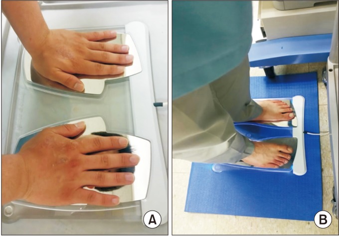
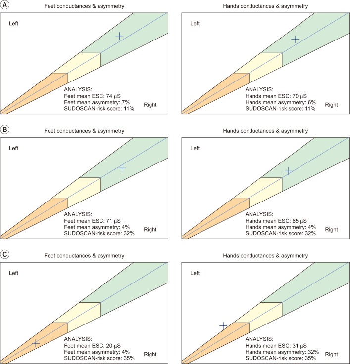
SUDOSCAN was performed on the same day by a separate examiner blind to patient's clinical symptoms and history before NCS and EMG evaluation. Sudomotor function was assessed as ESC values and SUDOSCAN-risk score using two pairs of electrodes for the feet and hands, and connected to a computer for data recording and modulation [11]. Subjects maintained contact with the electrodes for 3 minutes after their age, height, and weight had been recorded (Fig. 2). ESC values were calculated by analysis of electrochemical currents generated in response to low voltage stimulation and expressed in microsiemens (µS) [12] (Fig. 3).
Lower ESC values indicated dysfunction of the sweat glands. SUDOSCAN-risk scores were calculated automatically from ESC values, BMI, and age using an algorithm included in the device software. Scores are presented as percentages. Higher SUDOSCAN-risk scores have been related to increased risk of cardiac autonomic abnormalities. Both ESC values and SUDOSCAN-risk scores were displayed numerically with graphs on the device monitor.

Statistical analyses

Descriptive statistics are expressed numerically and as percentages for categorical variables, and as mean±standard deviation for continuous variables. Demographic characteristics and NCS values were compared by Kruskal-Wallis test followed by Man-Whitney U-test to identify differences among the three groups. Analysis of covariance (ANCOVA) followed by least significant difference (LSD) analysis were used to compare differences of SUDOSCAN-risk scores, hands-ESC, feet-ESC, and asymmetry of ESC on hands and feet, among the three groups with adjustment for age and DM history, which was confirmed showing normal distribution by Kolmogorov-Smirnov test [23].
Receiver operating characteristic (ROC) curves were calculated to estimate the effectiveness of foot-ESC values to predict PPNP and SUDOSCAN-risk scores in the abnormal electrophysiological findings of groups B and C. Spearman correlation coefficients were calculated to correlate NCS parameters (SNAP latencies and amplitude) with hands-ESC, feet-ESC, and SUDOSCAN-risk score. Two-sided p-values <0.05 were considered significant. Statistical analyses were performed using SPSS ver. 19.0 (SPSS Inc., Chicago, IL, USA).

RESULTS

Enrollment and demographic characteristics of outpatients

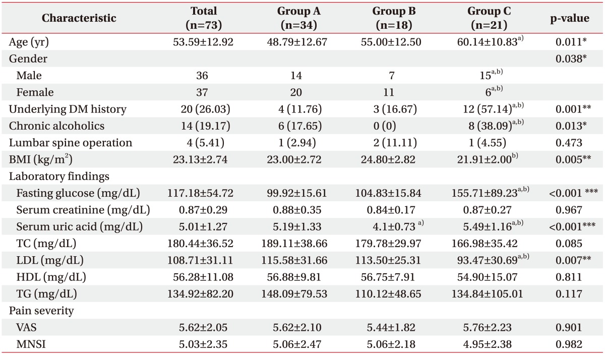
Out of the 401 patients who visited the electromyography clinic, 73 outpatients met the inclusion criteria. Thirty-four patients had normal NCS and EMG results (group A), 18 were diagnosed with LS radiculopathy (group B), and 21 with PPNP (group C). The patient demographic and clinical characteristics are shown in Table 1. There were no significant differences in MNSI and VAS scores among the three groups. There were significant baseline differences in age, sex, history of DM and alcohol abuse, BMI, serum fasting glucose, serum uric acid, and LDL level.

NCS and EMG results

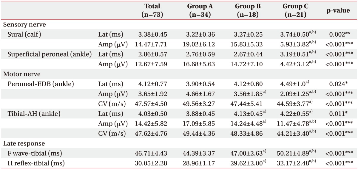
NCS did not reveal right to left side difference in any group. As expected, the latencies and amplitudes of SNAPs and CMAPs in all tested nerves of group C were significantly lower than in other groups (Table 2). The EMG profiles in group B showed denervation potentials in the lumbar paraspinalis muscles with multiple level involvement in 7 patients (38.9%), single level involvement in 8 (44.4%) patients, and in the lower limbs in 7 patients (38.9%). PPNP was associated with underlying alcohol consumption in 4 patients, uremia in 2, DM in 12, and was idiopathic in 3.
Low feet-ESCs were significantly correlated with decreased amplitudes of SNAPs of sural and superficial peroneal nerves (r=0.274 to 0.358, p<0.002 to 0.019). Hands-ESCs showed no correlation with amplitudes of sural and superficial peroneal nerves (r=0.133 to 0.198, p=0.126 to 0.263). High SUDOSCAN-risk score was significantly correlated with decreased SNAP amplitudes in both sural and superficial peroneal nerves (r=−0.352 to −0.415, p<0.001 to 0.002). ESC values and SUDOSCAN-risk score were not statistically correlated with latencies in both sural and superficial peroneal nerves.

Differences of SUDOSCAN values

ESC values and its diagnostic validity

Comparison of ESC values among the three groups was done using ANCOVA followed by LSD. The results indicated that feet-ESC values and asymmetry of group C were statistically different from those in groups A and B (Table 3).
Feet-ESC had a 57.1% sensitivity and 94.2% specificity at 48 µS to detect PPNP (95% confidence interval [CI], 0.646–0.915) with 80.0% positive predictive value and 84.5% negative predictive value. Hands-ESC had 71.4% sensitivity and 78.8% specificity at 55 µS (95% CI, 0.577–0.860) with a 57.7% positive predictive value and 87.2% negative predictive value.

SUDOSCAN-risk score

ANCOVA followed by LSD revealed significantly higher SUDOSCAN-risk scores were in groups B and C than group A (Table 3). The sensitivity and specificity of the SUDOSCAN-risk score for detecting abnormal electrophysiological findings (LS radiculopathy and PPNP) were 64.1% and 82.4%, respectively, at a cut-off on 29% and area under the curve (AUC) of 0.780 (95% CI, 0.674–0.886) with a positive predictive value of 80.65% and negative predictive value of 66.67%.

DISCUSSION

The results demonstrate that sudomotor evaluation through SUDOSCAN can help discriminate outpatients with dysesthesia of the lower extremities attributable to LS radiculopathy or PPNP. Although similar in clinical pain intensity, ESC values in the feet were significantly lower in patients diagnosed with PPNP, whereas the SUDOSCAN-risk score was higher in patients with LS radiculopathy or PPNP. The feet-ESC values at cut-off value of 48 µS, showed high specificity of 94.2% with 57.1% sensitivity to discriminate those with normal electrophysiologic findings and without PPNP. The sensitivity and specificity of the SUDOSCAN-risk score to detect abnormal electrophysiological findings (LS radiculopathy or PPNP) was 64.1% and 82.4%, respectively, at a 29% cut-off value. Overall, lower feet-ESC and higher SUDOSCAN-risk scores were found in those with PPNP, whereas higher SUDOSCAN-risk scores and normal feet-ESC values were found in those with LS radiculopathies. Findings of this study are relevant that SUDOSCAN may be a rapid, objective, and noninvasive method to evaluate sudomotor dysfunction and discriminate patients with LS radiculopathies and PPNP.
Recent studies have confirmed the diagnostic validity of ESC values, with a sensitivity of 73% to 78%, and specificity of 62% to 100% to screen PPNP in patients with diabetes as well as idiopathic and chemotherapy-induced PPNP [1213141524]. Previous studies evaluating the SUDOSCAN-risk score reported 65%–92% sensitivity and 49%–80% specificity to detect cardiac autonomic neuropathy [1425]. But, whether this score is helpful in radiculopathy has not been studied. Presently, we decided to determine whether patients with LS radiculopathies would have abnormal ESC values. The rationale was based on the possible association of sympathetic nerve dysfunction and LS radiculopathy previously suggested in studies that used SSR, an assessment tool similar to the SUDOSCAN, which evaluates sudomotor function [1161718].
Immunostaining revealed an increase of sympathetic fibers in radiculopathy lesions of dorsal root ganglia (DRG) at L4 and L5 in an animal model of spinal root-constriction [26]. Possible mechanisms involve inflammatory neuropeptides, such as substance P, calcitonin, and neuropeptide Y, which may spread from the spinal roots to the DRG at each spinal nerve level. Interactions in the vicinity of sinuvertebral nerves formed by sympathetic postganglionic and afferent sensory fibers [27] would result in sympathetic dysfunction in patients with LS radiculopathies. The abnormal stimuli would then be conveyed to the central nervous system from the dorsal horns of the spinal cord to generate hypersensitive autonomic reflex arcs resulting in low back and referred pain in the involved dermatomes [2728]. The role of abnormal autonomic reflexes in pain generation has also been confirmed in previous studies of the clinical benefits of spinal cord stimulators that help stabilize aberrant sympathetic responses in FBSS [2930].
Of clinical interest, our results revealed that SUDOSCAN-risk score in those with LS radiculopathy was increased although ESC values showed no differences compared to the normal group in NCS and EMG. This interesting result might be explained by past SSR studies that showed abnormal sudomotor functions in patients with lumbar lesions involving only they latency but not involving SSR amplitudes [1617]. SSR latency reflects peripheral C fiber function and conduction in long multi-neuronal pathways and SSR amplitude reflects the density of sweat glands [31]. Considering that ESC value reflects only the density of sweat glands, but not the latencies, involved in multi-neuronal pathways with the autonomic system, it is reasonable to conclude that the abnormal autonomic dysfunction involved in LS radiculopathies, related to delayed latencies of SSR, were reflected only in the SUDOSCAN-risk score . Therefore, the exception of abnormal SUDOSCAN-risk score with normal ESC values in patients with lumbar lesions may be a useful marker to detect abnormal sympathetic dysfunction. The clinical significance of SUDOSCAN-risk score abnormalities related with SSR latency is a topic that warrants more rigorous research.
The results can be applied meaningfully to clinical approaches to outpatients with dysesthesia in lower extremities. First, while previous studies focused on patients who had already been diagnosed with PPNP, this is the first study to evaluate SUDOSCAN in a cross-sectional study of outpatients with dysesthesia of the lower extremities. Our results show that sudomotor assessment may be used as a complimentary test to standard NCS and EMG studies. Secondly, previous studies on the diagnostic validity of ESC compared it with the vibration potential threshold, quantitative sudomotor axon reflex testing, and symptom scales like MNSI as references [32]. But, there is no report about SUDOSCAN with direct NCS and EMG results, which are the gold standard tools to detect PPNP [5678]. Our study validates ESC values and SUDOSCAN-risk scores to NCS and EMG values, and shows the high specificity diagnostic power in PPNP.
Unlike past studies on the diagnostic validity of SUDOSCAN, our results revealed lower levels of sensitivity but higher levels of specificity. This discrepancy may be attributable to different patient populations with the main objective to validate it in mostly outpatients with lower extremities dysesthesia due to more heterogenic causes. The 94.2% specificity level, which is indicative of low false negativity rates, is preferable to diagnose those without the disorder [3334]. High levels of specificity are necessary for the gold standard tests that may be expensive, time-consuming, and invasive such like NCS and EMG [35]. Because SUDOCAN exclusively assesses the small fibers function of the sweat glands and reflects sudomotor dysfunction, it cannot in principle reflect the large fibers dysfunction and denervation potentials obtained from NCS and needle EMG, and so cannot replace these two tests. However, considering the high level of specificity with the minimal false positive rate, it may help expedite the diagnosis of those who may show negative results in NCS and needle EMG in a rapid, low-cost, and non-invasive manner.
From this point of view, SUDOSCAN, which is a simple, quick, and non-invasive test, can be advantageously and effectively used in outpatient clinical situations. Especially, our SUDOSCAN-risk score results suggest the appropriate applicability of LS radiculopathy as well as PPNP. In addition, considering that normalized SSR latency in FBSS was reported in the use of SCS to stabilize sympathetic dysfunctions [29], the clinical utility of sudomotor evaluation as a measure of pain improvement or therapeutic response would be an interesting topic to investigate. Future studies of the relationship between LS radiculopathy and sudomotor dysfunction will be wise to expand the scope of the SUDOSCAN test.
There are several limiting factors of this study. This study is limited by its small size, despite statistically reliable and meaningful results. Small sample sizes can produce statistically unintentional errors because of heterogeneous patient populations. Second, group B with LS radiculopathy included uneven involved levels of distribution. Almost all subjects in group B were diagnosed as lower LS radiculopathy involving L4, L5 or/and S1, except for one involving L2 nerve root with 55 µS on feet ESC, which was lower by 10 points than the average in the LS radiculopathy group. Therefore, further study of sudomotor dysfunction using SUDOSCAN for LS radiculopathy undoubtedly needs to be done with a large population including upper lumbar radiculopathy. Third, patients with lower extremity dysesthesia may have the sudomotor abnormality even if they are not diagnosed with peripheral neuropathy or radiculopathy in NCS and EMG. Also, SUDOSCAN may not detect subtle sudomotor changes depending on the severity of radiculopathic lesions. Since group A as a control group did not exclude other causes of pain, it is necessary to compare group B with the healthy control group without pain symptom and patients with herniated disc lesion or spinal stenosis of group A.
In conclusion, this cross-sectional study is the first report of the usefulness of SUDOSCAN in outpatients with lower extremities dysesthesia including those with lumbar lesions and PPNP. Results obtained from preliminary SUDOSCAN evaluation may help complement NCS and EMG testing. Abnormal ESC and SUDOSCAN-risk scores indicate not only involvement of sudomotor in these patients but may also reflect the need of additional NCS and EMG to further confirm the presence of LS radiculopathy or PPNP. Normal ESC and SUDOSCAN-risk scores increase the probability of normal NCS and EMG results. Subsequent studies that expand the application of sudomotor dysfunction using SUDOSCAN testing and how this new assessment tool could complement with current NCS and EMG evaluations are warranted.

ACKNOWLEDGMENTS

We thank Ji Hyun Jun for her valuable contribution to statistical analysis and methods.

Notes

CONFLICT OF INTEREST: No potential conflict of interest relevant to this article was reported. This study was carried out with aid from Impeto Medical, but the authors did not receive any financial aid from the company during the study period.

References

1. Gunn CC. Radiculopathic pain: diagnosis and treatment of segmental irritation or sensitization. J Musculoskelet Pain. 1997; 5:119–134.
crossref
2. Spijker-Huiges A, Groenhof F, Winters JC, van Wijhe M, Groenier KH, van der Meer K. Radiating low back pain in general practice: incidence, prevalence, diagnosis, and long-term clinical course of illness. Scand J Prim Health Care. 2015; 33:27–32. PMID: 25693788.
crossref
3. Beith ID, Kemp A, Kenyon J, Prout M, Chestnut TJ. Identifying neuropathic back and leg pain: a cross-sectional study. Pain. 2011; 152:1511–1516. PMID: 21396774.
crossref
4. Mete T, Aydin Y, Saka M, Cinar Yavuz H, Bilen S, Yalcin Y, et al. Comparison of efficiencies of michigan neuropathy screening instrument, neurothesiometer, and electromyography for diagnosis of diabetic neuropathy. Int J Endocrinol. 2013; 2013:821745. PMID: 23818897.
crossref
5. Dyck PJ, O'Brien PC, Litchy WJ, Harper CM, Klein CJ, Dyck PJ. Monotonicity of nerve tests in diabetes: subclinical nerve dysfunction precedes diagnosis of polyneuropathy. Diabetes Care. 2005; 28:2192–2200. PMID: 16123489.
6. Weber F, Albert U. Electrodiagnostic examination of lumbosacral radiculopathies. Electromyogr Clin Neurophysiol. 2000; 40:231–236. PMID: 10907601.
7. Chouteau WL, Annaswamy TM, Bierner SM, Elliott AC, Figueroa I. Interrater reliability of needle electromyographic findings in lumbar radiculopathy. Am J Phys Med Rehabil. 2010; 89:561–569. PMID: 20567137.
crossref
8. Cho SC, Ferrante MA, Levin KH, Harmon RL, So YT. Utility of electrodiagnostic testing in evaluating patients with lumbosacral radiculopathy: an evidence-based review. Muscle Nerve. 2010; 42:276–282. PMID: 20658602.
crossref
9. Vinik AI, Nevoret ML, Casellini C. The new age of sudomotor function testing: a sensitive and specific biomarker for diagnosis, estimation of severity, monitoring progression, and regression in response to intervention. Front Endocrinol (Lausanne). 2015; 6:94. PMID: 26124748.
crossref
10. Chan AC, Wilder-Smith EP. Small fiber neuropathy: getting bigger! Muscle Nerve. 2016; 53:671–682. PMID: 26872938.
crossref
11. Yajnik CS, Kantikar VV, Pande AJ, Deslypere JP. Quick and simple evaluation of sudomotor function for screening of diabetic neuropathy. ISRN Endocrinol. 2012; 2012:103714. PMID: 22830040.
crossref
12. Casellini CM, Parson HK, Richardson MS, Nevoret ML, Vinik AI. Sudoscan, a noninvasive tool for detecting diabetic small fiber neuropathy and autonomic dysfunction. Diabetes Technol Ther. 2013; 15:948–953. PMID: 23889506.
crossref
13. Calvet JH, Dupin J, Winiecki H, Schwarz PE. Assessment of small fiber neuropathy through a quick, simple and non invasive method in a German diabetes outpatient clinic. Exp Clin Endocrinol Diabetes. 2013; 121:80–83. PMID: 23073917.
crossref
14. Selvarajah D, Cash T, Davies J, Sankar A, Rao G, Grieg M, et al. Sudoscan: a simple, rapid, and objective method with potential for screening for diabetic peripheral neuropathy. PLoS One. 2015; 10:e0138224. PMID: 26457582.
crossref
15. Smith AG, Lessard M, Reyna S, Doudova M, Singleton JR. The diagnostic utility of Sudoscan for distal symmetric peripheral neuropathy. J Diabetes Complications. 2014; 28:511–516. PMID: 24661818.
16. El-Badawy MA, El Mikkawy DM. Sympathetic dysfunction in patients with chronic low back pain and failed back surgery syndrome. Clin J Pain. 2016; 32:226–231. PMID: 25968450.
crossref
17. Erdem Tilki H, Coskun M, Unal Akdemir N, Incesu L. Axon count and sympathetic skin responses in lumbosacral radiculopathy. J Clin Neurol. 2014; 10:10–16. PMID: 24465257.
crossref
18. Andary MT, Stolov WC, Nutter PB. Sympathetic skin response in fifth lumbar and first sacral radiculopathies. Electromyogr Clin Neurophysiol. 1993; 33:91–99. PMID: 8383595.
19. Coster S, de Bruijn SF, Tavy DL. Diagnostic value of history, physical examination and needle electromyography in diagnosing lumbosacral radiculopathy. J Neurol. 2010; 257:332–337. PMID: 19763381.
crossref
20. Dumitru D, Amato A, Zwarts M. Electrodiagnostic medicine. 2nd ed. Philadelphia: Hanley & Belfus;2002.
21. England JD, Gronseth GS, Franklin G, Miller RG, Asbury AK, Carter GT, et al. Distal symmetric polyneuropathy: a definition for clinical research: report of the American Academy of Neurology, the American Association of Electrodiagnostic Medicine, and the American Academy of Physical Medicine and Rehabilitation. Neurology. 2005; 64:199–207. PMID: 15668414.
crossref
22. Kong X, Lesser EA, Potts FA, Gozani SN. Utilization of nerve conduction studies for the diagnosis of polyneuropathy in patients with diabetes: a retrospective analysis of a large patient series. J Diabetes Sci Technol. 2008; 2:268–274. PMID: 19885354.
crossref
23. Norman GR, Streiner DL. Biostatistics: the bare essentials. 3rd ed. Hamilton: BC Decker Inc;2008. p. 80.
24. Saad M, Psimaras D, Tafani C, Sallansonnet-Froment M, Calvet JH, Vilier A, et al. Quick, non-invasive and quantitative assessment of small fiber neuropathy in patients receiving chemotherapy. J Neurooncol. 2016; 127:373–380. PMID: 26749101.
crossref
25. Yajnik CS, Kantikar V, Pande A, Deslypere JP, Dupin J, Calvet JH, et al. Screening of cardiovascular autonomic neuropathy in patients with diabetes using non-invasive quick and simple assessment of sudomotor function. Diabetes Metab. 2013; 39:126–131. PMID: 23159130.
crossref
26. Chung K, Kim HJ, Na HS, Park MJ, Chung JM. Abnormalities of sympathetic innervation in the area of an injured peripheral nerve in a rat model of neuropathic pain. Neurosci Lett. 1993; 162:85–88. PMID: 7907173.
crossref
27. Bernini PM, Simeone FA. Reflex sympathetic dystrophy associated with low lumbar disc herniation. Spine (Phila Pa 1976). 1981; 6:180–184. PMID: 7280819.
crossref
28. Jinkins JR, Whittemore AR, Bradley WG. The anatomic basis of vertebrogenic pain and the autonomic syndrome associated with lumbar disk extrusion. AJR Am J Roentgenol. 1989; 152:1277–1289. PMID: 2718865.
crossref
29. de Andrade DC, Bendib B, Hattou M, Keravel Y, Nguyen JP, Lefaucheur JP. Neurophysiological assessment of spinal cord stimulation in failed back surgery syndrome. Pain. 2010; 150:485–491. PMID: 20591571.
crossref
30. North RB, Kidd DH, Farrokhi F, Piantadosi SA. Spinal cord stimulation versus repeated lumbosacral spine surgery for chronic pain: a randomized, controlled trial. Neurosurgery. 2005; 56:98–106.
crossref
31. Kimura J. Electrodiagnosis in disease of nerve and muscle: principles and practice. 3rd ed. New York: Oxford University Press;2001. p. 130.
32. Low PA. Evaluation of sudomotor function. Clin Neurophysiol. 2004; 115:1506–1513. PMID: 15203051.
crossref
33. Akobeng AK. Understanding diagnostic tests 1: sensitivity, specificity and predictive values. Acta Paediatr. 2007; 96:338–341. PMID: 17407452.
crossref
34. Bhandari M, Guyatt GH. How to appraise a diagnostic test. World J Surg. 2005; 29:561–566. PMID: 15827842.
crossref
35. Kanchanaraksa S. Evaluation of diagnostic and screening tests: validity and reliability. Baltimore: John Hopkins University;2008.
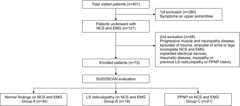
Fig. 1

Flow chart of participants. NCS, nerve conduction study; EMG, electromyography; LS, lumbosacral; PPNP, peripheral polyneuropathy.

arm-41-1028-g001
Fig. 2

Position of a subject undergoing a typical SUDOSCAN examination performed in standing position with full contact of electrodes on the hands (A) and the feet (B).

arm-41-1028-g002
Fig. 3

Representative figures from each group obtained from SUDOSCAN. (A) Results obtained in a patient who showed normal NCS and needle EMG findings with normal mean feet ESC with low SUDOSCAN risk score. (B) Results obtained in a patient with needle EMG findings compatible with lumbosacral radiculopathies with normal mean feet ESC but increased SUDOSCAN-risk score. (C) Results obtained in a patient with NCS findings compatible with peripheral polyneuropathy with low mean feet ESC and increased SUDOSCAN-risk score. NSC, nerve conduction study; EMG, electromyography; ESC, electrochemical skin conductance.

arm-41-1028-g003
Table 1

Baseline characteristics of the enrolled patients

arm-41-1028-i001

Values are presented as mean±standard deviation or number (%).

The patients were classified to the normal (group A), lumbosacral radiculopathy (group B), or peripheral polyneuropathy group (group C) according to nerve conduction study and electromyography examinations.

BMI, body mean index; DM, diabetes mellitus; TC, total cholesterol; LDL, low density lipoprotein; HDL, high-density lipoprotein; TG, total triglyceride; VAS, visual analogue scale; MNSI, Michigan Neuropathy Screening Instrument.

*p<0.05, **p<0.01, ***p<0.001.

a)p<0.05 in comparison with group A, b)p<0.05 in comparison with group B.

Table 2

Nerve conduction study findings in three groups (left side)

arm-41-1028-i002

Values are presented as mean±standard deviation.

The patients were classified to the normal (group A), lumbosacral radiculopathy (group B), or peripheral polyneuropathy group (group C) according to nerve conduction study and electromyography examinations.

EDB, extensor digitorum brevis; AH, abductor halluces; Lat, latency (motor, onset latency; sensory, peak latency); Amp, amplitude; CV, conduction velocity.

*p<0.05, **p<0.01, ***p<0.001.

a)p<0.05 in comparison with group A, b)p<0.05 in comparison with group B.

Table 3

ANCOVA analysis for the differences of ESC and SUDOSCAN risk score values in three groups

arm-41-1028-i003

Values are presented as mean±standard deviation.

The patients were classified to the normal (group A), lumbosacral radiculopathy (group B), or peripheral polyneuropathy group (group C) according to nerve conduction study and electromyography examinations.

ESC, electrochemical skin conductance.

*p<0.05, **p<0.01.

a)p<0.05 in comparison with group A, b)p<0.05 in comparison with group B.

TOOLS
Similar articles