Journal List > Ann Rehabil Med > v.40(5) > 1150156

Choi, Yu, Yoon, Kim, and Jeon: Relationship Between HbA1c and Complex Regional Pain Syndrome in Stroke Patients With Type 2 Diabetes Mellitus

Abstract

Objective

To investigate the relationship between glycosylated hemoglobin A (HbA1c) and complex regional pain syndrome (CRPS) in stroke patients with type 2 diabetes mellitus (T2DM).

Methods

A retrospective chart review was performed of stroke patients from January 2012 to December 2013. We reviewed 331 patients and included 200 in the analysis. We divided them into CRPS and non-CRPS groups and compared them by age, gender, stroke lesion, cause of stroke, duration of T2DM, HbA1c (%), National Institutes of Health Stroke Scale score, affected shoulder flexor muscle strength, Fugl-Meyer Assessment score, motricity index, Functional Independence Measure, Korean version of Modified Barthel Index, blood glucose level on admission day, duration from stroke onset to HbA1c check, and duration from stroke onset to three-phase bone scan for CRPS diagnosis. Thereafter, we classified the patients into five groups by HbA1c level (group 1, 5.0%–5.9%; group 2, 6.0%–6.9%; group 3, 7.0%–7.9%; group 4, 8.0%–8.9%; and group 5, 9.0%–9.9%) and we investigated the difference in CRPS prevalence between the two groups.

Results

Of the 200 patients, 108 were in the CRPS group and 92 were in the non-CRPS group. There were significant differences in HbA1c (p<0.05) between the two groups but no significant differences in any other factors. Across the five HbA1c groups, there were significant differences in CRPS prevalence (p<0.01); specifically, it increased as HbA1c increased.

Conclusion

This study suggests that higher HbA1c relates to higher CRPS prevalence and thus that uncontrolled blood glucose can affect CRPS occurrence in stroke patients with diabetes.

INTRODUCTION

Complex regional pain syndrome (CRPS) is characterized by disproportionate pain, allodynia, vasomotor changes, decreased range of motion, hyperalgesia, edema, and skin temperature or color change [1]. In the past, it was called reflux sympathetic dystrophy in acknowledgement that it was primarily an abnormal sympathetic nervous reflex that was involved. However, both sympathetically maintained pain and sympathetically independent pain have been found to exist, and thus the name was ultimately changed to CRPS by the International Association for the Study of Pain (IASP) in 1994. CRPS is divided into type I and type II by absence or presence of peripheral nerve injury [12]; type II generally occurs more frequently, but type I occurs more frequently in stroke patients [3].
CRPS occurs in the affected limbs of stroke patients in the recovery phase. Prevalence differs widely according to diagnostic standards and methods [4] and has been reported to vary from 12.5% to 70% depending on clinical signs and symptoms [56]. CRPS onset time is known to be from 1 to 5 months after stroke, and if it is not treated properly, there is a high possibility of irreversible change [4].
The pathophysiology of CRPS is unclear, but it is usually considered a multifactorial disease with clinical features that range from abnormal reflection to tissue damage causes such as neurogenic inflammation, nociceptive sensitization, vasomotor dysfunction, and maladaptive neuroplasticity [7].
Diabetes mellitus (DM) is a multisystem disease characterized by persistent hyperglycemia that has both acute and chronic biochemical and anatomical sequelae [8]. Glycosylated hemoglobin A (HbA1c) is an index of blood glucose control, and it reflects the average blood glucose level for 2 to 3 months [910]. Normal HbA1c is below 6%, but it exceeds 6% in diabetes and can increase up to 10%–12% [11].
A number of studies have reported that CRPS is one of the musculoskeletal complications in patients with DM [81213], but there are no studies on the relationship between HbA1c and CRPS prevalence. Thus, the purpose of this study was to investigate the relationship between HbA1c and CRPS prevalence in stroke patients with diabetes and to understand how uncontrolled hyperglycemia can affect CRPS occurrence in these patients.

MATERIALS AND METHODS

Subjects

We retrospectively reviewed the medical records of 331 patients who had been admitted to Jesus Hospital (Presbyterian Medical Center) from January 2012 to December 2013. The inclusion criteria for the study were (1) stroke confirmed through either cerebral infarction or cerebral hemorrhage and (2) a diagnosis of diabetes. The following were the exclusion criteria for this study: history of injury in an upper extremity, hyperthyroidism, hyperparathyroidism, peripheral neuropathy (including from diabetes), or previous CRPS.

Methods

Diagnosis of CRPS

The patients were diagnosed using the IASP's proposed revised CRPS clinical diagnostic criteria [14] and diagnosed by a physician in the Department of Nuclear Medicine based on a three-phase bone scan (TPBS) [15]. We divided the patients into two groups, with or without CRPS, and compared them by age, gender, stroke lesion, DM duration, HbA1c (%), National Institutes of Health Stroke Scale (NIHSS) score, affected shoulder flexor muscle strength, Fugl-Meyer Assessment (FMA) score, motricity index (MI), Functional Independence Measure (FIM), Korean version of Modified Barthel Index (K-MBI) score, blood glucose level on admission day, duration from stroke onset to HbA1c check, and duration from stroke onset to the TPBS for CRPS diagnosis (Fig. 1).

HbA1c

Among the 200 stroke patients with DM, we classified them into five groups by HbA1c value: group 1, 5.0%–5.9%; group 2, 6.0%–6.9%; group 3, 7.0%–7.9%; group 4, 8.0%–8.9%; and group 5, 9.0%–9.9%. Specifically, we compared the CRPS prevalence in each HbA1c group (Fig. 2).

Statistical analysis

Statistical analyses were performed using SPSS ver. 20.0 (IBM SPSS Inc., Armonk, NY, USA). We conducted independent t-tests to compare the CRPS and non-CRPS groups and chi-squared tests to compare CRPS prevalence in the five HbA1C groups. Statistical significance was set at p<0.05 for both sets of tests.

RESULTS

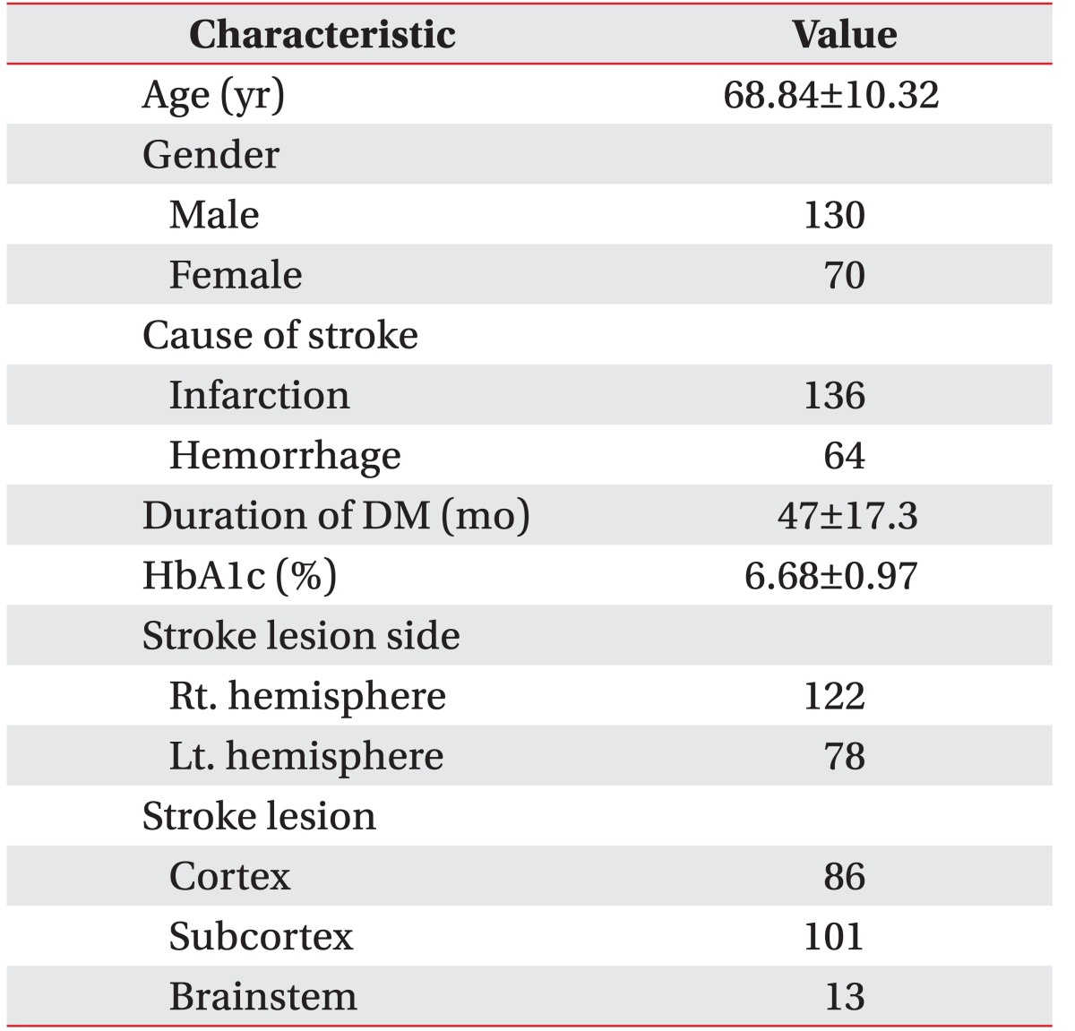
We analyzed the data from a total of 200 stroke patients with DM, 130 males and 70 females; mean patient age was 68.84±10.32 years. The patients comprised 136 with cerebral infarction and 64 with hemorrhagic stroke; mean DM duration was 47±17.3 months and mean HbA1c was 6.68±0.97% (Table 1).
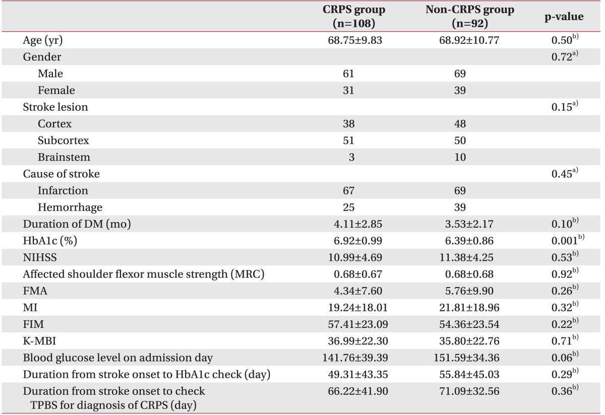
One hundred eight of the 200 subjects were diagnosed with CRPS, with the remaining 92 comprising the non-CRPS group. There were significant differences in HbA1c (p<0.05) but no significant differences between the CRPS groups in age, gender, stroke lesion, cause of stroke, DM duration, NIHSS score, affected shoulder flexor muscle strength, FMA score, MI, FIM, K-MBI score, blood glucose level duration from stroke onset to HbA1c check, or duration from stroke onset to the TPBS for CRPS diagnosis (p>0.05) (Table 2).
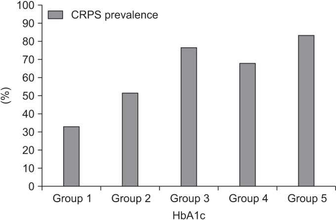
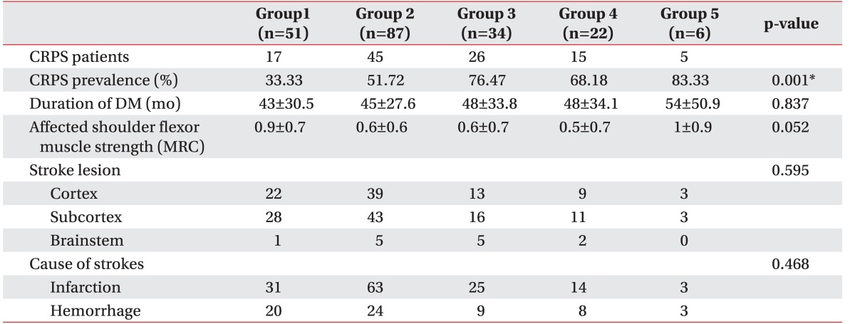
By HbA1c level, CRPS prevalence differed significantly (p<0.01)—group 1, 33%; group 2, 51%; group 3, 76%; group 4, 68.18%; and group 5, 83.33% (Table 3); that is, as HbA1c increased, CRPS prevalence increased as well (Fig. 3).

DISCUSSION

DM is considered highly related to CRPS occurrence [81213], and thus we attempted to investigate how uncontrolled hyperglycemia can affect CRPS occurrence in stroke patients with diabetes by analyzing the relationship between HbA1c and CRPS prevalence in these patients. In this study, as HbA1c values increased, CRPS prevalence increased as well, and thus we consider that maintaining adequate blood glucose levels is important for reducing CRPS occurrence in stroke patients with diabetes.
CRPS is characterized by severe pain, allodynia, hyperalgesia, and motor and autonomic signs and symptoms [7]. With regard to the pain itself, Calder et al. [16] and Van der Laan and Goris [17] reported that inflammatory response is the primary pain mechanism in CRPS patients. In addition, the activation of skin nociceptor that we can see in these patients induces retrograde depolarization of primary afferent nerves with small diameters, increasing the secretion of neuropeptides such as substance P and calcitonin gene-related peptide (CGRP) in the sensory endings of the skin [18]. These neuropeptides evoke vasodilatation and protein extravasations in the tissue, and the resulting signs such as reddening, warming, and edema are called neurogenic inflammation [18]; most inflammatory changes from CRPS are mediated by CGRP and substance P. Serum concentrations of both of these substances are higher in CRPS patients than in healthy control individuals [1920].
The concentrations of cytokines also show differences. In skin biopsy results, the concentrations of tumor necrosis factor-alpha (TNF-α) and interleukin-6 (IL-6) were much higher in upper extremities affected by CRPS than in normal upper extremities [2122]. In early-stage CRPS (3 months on average), water-soluble TNF receptor and pro-inflammatory cytokines (TNF), IL-1, and IL-8 increased in blood serum whereas anti-inflammatory cytokines (IL-4, IL-10, and transforming growth factor-beta1 [TGF-β1]) decreased [2324]. It was reported that these pro-inflammatory cytokines caused sensitization and hyperalgesia [25], which indicates that neurological inflammatory response plays an important role in the occurrence of CRPS.
Type 2 diabetes mellitus (T2DM) is an inflammatory disorder that develops when there is a pathologic problem in the immune system [26], and it features high blood concentrations of congenital immune mediators due to the occurrence of pathologic consequences [927]. It has been reported that compared with control groups, the serum levels of pro-inflammatory cytokines (IL-1β, IL-6 and TNF-α), sialic acid and C-reactive protein, and malondialdehyde were significantly higher in T2DM patients [92627282930313233]. As glycemic control in these patients worsens (HbA1c >6.5%), serum TNF-α appears to increase proportionally [28].
These secretions of pro-inflammatory cytokines in diabetes play an important role and could be associated with neurogenic inflammation and vasomotor dysfunction in CRPS. Classically, CRPS is subdivided into three phases (acute, atrophic, and dystrophic). Among these, the primary acute inflammatory phase can last for weeks up to 6 months and is characterized by painful range of motion, edema, warmth, and erythema of the hand and wrist [34353637]. Therefore, we consider that worsening glycemic control is associated with the acute inflammatory phase of CRPS.
According to the result of this study, the higher the HbA1c in stroke patients with diabetes, the higher their CRPS prevalence. HbA1c values reflect hyperglycemia, during which pro-inflammatory cytokines and acute-phase reactive proteins increase, resulting in increased neuroinflammatory reactions and CRPS.
However, our study was limited by a number of important factors. The study was cross-sectional and thus it was limited to inferring causal relationships. Second, shoulder joint subluxation, which is known a factor in CRPS occurrence, was not included in the study; instead we substituted shoulder muscle flexor strength and motricity index, neither of which controlled for the shoulder subluxation. Finally, the number of CRPS patients was small for the comparison between CRPS prevalence and HbA1c. These limitations suggest possible directions for future study. Furthermore, long-term follow-up research on the relationship between decreased HbA1c and improved CRPS is considered necessary. Then, we can suggest physiological grounds for any causal relationships between blood glucose level and CRPS through studies about improved CRPS symptoms or reduced absorption ratio on TPBS with lower HbA1c.
In this study, we concluded that uncontrolled hyperglycemia is related to CRPS occurrence in stroke patients with DM by comparing their HbA1c values with their CRPS prevalence; because CRPS prevalence increased as HbA1c increased, we considered that uncontrolled hyperglycemia affects CRPS occurrence. Therefore, it is necessary to maintain adequate blood glucose control in stroke patients with diabetes in addition to providing range of motion exercises and medical, physical, and occupational therapy.

Notes

CONFLICT OF INTEREST: No potential conflict of interest relevant to this article is reported.

References

1. Stanton-Hicks M, Janig W, Hassenbusch S, Haddox JD, Boas R, Wilson P. Reflex sympathetic dystrophy: changing concepts and taxonomy. Pain. 1995; 63:127–133. PMID: 8577483.
crossref
2. Merskey H, Bogduk N. Classification of chronic pain: descriptions of chronic pain syndromes and definitions of pain terms. Seattle: IASP Press;1994.
3. Pertoldi S, Di Benedetto P. Shoulder-hand syndrome after stroke: a complex regional pain syndrome. Eura Medicophys. 2005; 41:283–292. PMID: 16474282.
4. Tepperman PS, Greyson ND, Hilbert L, Jimenez J, Williams JI. Reflex sympathetic dystrophy in hemiplegia. Arch Phys Med Rehabil. 1984; 65:442–447. PMID: 6466074.
5. Cheng PT, Hong CZ. Prediction of reflex sympathetic dystrophy in hemiplegic patients by electromyographic study. Stroke. 1995; 26:2277–2280. PMID: 7491650.
crossref
6. Daviet JC, Preux PM, Salle JY, Lebreton F, Munoz M, Dudognon P, et al. Clinical factors in the prognosis of complex regional pain syndrome type I after stroke: a prospective study. Am J Phys Med Rehabil. 2002; 81:34–39. PMID: 11807329.
7. Marinus J, Moseley GL, Birklein F, Baron R, Maihofner C, Kingery WS, et al. Clinical features and pathophysiology of complex regional pain syndrome. Lancet Neurol. 2011; 10:637–648. PMID: 21683929.
crossref
8. Wyatt LH, Ferrance RJ. The musculoskeletal effects of diabetes mellitus. J Can Chiropr Assoc. 2006; 50:43–50. PMID: 17549168.
9. Pickup JC, Crook MA. Is type II diabetes mellitus a disease of the innate immune system? Diabetologia. 1998; 41:1241–1248. PMID: 9794114.
crossref
10. Malkani S, Mordes JP. Implications of using hemoglobin A1C for diagnosing diabetes mellitus. Am J Med. 2011; 124:395–401. PMID: 21531226.
crossref
11. Graf RJ, Halter JB, Pfeifer MA, Halar E, Brozovich F, Porte D Jr. Glycemic control and nerve conduction abnormalities in non-insulin-dependent diabetic subjects. Ann Intern Med. 1981; 94:307–311. PMID: 7013592.
crossref
12. Marshall AT, Crisp AJ. Reflex sympathetic dystrophy. Rheumatology (Oxford). 2000; 39:692–695. PMID: 10908684.
crossref
13. Kim RP. The musculoskeletal complications of diabetes. Curr Diab Rep. 2002; 2:49–52. PMID: 12643122.
crossref
14. Harden RN, Bruehl S, Stanton-Hicks M, Wilson PR. Proposed new diagnostic criteria for complex regional pain syndrome. Pain Med. 2007; 8:326–331. PMID: 17610454.
crossref
15. Stanton-Hicks MD, Burton AW, Bruehl SP, Carr DB, Harden RN, Hassenbusch SJ, et al. An updated interdisciplinary clinical pathway for CRPS: report of an expert panel. Pain Pract. 2002; 2:1–16. PMID: 17134466.
crossref
16. Calder JS, Holten I, McAllister RM. Evidence for immune system involvement in reflex sympathetic dystrophy. J Hand Surg Br. 1998; 23:147–150. PMID: 9607647.
crossref
17. van der Laan L, Goris RJ. Reflex sympathetic dystrophy: an exaggerated regional inflammatory response. Hand Clin. 1997; 13:373–385. PMID: 9279543.
18. Holzer P. Neurogenic vasodilatation and plasma leakage in the skin. Gen Pharmacol. 1998; 30:5–11. PMID: 9457475.
crossref
19. Birklein F, Schmelz M, Schifter S, Weber M. The important role of neuropeptides in complex regional pain syndrome. Neurology. 2001; 57:2179–2184. PMID: 11756594.
crossref
20. Schinkel C, Gaertner A, Zaspel J, Zedler S, Faist E, Schuermann M. Inflammatory mediators are altered in the acute phase of posttraumatic complex regional pain syndrome. Clin J Pain. 2006; 22:235–239. PMID: 16514322.
crossref
21. Huygen FJ, Ramdhani N, van Toorenenbergen A, Klein J, Zijlstra FJ. Mast cells are involved in inflammatory reactions during Complex Regional Pain Syndrome type 1. Immunol Lett. 2004; 91:147–154. PMID: 15019283.
crossref
22. Groeneweg JG, Huygen FJ, Heijmans-Antonissen C, Niehof S, Zijlstra FJ. Increased endothelin-1 and diminished nitric oxide levels in blister fluids of patients with intermediate cold type complex regional pain syndrome type 1. BMC Musculoskelet Disord. 2006; 7:91. PMID: 17137491.
crossref
23. Uceyler N, Eberle T, Rolke R, Birklein F, Sommer C. Differential expression patterns of cytokines in complex regional pain syndrome. Pain. 2007; 132:195–205. PMID: 17890011.
24. Wesseldijk F, Huygen FJ, Heijmans-Antonissen C, Niehof SP, Zijlstra FJ. Six years follow-up of the levels of TNF-alpha and IL-6 in patients with complex regional pain syndrome type 1. Mediators Inflamm. 2008; 2008:469439. PMID: 18596918.
25. Parkitny L, McAuley JH, Di Pietro F, Stanton TR, O'Connell NE, Marinus J, et al. Inflammation in complex regional pain syndrome: a systematic review and meta-analysis. Neurology. 2013; 80:106–117. PMID: 23267031.
crossref
26. Pickup JC. Inflammation and activated innate immunity in the pathogenesis of type 2 diabetes. Diabetes Care. 2004; 27:813–823. PMID: 14988310.
crossref
27. Crook MA, Tutt P, Pickup JC. Elevated serum sialic acid concentration in NIDDM and its relationship to blood pressure and retinopathy. Diabetes Care. 1993; 16:57–60. PMID: 8422833.
crossref
28. Mirza S, Hossain M, Mathews C, Martinez P, Pino P, Gay JL, et al. Type 2-diabetes is associated with elevated levels of TNF-alpha, IL-6 and adiponectin and low levels of leptin in a population of Mexican Americans: a cross-sectional study. Cytokine. 2012; 57:136–142. PMID: 22035595.
crossref
29. Alexandraki KI, Piperi C, Ziakas PD, Apostolopoulos NV, Makrilakis K, Syriou V, et al. Cytokine secretion in long-standing diabetes mellitus type 1 and 2: associations with low-grade systemic inflammation. J Clin Immunol. 2008; 28:314–321. PMID: 18224429.
crossref
30. Bastard JP, Pieroni L, Hainque B. Relationship between plasma plasminogen activator inhibitor 1 and insulin resistance. Diabetes Metab Res Rev. 2000; 16:192–201. PMID: 10867719.
crossref
31. Donath MY, Shoelson SE. Type 2 diabetes as an inflammatory disease. Nat Rev Immunol. 2011; 11:98–107. PMID: 21233852.
crossref
32. Pickup JC, Chusney GD, Thomas SM, Burt D. Plasma interleukin-6, tumour necrosis factor alpha and blood cytokine production in type 2 diabetes. Life Sci. 2000; 67:291–300. PMID: 10983873.
33. Dandona P, Aljada A, Chaudhuri A, Bandyopadhyay A. The potential influence of inflammation and insulin resistance on the pathogenesis and treatment of atherosclerosis-related complications in type 2 diabetes. J Clin Endocrinol Metab. 2003; 88:2422–2429. PMID: 12788837.
crossref
34. Veldman PH, Reynen HM, Arntz IE, Goris RJ. Signs and symptoms of reflex sympathetic dystrophy: prospective study of 829 patients. Lancet. 1993; 342:1012–1016. PMID: 8105263.
crossref
35. Kozin F. Reflex sympathetic dystrophy syndrome: a review. Clin Exp Rheumatol. 1992; 10:401–409. PMID: 1395224.
36. Schwartzman RJ, McLellan TL. Reflex sympathetic dystrophy: a review. Arch Neurol. 1987; 44:555–561. PMID: 3495254.
37. Shelton RM, Lewis CW. Reflex sympathetic dystrophy: a review. J Am Acad Dermatol. 1990; 22:513–520. PMID: 2097997.
crossref
Fig. 1

Flow diagram of patients included in this study by CRPS versus non-CRPS groups. DM, diabetes mellitus; CRPS, complex regional pain syndrome; PPN, peripheral polyneuropathy.

arm-40-779-g001
Fig. 2

Flow diagram of the five HbA1c groups. DM, diabetes mellitus; HbA1c, glycosylated hemoglobin A.

arm-40-779-g002
Fig. 3

CRPS prevalence by HbA1c; as HbA1c increased, CRPS prevalence increased. CRPS, complex regional pain syndrome; HbA1c, glycosylated hemoglobin A.

arm-40-779-g003
Table 1

General characteristics of subjects (n=200)

arm-40-779-i001

Values are presented as mean±standard deviation or number of subjects.

DM, diabetes mellitus; HbA1c, glycosylated hemoglobin A.

Table 2

Comparisons between the CPRS and non-CRPS groups

arm-40-779-i002

Values are presented as mean±standard deviation or number of subjects.

DM, diabetes mellitus; HbA1c, glycosylated hemoglobin A; NIHSS, National Institutes of Health Stroke Scale; MRC, Medical Research Council; FMA, Fugl-Meyer Assessment scale; MI, motricity index; FIM, Functional Independence Measure; K-MBI, Korean version of Modified Barthel Index; TPBS, three-phase bone scan; CRPS, complex regional pain syndrome.

Statistically significant as p<0.05. a)Chi-square test, b)independent t-test.

Table 3

Correlations between CRPS prevalence, age, DM duration, affected shoulder flexor muscle strength, stroke lesion, and stroke cause by HbA1c

arm-40-779-i003

Values are presented as number of subjects or mean±standard deviation.

CRPS, complex regional pain syndrome; MRC, Medical Research Council; FMA, Fugl-Meyer Assessment scale; MI, motricity index; NIHSS, National Institutes of Health Stroke Scale score; FIM, Functional Independence Measure; K-MBI, Korean version of Modified Barthel Index.

HbA1c level: group 1, 5.0%–5.9%; group 2, 6.0%–6.9%, group 3, 7.0%–7.9%; group 4, 8.0%–8.9%; group 5, 9.0%–9.9%. *p<0.05 by chi-square test.

TOOLS
Similar articles