Abstract
Objective
To investigate the predictive value of enhanced-magnetic resonance imaging (MRI) and fluoroscopic factors regarding the effects of transforaminal epidural steroid injections (TFESIs) in low back pain (LBP) patients with lumbosacral radiating pain.
Methods
A total of 51 patients who had LBP with radiating pain were recruited between January 2011 and December 2012. The patient data were classified into the two groups ‘favorable group’ and ‘non-favorable group’ after 2 weeks of follow-up results. The favorable group was defined as those with a 50%, or more, reduction of pain severity according to the visual analogue scale (VAS) for back or leg pain. The clinical and radiological data were collected for univariate and multivariate analyses to determine the predictors of the effectiveness of TFESIs between the two groups.
Results
According to the back or the leg favorable-VAS group, the univariate analysis revealed that the corticosteroid approach for the enhanced nerve root, the proportion of the proximal flow, and the contrast dispersion of epidurography are respectively statistically significant relative to the other factors. Lastly, the multiple logistic regression analysis showed a significant association between the corticosteroid approach and the enhanced nerve root in the favorable VAS group.
Low back pain (LBP) is extremely common, whereby a lifetime prevalence as high as 80% has been recorded [123]. In some patients, the pain associated with an initial acute episode does not resolve and persists for more than 3 months, leading to the development of chronic LBP [4]. Most chronic LBP patients do not respond to medication or physical therapy, with complaints of continuous LBP, and it is eventually the cause of muscle weakness in the low back area that is secondary to a lack of exercise.
The use of lumbosacral transforaminal epidural steroid injections (TFESIs), which are an alternative diagnostic and therapeutic spinal intervention for LBP, radicular pain, and spinal stenosis, have increased over the past few years [5]. Even though a great amount of debate has occurred regarding the evidence of lumbosacral TFESIs, lumbosacral TFESIs are a helpful treatment for lumbosacral radicular pain [367].
Most patients can benefit from lumbosacral TFESIs, but not all patients receive the benefits from such methods. Because a lot of factors contribute to the effectiveness of TFESIs, some researchers have studied the predictors of the effectiveness of TFESIs [89]. One study revealed clinical data such as an initially higher results for the visual analogue scale (VAS) and the McGill Pain Inventory, a lack of worsening pain during walking, and a positive femoral stretch test for the prediction of the pain reduction after TFESIs [8]. The other study showed low-grade nerve root compression with a prognostic value after the administration of TFESIs using magnetic resonance imaging (MRI) [9].
So far, only a few studies have used radiological parameters including MRI and clinical parameters to predict the effectiveness of TFESIs in terms of lumbosacral radiating pain. Also, the radiological factors for the predicting of successful TFESI outcomes has not been well investigated. To the best of the authors’ knowledge, the relationships between TFESIs and radiological factors, including contrast-enhanced MRI and fluoroscopic findings, have never been investigated.
The aim of the present study is a determination of the favorable-outcome predictive value for radiological factors such as MRI or fluoroscopic variables in terms of lumbosacral radiating pain.
This study is a retrospective study conducted from January 2011 to December 2012. A total of 67 patients who had been complaining of LBP with radiating pain are included in this analysis.
The patient-inclusion criteria were as follows: (1) those with a diagnosed herniated nucleus pulposus or spinal stenosis from a physical examination and imaging studies; (2) those who received spinal MRI with/without an enhancement; and (3) those who had been given first time TFESIs. The exclusion criteria were as follows: (1) patient’s refusal of spinal MRI study; (2) patient who had not received TFESIs; (3) patient who had an allergic response to steroids or lidocaine; (4) follow-up loss; (5) patient who had been given physical therapy; (6) patient who had received a surgical operation; and (7) individuals with previous or current respiratory, cardiovascular, neurological, or psychological diseases. The study protocol was approved by the Institutional Review Board (IRB No. BD2014-081), and all of the participants were provided the purpose of the study on the written informed consent.
The medical records of the patients who met the inclusion and exclusion criteria were reviewed, and the clinical and radiological parameters were subsequently set.
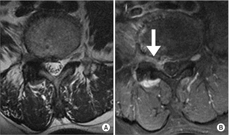
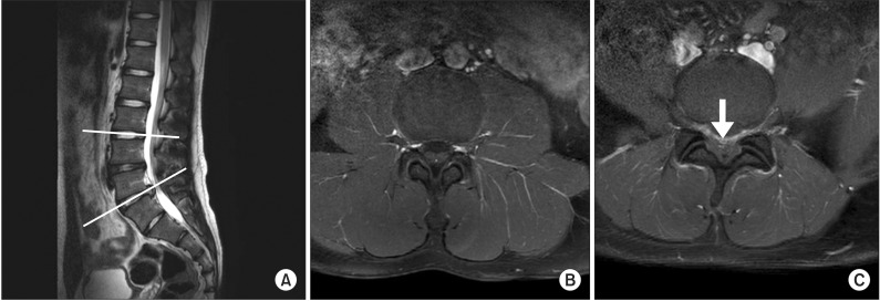
The authors assessed the radiological features including MRI to arrange the level of the herniated nucleus pulposus (HNP), the type of HNP, the grade of the foraminal stenosis, the location of the herniated intervertebral disc, the existence of the contrast-enhanced nerve root, and the block level of the nerve root. The 1.5T MRI was used, and it included sagittal and axial T1- and T2-weighted gadolinium-enhanced images. The types of HNP were categorized as bulging, protrusion, extrusion, and sequestration [10]. There are four grades according to Wildermuth’s MRI grading system for the lumbar spine foraminal stenosis, as follows: (1) grade 0 (normal), normal dorsolateral border of the intervertebral disc and normal form at the foraminal epidural fat; (2) grade 1 (slight foraminal stenosis), deformity of the epidural fat, with the remaining fat still completely surrounding the exiting nerve; (3) grade 2 (marked foraminal stenosis), epidural fat only partially surrounding the nerve root; and (4) grade 3 (advanced foraminal stenosis), obliteration of the epidural fat [111213]. An enhancement of a nerve root in the MRI was firstly evaluated by one of the authors, who was a physician trained in musculoskeletal pain and intervention, from an analysis of the pre- and post-contrast images stored at the same level settings. And then, the nerve root and the epidural enhancement were investigated by comparing the same nerve root of the pre-contrast images with the unaffected side or the newly detected root or epidural enhancement in the post-contrast images, which was differentiated from the opposite nerve root of the same level (Figs. 1, 2). This is similar to the method of the previously described literature [14]. According to the patient’s clinical presentation with the MRI studies, the authors decided the suitable injection level. When the patient had multilevel HNP, the authors determined the single target root level depending on the severity. According to the presence of the nerve root enhancement in the MRI, two groups were formed as follows: enhancement and non-enhancement. The authors injected an enhanced nerve root level in the enhancement group. In the case of the non-enhancement group, the patients were given an injection according to the severity of their symptoms and the imaging correlation.
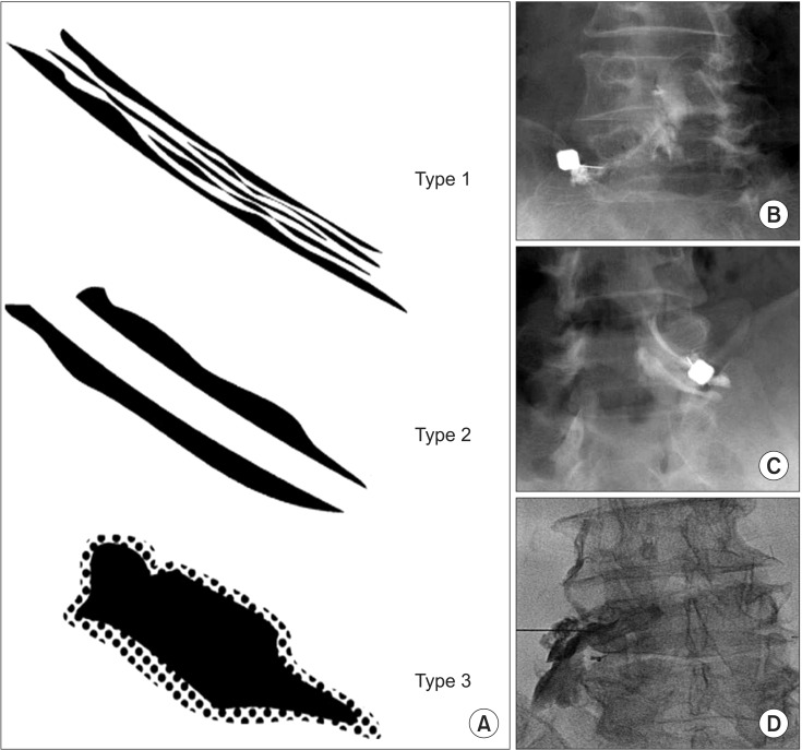
In addition to the MRI findings, the fluoroscopic findings such as the contrast dispersion of the epidurography was defined in the previous study [1516], the proportion of the proximal-flow pattern, the number of the flow level distribution, and the drug for the enhanced nerve root found in the MRI were also evaluated. The contrast distribution of the epidurographic findings were categorized into three distinct types, as defined by Pfirrmann et al. [16], as follows: intraepineural, extraepineural, and paraneural patterns (Fig. 3). These three contrast patterns are as follows: (1) the intraepineural pattern appears to be a tubular outline of the nerve root or within the nerve root sheath, (2) the extraepineural pattern was defined as a contrast filling around the nerve root sheath that was like a tubular filling defect, and (3) the paraneural pattern is explained by a cloudlike appearance, which is located beneath the pedicle in the lateral neural foramen and was not delineated by the contrast. The contrast flow patterns were evaluated among the groups according to whether the extent of the proximal was greater than 75%, 75% to 25%, or less than 25%, based on the final needle-tip position in the anterior-posterior fluoroscopic view; that is, the authors evaluated the fluoroscopic images for the spreading of the injectate to either the superior and/or inferior level after the needle was placed. Before the corticosteroid was administered to the target, the contrast material was injected for the epidural flow pattern. In this article, a volume of 0.5 mL was first injected, and an additional 0.5 mL contrast was injected as a second injection for the checking of the number of the distributed levels. It was confirmed whether the reach of the injected contrast material is more proximal to the target nerve root under fluoroscopy when the patients had the enhanced nerve root lesion in the contrast-enhanced MRI.
The clinical parameters were as follows: (1) duration of LBP, (2) duration of radiating pain, (3) intensity of low back and radiating pains, and (4) presence of sensory and motor abnormalities. The VAS, a scale from 0 (no pain) to 10 (worst pain imaginable), was used to evaluate the pain at the time of the initial visit and again 2 weeks after the intervention according to the back and leg pains, respectively. Because of the minimal therapeutic effect of the TFESIs, the pain intensity was evaluated 2 weeks after the injection [16]. The collected patient data were classified into two groups according to the VAS change before and at 2 weeks after the intervention. According to the back- or leg-pain severity change from the VAS, the patients were divided into the ‘favorable-VAS back or leg TFESIs group’ and the ‘non-favorable-VAS back or leg TFESIs group,’ respectively. The favorable TFESIs group represents a VAS decline of at least 50% at 2 weeks after the intervention.
All of the patients were given TFESIs using fluoroscopic guidance. The patient was positioned on a fluoroscopic table with a cushion under the lower abdomen in a C-arm room. The level of interest was prepped in a sterile manner, and the C-arm was tilted obliquely and rotated into an arrangement to identify the target root. After a 22-gauge, 3.5-in-length spinal needle was placed at the appropriate level, the needle was advanced toward the target nerve root. The needle position was confirmed using the axial and lateral fluoroscopic views. Before the corticosteroid injection, 0.5 mL of the contrast dye was injected initially, and the X-ray image was stored. After the 0.5 mL of contrast dye (a total dose of 1.0 mL) was administered, the image was stored consecutively to check for the epidural flow distribution. Lastly, all of the patients were given unilateral single level procedures and 20 mg (40 mg/mL) of triamcinolone and 1.5 mL of 1% lidocaine with 1,500 IU of hyaluronidase H-lase for each treatment. And there were no acute complications after the TFESIs in this study.
Primary comparisons were performed between the favorable and non-favorable groups. The data were expressed as the mean±standard deviation or as percentages. The categorical variables were compared using the χ2 and the Fisher exact test. The continuous variables were compared using the two-tailed unpaired Student t-test for the variables with normal distributions, and the two-tailed Mann-Whitney U test was used for the variables with non-normal distributions. The multivariate logistic regression analysis was then performed. The logistic regression coefficients were used to assess the odds ratios for each independent variable. The entire statistical analysis was performed using the SPSS ver. 19 for Windows (IBM SPSS Inc., Armonk, NY, USA). A p-value smaller than 0.05 indicates a statistically significant difference.
Of the total of 67 patients, 51 patients met the inclusion criteria (the excluded patients comprise 7 follow-up loss, 4 incomplete-MRI study, and 5 repeated TFESIs). Of these patients, 19 were men and the other 32 were women. The subjects are all Korean people with a mean age of 53.10 years±16.54 years (range, 16–83 years). The mean durations of the back and leg pains are 15.09±52.18 and 15.00±52.20, respectively. The numbers of patients with sensory and motor abnormalities were 38 (74.5%) and 48 (94.1%), respectively. All of the interventions were executed at the L4 level in 20 subjects (39.2%), the L5 level in 25 subjects (49.0%), and the S1 level in 6 subjects (11.8%). The initial VAS score is 7.42±2.12, and the VAS score 2 weeks after the intervention is 3.13±2.51.
In the radiological data, 23 patients had the unilevel HNP and the other 28 had the multilevel HNP. None of the patients had the L2-3 HNP, one (2.0%) had the L3-4, 28 (54.9%) had the L4-5, and 22 (43.1%) had the L5-S1. Depending on the HNP severity, 7 patients (13.7%) had disc bulging, 16 (31.4%) had a disc protrusion, 21 (41.2%) had a disc extrusion, and 7 (13.7%) had sequestration. Most of the patients had grade 0 or 2 foraminal stenosis (41.2% and 31.4%, respectively). According to the location of the HNP, most of the patients (49.0%) had a central protrusion. The enhancement of a nerve root in the MRI was shown in 44 (86.3%) of the patients. During the TFESIs, the corticosteroid reached the enhanced nerve root in 35 (68.6%) cases. On the epidurographic contrast dispersion findings, 7 (13.7%) were intraepineural, 32 (62.7%) were extraepineural, and 12 (23.5%) were paraneural. The proximal flow patterns from immediately after the injection of the corticosteroid were recorded in this study, depending on the proportion, and 31 (60.8%) cases approached a proximal of more than 75 %. The other 18 (35.3%) cases were between the proximals of 75% and 25 %, and the other three (3.9 %) cases were smaller than a proximal of 25%. The 0.5 mL volume of the contrast reached only the 1 level, while 19 (37.3 %) cases covered the 2 level when 1 mL of the contrast was infused.
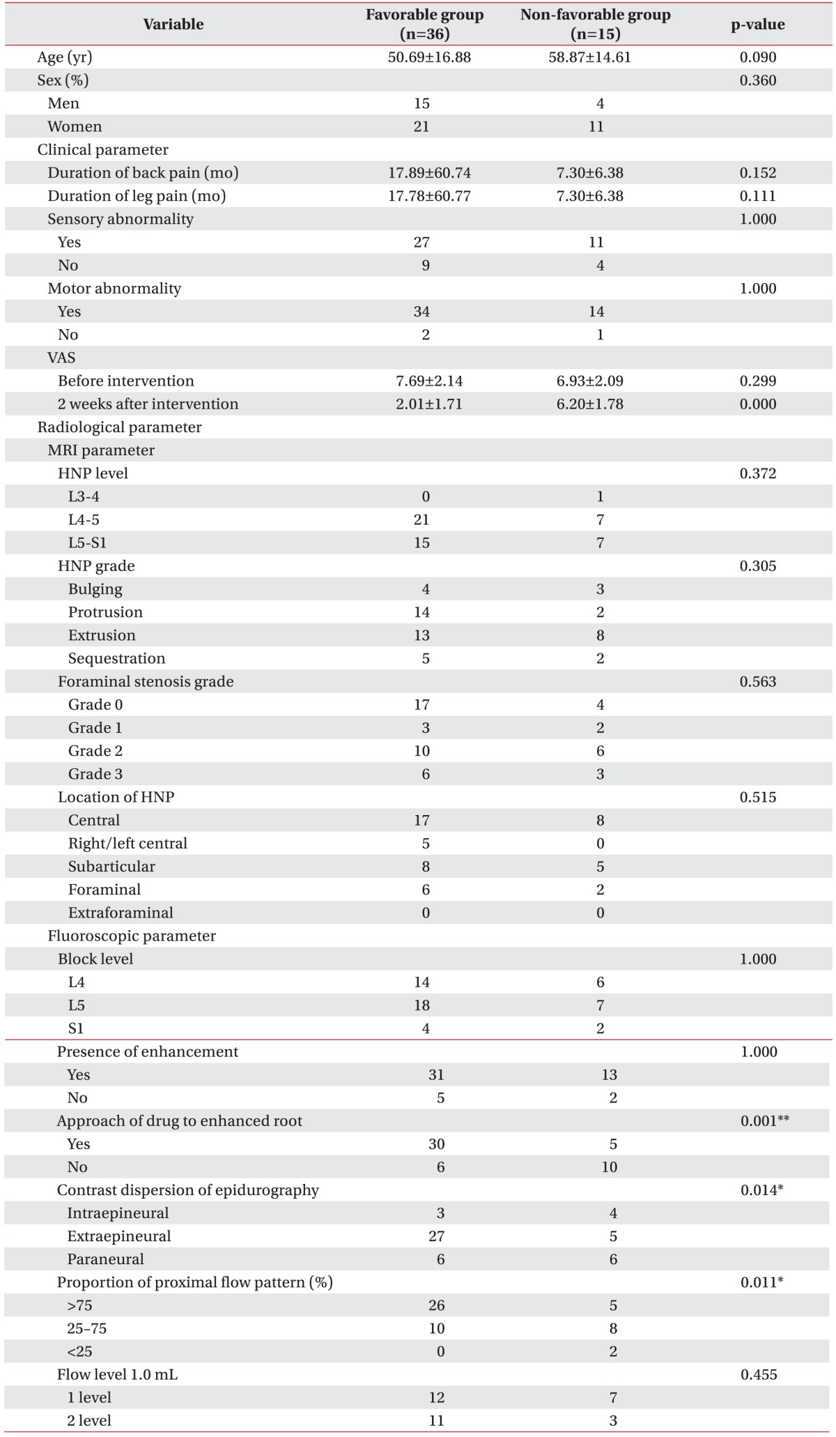
A significant correlation was observed between several of the predictive parameters and the favorable TFESIs. The results of the univariate analysis are listed in Table 1. By using the univariate analysis, the injected material approach for the enhancement of the nerve root, the distribution of the contrast pattern, and the proportion of the proximal flow pattern are significantly associated with the favorable VAS group. Otherwise, the clinical factors such as age, sex, duration of LBP, duration of radiating pain, and the presence of a neurological deficit, and the radiological factors such as the degrees of the HNP and the foraminal stenosis, the type of herniated intervertebral disc, the block level, and the number of flow levels did not show significant differences.
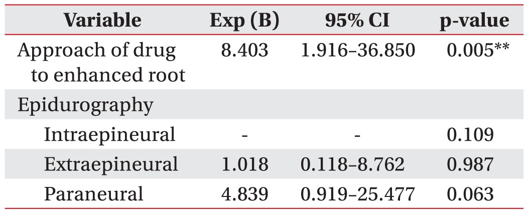
Multiple logistic regression models were used to control for the confounding variables that were significant from the univariate analysis, and the results are summarized in Table 2. Due to a multicollinearity among the independent variables, the proportion of the proximal flow pattern was excluded from the multiple logistic regression analysis. The corticosteroid approach for the enhanced nerve root was the only significant outcome prognostic factor that was associated with the favorable TFESIs group (odds ratio, 8.403; 95% confidence interval, 1.916–36.850; p=0.005).
This retrospective study was designed to elucidate the meaningful clinical and radiological parameters of successful TFESIs in lumbosacral radicular pain patients. The univariate analysis results demonstrated three significant different variables, including the corticosteroid approach for the enhanced nerve root, the contrast dispersion pattern, and the pattern of the proximal flow between the favorable VAS group and the non-favorable VAS group after the intervention. After the multiple logistic regression analysis was conducted, this study revealed that the corticosteroid approach for the enhanced nerve root found in MRI is the most important prognostic factor of TFESIs; that is, the nearer that the administered corticosteroid is to the target nerve root, the more effective the pain reduction will be.
The contrast-enhanced spine MRI can display the inflammatory nerve pathology, and it is a useful method for diagnosing the herniated nucleus pulposus associated with radiculitis and the pain related to neural inflammation [171819]; that is, the nerve root enhancement on the contrast spine MRI suggests neurological deficits because it is related to granulation-tissue accumulation, endoneurial capillary interruption, and inflammatory cytokines [202122]. So, the nature of the TFESIs, which is a procedure for the distribution of a corticosteroid to the area of the target nerve root, enables the formulation of an explanation regarding the reduced radiating pain of the patients [23]. Also, for the effective TFESIs, an identification of the precise target nerve with the use of the contrast material and the injecting of the corticosteroid into the superior portion could be helpful [16]. This finding is in accordance with the univariate analysis results of this study, but the enhanced spine MRI was also reviewed to find the precise target nerve root that was causing the radicular pain. The findings of Tak et al. [24] resemble the results of this study, in that they found improvements of the NRS and the ODI in the nerve root enhancement groups from the contrast-enhanced MRI; however, they did not evaluate the effectiveness at the L5-S1 HNP level and other factors such as the clinical or the radiological factors that are included in this study.
In the favorable VAS TFESIs group, the extraepineural dispersion was observed in 32 (62.7%) patients in this study. The association between the contrast distributed pattern and a clinical improvement after TFESIs is the focus of a number of studies [151625], and one study showed results that are similar to those of this study. Lee et al. [25] revealed that the extraepineural injection may be a factor in the achievement of improved results for the radicular symptom after TFESIs. They hypothesized that an extraepineural injection was a more successful outcome because the corticosteroid was delivered adjacent to the inflammatory epineurium of the nerve root. In contrast, Paidin et al. [15] revealed that the intraepineural and paraneural patterns were beneficial for the relieving of pain. Also, Pfirrmann et al. [16] reported no significant differences according to the contrast pattern, but they suggested that the intraepineural type would be a more effective response compared with the others. But, the intraepineural pattern should be avoided because it may cause more pain to the patients during the intervention [26]. Although they anticipated that the paraneural and extraepineural patterns would produce a similar result if the drug enters into the epidural space, the extraepineural pattern did not show a significant pain reduction. The results of these two studies are in contrast to the results of the present study, as this study revealed that the extraepineural pattern was significantly different from the others. Although both the extraepineural and paraneural patterns enable the entrance of the drug into the epidural space, the extraepineural pattern more precisely encircles the sheath of the target nerve root according to the classification of Pfirrmann et al. [16]. It is therefore possible to infer that the extraepineural pattern is more effective than the paraneural pattern because the injected material can be more adjacent to the nerve in accordance with the overall results of the present study. In this study, the authors not only evaluated the contrast dispersion pattern, but other prognostic factors such as the proximal flow pattern under fluoroscopy and specific inflamed neural structures under an enhanced MRI were also examined.
In general, radicular pains are the result of an inflammatory response within the proximal area of a nerve root [27]. In this study, the proportion of the proximal flow after the injection was another predictive value of successful TFESIs; that is, the more available the administered corticosteroid is regarding the proximal reach, the more effective the outcome will be. For the sake of a proximal injection, it would be more favorable to position the needle more adjacent to the target nerve, to administer a sufficient volume, and to apply force during the injection of the drug materials.
In lumbar spine foraminal stenosis patients, TFESIs are not always beneficial because the administered material cannot reach the target nerve due to perineural and epineural adhesions; therefore, the degree of the spine foraminal stenosis was considered for the investigation of this study. According to this study’s results, a significant difference of the therapeutic effect owing to the degree of the spine foraminal stenosis was not found. The results of the present study correspond well with those of the earlier research regarding scoliosis patients [28]. In [28], the degree of the lateral canal stenosis in the MRI did not correlate with the initial TFESI-induced pain relief of the patients with the degenerative lumbar scoliosis stenosis. Likewise, a number of articles discuss the effectiveness of TFESIs in patients with spinal stenosis [29303132]; according to these articles, there is a dearth of evidence regarding the effectiveness of TFESIs for the radicular pain that is caused by the spinal stenosis. In addition, in a previous study, the severity of the spinal stenosis is not significantly different for the patients with the improvement compared with the patients without the improvement [33].
The findings of the present study are distinguished from those of other research in that the meaningful prognostic factors are different. Choi et al. [9] evaluated the prognostic parameters of TFESIs according to the location of the HNP and the degree of the nerve root compression. They concluded that the centrally or extraforaminally herniated discs and the low-grade nerve root compression were different between the responder and non-responder groups. Another study investigated the clinical and radiological factors like the present research [8]. In their study, only the presence of the HNP, central stenosis, foraminal stenosis and spondylolisthesis was checked for in the radiological findings; among these, there were no significant predictive factors. Instead, they found meaningful clinical predictive factors including higher baseline scores for tools such as the VAS and the McGill Pain Inventory, a positive femoral stretch test, and a reduction of the worsening of the pain during walking. By inferring from an incorporation of the previous findings with those of the present study, the best way to achieve favorable TFESIs could be a precise administration of a corticosteroid to the target nerve root that is found in an enhanced spine MRI.
The limitations of the present study are as follows. First, this study was a retrospective research undertaking, and therefore, a selection bias may exist. Second, the small sample size is another limitation, and a larger sample size may clarify significant prognostic factors; therefore, future research is required to confirm the results of this study. Third, a relatively short-term follow-up period was the other limitation of this study. The follow-up period of the 2 weeks after the intervention was selected to achieve a corticosteroid effect according to the previously described articles [1634]. The follow-up was so brief that the mid- or long-term therapeutic effects could not be evaluated sufficiently.
To the authors’ knowledge, this is the first research study wherein a simultaneous investigation of enhanced-MRI and fluoroscopic parameters, including the lumbar foraminal stenosis according to the degree shown in MRI, has been conducted for the prediction of the effectiveness of TFESIs.
In summary, the results of this study could provide practitioners with useful pain-management information. When TFESIs are administered, the patients with lumbosacral radiating pain will experience a more-favorable outcome if the corticosteroid is injected more proximally, if the corticosteroid reaches the targeted enhanced nerve root, and if the extraepineural pattern is distributed. Above all, the corticosteroid approach regarding the target enhanced nerve shown in MRI is the most predictive of a favorable post-TFESI outcome.
ACKNOWLEDGMENTS
The authors thank the Medical Research Collaborating Center at Seoul National University Bundang Hospital for statistical analyses.
References
1. Waddell G. 1987 Volvo award in clinical sciences: a new clinical model for the treatment of low-back pain. Spine (Phila Pa 1976). 1987; 12:632–644. PMID: 2961080.
3. Abdi S, Datta S, Trescot AM, Schultz DM, Adlaka R, Atluri SL, et al. Epidural steroids in the management of chronic spinal pain: a systematic review. Pain Physician. 2007; 10:185–212. PMID: 17256030.
4. Koes BW, van Tulder M, Lin CW, Macedo LG, McAuley J, Maher C. An updated overview of clinical guidelines for the management of non-specific low back pain in primary care. Eur Spine J. 2010; 19:2075–2094. PMID: 20602122.
5. Eckel TS, Bartynski WS. Epidural steroid injections and selective nerve root blocks. Tech Vasc Interv Radiol. 2009; 12:11–21. PMID: 19769903.
6. Ghahreman A, Ferch R, Bogduk N. The efficacy of transforaminal injection of steroids for the treatment of lumbar radicular pain. Pain Med. 2010; 11:1149–1168. PMID: 20704666.
7. Roberts ST, Willick SE, Rho ME, Rittenberg JD. Efficacy of lumbosacral transforaminal epidural steroid injections: a systematic review. PM R. 2009; 1:657–668. PMID: 19627959.
8. McCormick Z, Cushman D, Casey E, Garvan C, Kennedy DJ, Plastaras C. Factors associated with pain reduction after transforaminal epidural steroid injection for lumbosacral radicular pain. Arch Phys Med Rehabil. 2014; 95:2350–2356. PMID: 25108099.
9. Choi SJ, Song JS, Kim C, Shin MJ, Ryu DS, Ahn JH, et al. The use of magnetic resonance imaging to predict the clinical outcome of non-surgical treatment for lumbar intervertebral disc herniation. Korean J Radiol. 2007; 8:156–163. PMID: 17420633.
10. Fardon DF. Nomenclature and classification of lumbar disc pathology. Spine (Phila Pa 1976). 2001; 26:461–462. PMID: 11242371.
11. Lee S, Lee JW, Yeom JS, Kim KJ, Kim HJ, Chung SK, et al. A practical MRI grading system for lumbar foraminal stenosis. AJR Am J Roentgenol. 2010; 194:1095–1098. PMID: 20308517.
12. Attias N, Hayman A, Hipp JA, Noble P, Esses SI. Assessment of magnetic resonance imaging in the diagnosis of lumbar spine foraminal stenosis: a surgeon’s perspective. J Spinal Disord Tech. 2006; 19:249–256. PMID: 16778658.
13. Wildermuth S, Zanetti M, Duewell S, Schmid MR, Romanowski B, Benini A, et al. Lumbar spine: quantitative and qualitative assessment of positional (upright flexion and extension) MR imaging and myelography. Radiology. 1998; 207:391–398. PMID: 9577486.
14. Toyone T, Takahashi K, Kitahara H, Yamagata M, Murakami M, Moriya H. Visualisation of symptomatic nerve roots: prospective study of contrast-enhanced MRI in patients with lumbar disc herniation. J Bone Joint Surg Br. 1993; 75:529–533. PMID: 8331104.
15. Paidin M, Hansen P, McFadden M, Kendall R. Contrast dispersal patterns as a predictor of clinical outcome with transforaminal epidural steroid injection for lumbar radiculopathy. PM R. 2011; 3:1022–1027. PMID: 21974903.
16. Pfirrmann CW, Oberholzer PA, Zanetti M, Boos N, Trudell DJ, Resnick D, et al. Selective nerve root blocks for the treatment of sciatica: evaluation of injection site and effectiveness: a study with patients and cadavers. Radiology. 2001; 221:704–711. PMID: 11719666.
17. Haijiao W, Koti M, Smith FW, Wardlaw D. Diagnosis of lumbosacral nerve root anomalies by magnetic resonance imaging. J Spinal Disord. 2001; 14:143–149. PMID: 11285427.
18. Jinkins JR. MR of enhancing nerve roots in the unoperated lumbosacral spine. AJNR Am J Neuroradiol. 1993; 14:193–202. PMID: 8427089.
19. Bradley WG. Use of contrast in MR imaging of the lumbar spine. Magn Reson Imaging Clin N Am. 1999; 7:439–457. viiPMID: 10494528.
20. Itoh R, Murata K, Kamata M, Mukubou N, Morita R. Lumbosacral nerve root enhancement with disk herniation on contrast-enhanced MR. AJNR Am J Neuroradiol. 1996; 17:1619–1625. PMID: 8896611.
21. Ross JS, Modic MT, Masaryk TJ, Carter J, Marcus RE, Bohlman H. Assessment of extradural degenerative disease with Gd-DTPA-enhanced MR imaging: correlation with surgical and pathologic findings. AJNR Am J Neuroradiol. 1989; 10:1243–1249. PMID: 2512790.
22. Vroomen PC, Van Hapert SJ, Van Acker RE, Beuls EA, Kessels AG, Wilmink JT. The clinical significance of gadolinium enhancement of lumbar disc herniations and nerve roots on preoperative MRI. Neuroradiology. 1998; 40:800–806. PMID: 9877135.
23. Bogduk N. Practice guidelines for spinal diagnostic and treatment procedures. San Francisco: International Spine Intervention Society;2004. p. 163–187.
24. Tak HJ, Jones R, Cho YW, Kim EH, Ahn SH. Clinical evaluation of transforaminal epidural steroid injection in patients with gadolinium enhancing spinal nerves associated with disc herniation. Pain Physician. 2015; 18:E177–E185. PMID: 25794217.
25. Lee JW, Kim SH, Lee IS, Choi JA, Choi JY, Hong SH, et al. Therapeutic effect and outcome predictors of sciatica treated using transforaminal epidural steroid injection. AJR Am J Roentgenol. 2006; 187:1427–1431. PMID: 17114531.
26. Yeom JS, Lee JW, Park KW, Chang BS, Lee CK, Buchowski JM, et al. Value of diagnostic lumbar selective nerve root block: a prospective controlled study. AJNR Am J Neuroradiol. 2008; 29:1017–1023. PMID: 18272560.
27. Popescu A, Gupta A. Lumbar transforaminal epidural injections. In : Gupta A, editor. Interventional pain medicine. New York: Oxford University Press;2012. p. 106–111.
28. Ploumis A, Transfeldt EE, Gilbert TJ, Mehbod AA, Pinto MR, Denis F. Radiculopathy in degenerative lumbar scoliosis: correlation of stenosis with relief from selective nerve root steroid injections. Pain Med. 2011; 12:45–50. PMID: 21087400.
29. Lee JH, Lee SH. Clinical effectiveness of percutaneous adhesiolysis and predictive factors of treatment efficacy in patients with lumbosacral spinal stenosis. Pain Med. 2013; 14:1497–1504. PMID: 23802996.
30. Lee JH, An JH, Lee SH. Comparison of the effectiveness of interlaminar and bilateral transforaminal epidural steroid injections in treatment of patients with lumbosacral disc herniation and spinal stenosis. Clin J Pain. 2009; 25:206–210. PMID: 19333170.
31. Park Y, Lee WY, Ahn JK, Nam HS, Lee KH. Percutaneous adhesiolysis versus transforaminal epidural steroid injection for the treatment of chronic radicular pain caused by lumbar foraminal spinal stenosis: a retrospective comparative study. Ann Rehabil Med. 2015; 39:941–949. PMID: 26798608.
32. Delport EG, Cucuzzella AR, Marley JK, Pruitt CM, Fisher JR. Treatment of lumbar spinal stenosis with epidural steroid injections: a retrospective outcome study. Arch Phys Med Rehabil. 2004; 85:479–484. PMID: 15031837.
33. Campbell MJ, Carreon LY, Glassman SD, McGinnis MD, Elmlinger BS. Correlation of spinal canal dimensions to efficacy of epidural steroid injection in spinal stenosis. J Spinal Disord Tech. 2007; 20:168–171. PMID: 17414988.
34. Jamison RN, VadeBoncouer T, Ferrante FM. Low back pain patients unresponsive to an epidural steroid injection: identifying predictive factors. Clin J Pain. 1991; 7:311–317. PMID: 1839719.