

 PDF
PDF Citation
 Print
Print


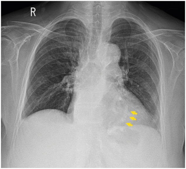
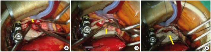
An 87-year-old female was transferred to the emergency department due to fever for three days. She has been treated for hypertension and diabetes mellitus. Physical examination revealed a clear breathing sound and regular heart sound without murmur. Chest X-ray demonstrated mild cardiomegaly and calcification in the position of the heart (Figure 1). Laboratory findings showed leukocytosis, high hs-C-reactive protein (3.65 mg/dL), and elevated erythrocyte sedimentation rate (32 mm) suggestive of infection. Computed tomography of chest and abdomen showed non-specific findings except prominent mitral annular calcification (MAC) (Figure 2). Transthoracic echocardiography (TTE) revealed a mobile, echogenic mass attached to MAC (Figure 3, Supplementary Video 1). Transesophageal echocardiography showed a 1.2×1.1 cm-sized, stippled cystic mass containing multiple sparkling dots suggestive of focal calcifications (Figure 4A-C, Supplementary Video 2). Based on the peculiar echocardiographic finding, we made a diagnosis of infective endocarditis on MAC and began empirical antibiotics therapy for the most common organism, staphylococci.1)2) Considering the large size of vegetation and the site of endocarditis despite very old age, minimally invasive mitral surgery was undergone to remove vegetation and repair posterior mitral annulus with bovine pericardium after partial removal of MAC (Figure 5). Postoperative TTE found normal mitral function and no evidence of newly developed echogenic mass on MAC (Figure 6). After 6 weeks of antibiotic treatment using ceftriaxone and vancomycin, the patient was discharged without a neurologic deficit. This case highlights typical findings of MAC endocarditis represented by large, stippled vegetation in the elderly.
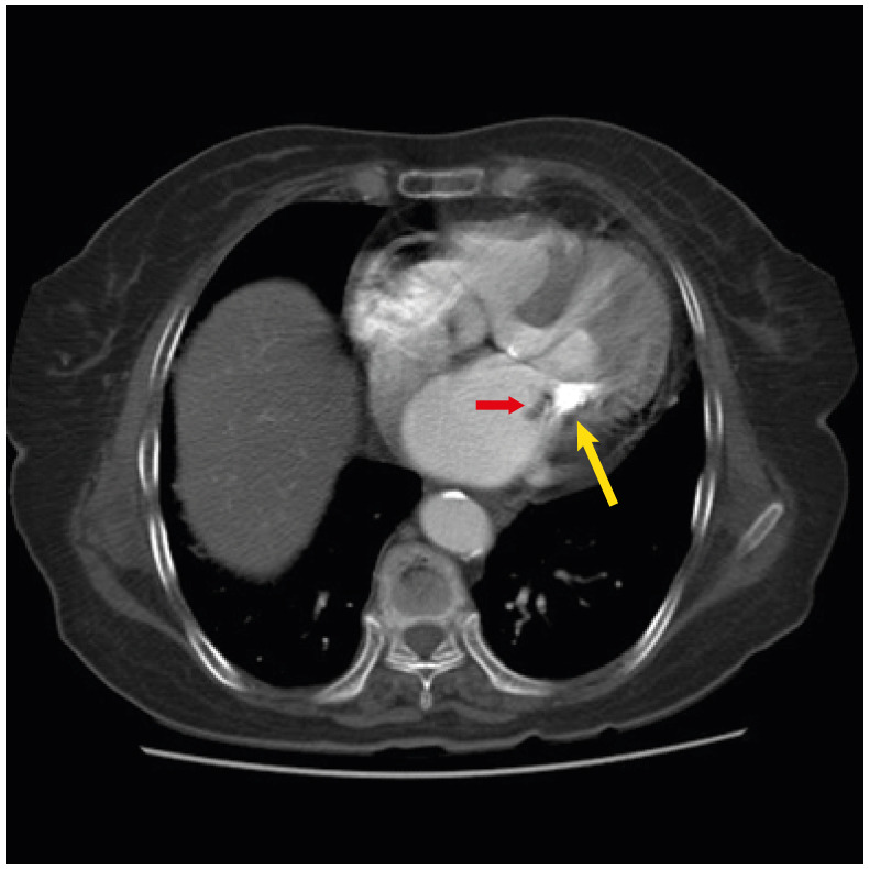 | Figure 2Chest computed tomography demonstrated massive calcification of posterolateral aspect of mitral annulus (yellow arrow) and low density mass on the left atrial side of calcification (red arrow).
|
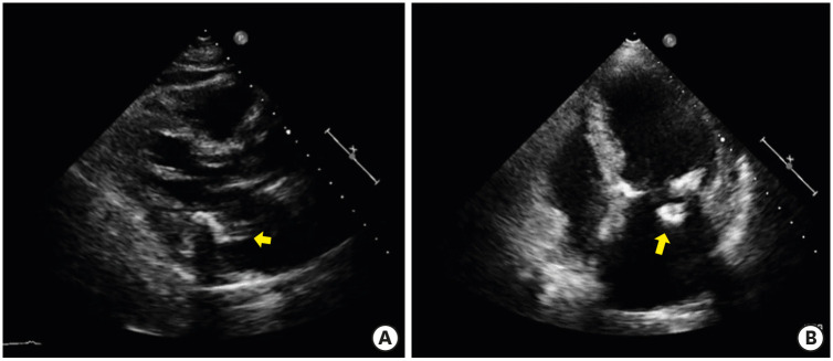 | Figure 3Transthoracic echocardiographic images. (A) Parasternal long axis view demonstrated MAC with vegetation. (B) Apical 4 chamber view demonstrated inhomogenous echogenic mass attached to MAC on left atrial side which was intermittently seen due to acoustic shadowing of MAC.MAC = mitral annular calcification.
|
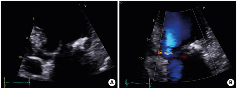
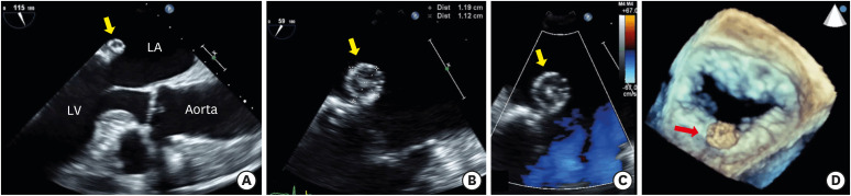 | Figure 4Transesophageal echocardiography images. (A) Mid-esophageal 115° view demonstrated round vegetation on poster MAC. (B) In mid-esophageal 59° view, size of vegetation was 1.2×1.1 cm. (C) Color flow imaging revealed no significant steno-insufficiency of mitral valve. (D) Three-dimensional image showed large vegetation attached to posterior mitral annulus.LA = left atrium; LV = left ventricle; MAC = mitral annular calcification.
|
The Institutional Review Board (IRB) of Pusan National University Yangsan Hospital approved this study and the patient's informed consent was waived (IRB number: 04-2021-047).

 XML Download