

 PDF
PDF Citation
 Print
Print


INTRODUCTION
Hepatic artery thrombosis (HAT) following living donor liver transplantation (LDLT) is a lethal complication, with incidence ranging from 2.5% to 9% [1,2]. The clinical manifestation of HAT varies greatly and includes liver abscess formation, bile duct stricture, graft necrosis, and graft failure [3]. In our institution, the right gastroepiploic artery is the mostly preferred inflow arterial source for primary and redo hepatic artery reconstruction if the native hepatic artery branches are not available [4,5]. If other nonanatomical arterial alternatives are not available, redo hepatic artery reconstruction using an aorto-hepatic jump graft can be considered, as in deceased donor liver transplantation (DDLT) [6,7]. However, in Korea, the availability of a fresh iliofemoral artery graft is very limited, thus redo hepatic artery reconstruction with an aorto-hepatic jump graft following LDLT is likely very limited [5]. We present the case of a patient who underwent salvage redo hepatic artery reconstruction with an aorto-hepatic jump graft because of HAT following LDLT. The patient has been doing well for 10 years without any vascular complications.
Go to : 
CASE REPORT
This study was approved by the Institutional Review Board of Asan Medical Center (IRB No. 2020-0822), which waived the requirement for informed consent due to the retrospective nature of this study.
A 64-year-old female patient diagnosed with hepatitis C virus (HCV)-associated liver cirrhosis and a 5-cm-sized hepatocellular carcinoma (HCC) underwent partial hepatectomy for HCC. Soon after hepatectomy, ascites and jaundice occurred because of post-hepatectomy hepatic decompensation. A small HCC was newly diagnosed at 3 months following hepatectomy (Fig. 1). To treat the liver cirrhosis and HCC concurrently, an LDLT operation was planned at 5 months after hepatectomy.
The donor was the 31-year-old son of the patient. The modified right liver graft from this donor weighed 850 g, making a graft-to-recipient weight ratio of 1.67%. The donor recovered uneventfully from the donor operation and was discharged 8 days after the operation.
During the recipient’s operation, hilar dissection was performed uneventfully although the upper abdominal cavity had heavy adhesions because of the previous hepatectomy. Recipient hepatectomy was done according to the standard procedure of LDLT. At the back table, the right hepatic vein orifice of the liver graft was enlarged with incision and patch venoplasty. The graft middle hepatic vein branches were interposed with a cryopreserved iliac vein conduit. Immediately after finishing the bench work for the liver graft, the modified right liver graft was implanted according to the standard procedure of LDLT. The graft right hepatic vein and the interposed middle hepatic vein conduit were separately reconstructed at the recipient’s right hepatic vein and left-middle hepatic vein trunk stumps, respectively. The recipient portal bifurcation was used for anastomosis with the graft portal vein. The recipient right hepatic artery was used for arterial reconstruction. Biliary reconstruction was performed with duct-to-duct anastomosis.
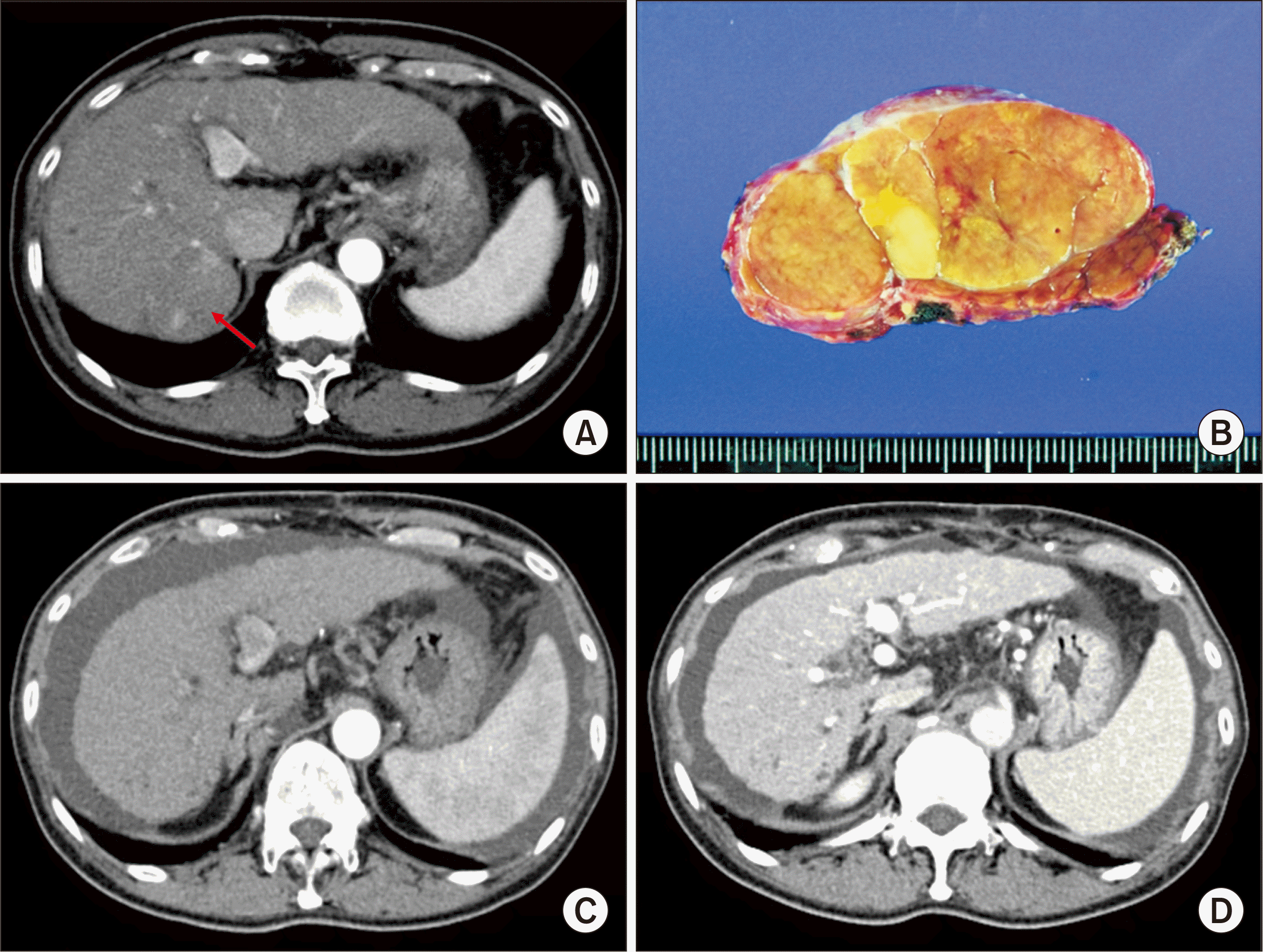
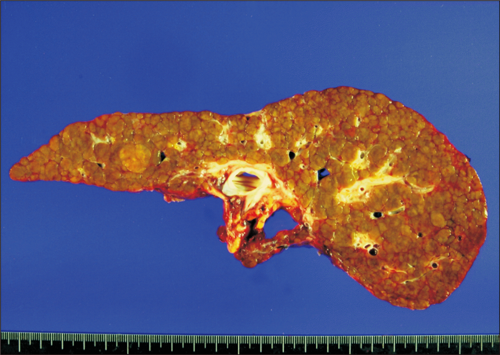
The pathology report of the explant liver showed mixed macronodular and micronodular cirrhosis associated with HCV infection. (Fig. 2). A 1.9-cm-sized HCC without microvascular invasion was identified. On posttransplant day 1, Doppler ultrasonography showed that the inferior hepatic vein and segment V vein were occluded. Therefore, wall stents were inserted through a transjugular approach on posttransplant day 3. On posttransplant day 4, liver dynamic computed tomography (CT) showed partial parenchymal infarct at the peripheral portion of the liver graft, but Doppler ultrasonography showed the hepatic artery flow to be maintained. Liver function profiles gradually deteriorated. On posttransplant day 9, liver dynamic CT showed an increase of the parenchymal infarct, suggesting hepatic arterial insufficiency (Fig. 3). On the same day, celiac arteriography showed complete occlusion of the graft hepatic artery (Fig. 4), in which the extent of HAT appeared to be wide with retrograde propagation of thrombus. The gastroepiploic artery was poorly developed, thus it appeared to be an unsuitable source for arterial inflow. At that time, the laboratory findings of the patient showed aspartate transaminase 440 IU/mL, alanine transaminase 1,211 IU/mL, total bilirubin 9.8 mg/dL and prothrombin time international normalized ratio 1.44. We seriously considered enrolling the patient in the DDLT waiting list for retransplantation.
 | Fig. 2Gross photograph of the explanted liver. There was mixed macro- and micronodular cirrhosis associated with hepatitis C virus infection. A 1.9-cm-sized hepatocellular carcinoma was identified.
|
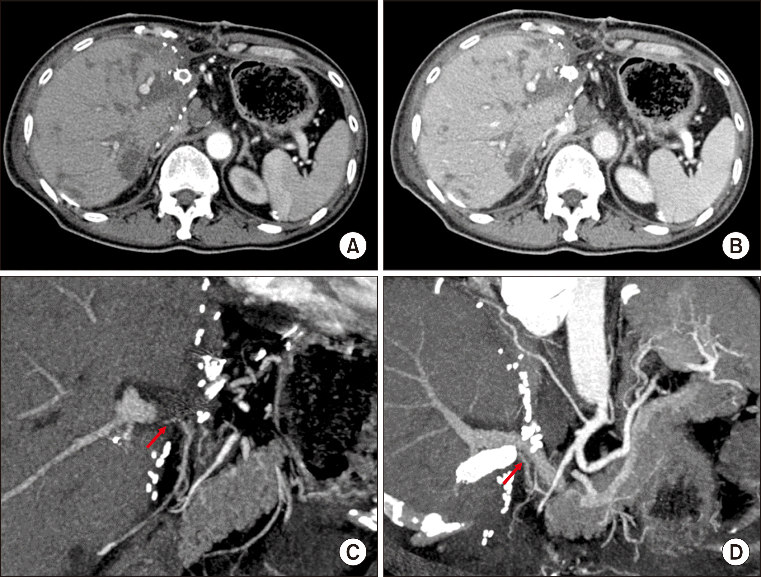
Fortunately, cold-stored fresh iliofemoral artery and superior mesenteric artery grafts were stored at our institutional tissue bank, which had been harvested from a deceased donor one day before. Thus, we decided to perform redo hepatic artery reconstruction using this artery homograft 10 days following the LDLT operation. The infrarenal aorta was dissected and an iliofemoral artery graft was anastomosed. The artery graft conduit was passed posterior to the stomach, and the distal femoral artery was anastomosed to the superior mesenteric artery segment for size matching. A size-matched branch of the superior mesenteric artery conduit was then anastomosed to the graft hepatic artery under surgical microscopy (Fig. 5). Soon after hepatic artery revascularization, liver function improved progressively and the infarct area at the liver graft was reduced (Fig. 6).
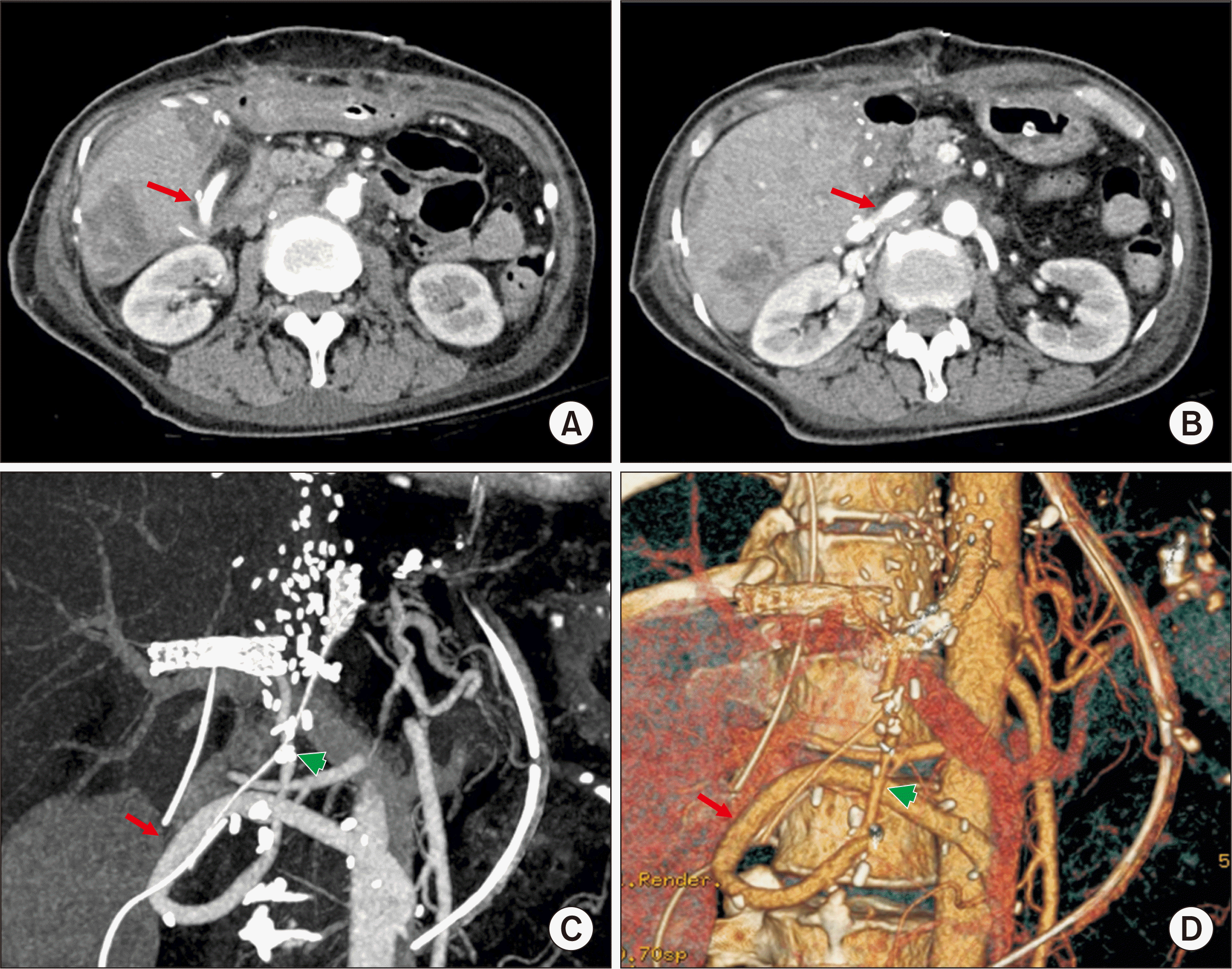 | Fig. 5Hepatic artery findings at 1 day after hepatic artery revascularization. (A, B) Computed tomography sectional images show the running course of the interposed iliofemoral artery homograft (red arrows). (C, D) Computed tomography arteriography shows the running courses of the interposed iliofemoral artery homograft from the infrarenal aorta (red arrows) and the interposed superior mesenteric artery homograft between the graft hepatic artery and iliofemoral artery homograft (green arrows).
|
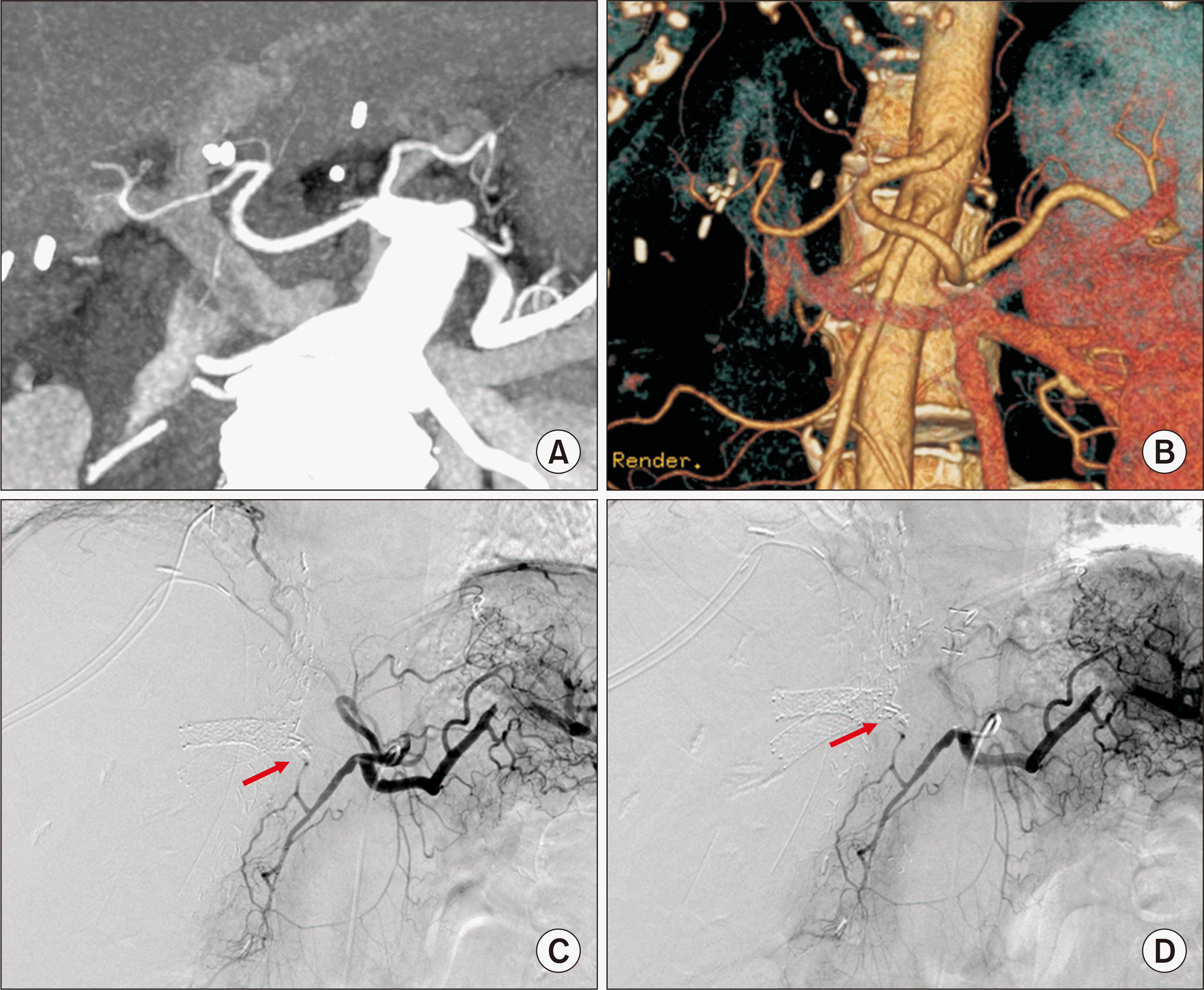
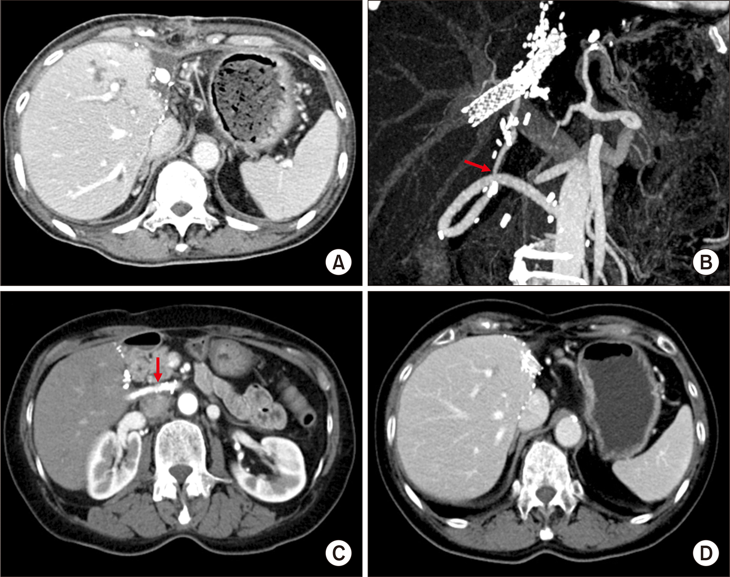 | Fig. 6Posttransplant computed tomography findings. (A, B) Images taken at 1 month after transplantation show marked reduction of the graft infarct area and patent hepatic artery inflow (arrow). (C, D) Images taken at 9 years 8 months following transplantation show the patent iliofemoral graft (arrow) and absence of perfusion abnormality of the liver graft.
|
The HCV was treated with interferon therapy at 6 months following LDLT and HCV RNA has been negative for 8 years since its negative conversion at 2 years posttransplantation. HCC recurrence has not been noted on follow-up studies. The patient has been doing well with administration of immunosuppressant and low-dose aspirin (Fig. 6).
Go to :
DISCUSSION
LDLT is more vulnerable to HAT than DDLT. In LDLT, the graft hepatic artery is smaller and its stump is shorter than a whole-liver graft, thus extensive dissection of the recipient hepatic arteries is necessary for matching the size for arterial reconstruction. This procedure may increase the risk of intimal dissection or transmural hematoma in the recipient hepatic artery in the situation of an enlarged and vulnerable hepatic artery caused by portal hypertension. Pretransplant transarterial chemoembolization, radiofrequency ablation, radiation therapy, and prior hepatic resection for HCC management may incur hepatic arterial injuries [5].
HAT is a fatal complication of LT, but the most efficient management of it is debated. Retransplantation is the treatment of choice in early HAT, but we reported that only one adult LDLT recipient underwent retransplantation because of graft dysfunction caused by HAT-induced ischemic graft damage [5]. Considering the real-world situation of a very small donor pool, urgent re-arterialization of HAT should be considered as the first therapeutic option to prevent retransplantation [3].
If HAT is detected early with minimal ischemic graft injury, hepatic artery revascularization may provide favorable results. Several studies report that the mortality rate after diagnosis of HAT was 33.3% (range, 0%–80%) [3,4,8], but we have noted zero mortality in 21 cases of HAT [5]. Early detection of HAT is essential to prevent graft or patient loss. Therefore, we have been evaluating hepatic artery flow with daily Doppler ultrasonography up to postoperative 5 days as well as dynamic liver CT at least once a week during admission. When HAT is suspicious on Doppler ultrasonography, we immediately performed dynamic CT and hepatic angiography. Before performing an urgent hepatic artery revision, splenic artery embolization is done to augment the artery diameter and flow during celiac angiography, when HAT is limited to the right or left hepatic artery and the right gastroepiploic artery is small.
As a substitute for the native hepatic arteries in recipients, several studies have suggested using the right gastroepiploic artery, splenic artery, left gastric artery, middle colic artery, or various interposition grafts [5,9-11]. If HAT had not propagated down to the proper hepatic artery level, reusing the recipient’s native hepatic artery appears to be feasible. Based on our experience, other alternative hepatic artery inflows may not be necessary if the native hepatic artery and the right gastroepiploic artery can be used for hepatic artery inflow.
However, if no source of hepatic artery inflow is available, a jump graft from the aorta using a fresh iliofemoral artery graft obtained from a deceased donor can be a viable option [5-7]. In Korea, the incidence of deceased donors has been low, thus the availability of a fresh iliofemoral artery graft is also very limited. It is not recommended to use a cryopreserved iliofemoral artery graft stored at the tissue bank because it is vulnerable to aneurysmal dilatation [12]. Fortunately, a cold-stored fresh iliofemoral artery graft was available at the time of hepatic artery revision in the present patient. We have rarely performed aorto-hepatic jump anastomosis using an iliofemoral artery graft for primary LDLT, especially salvage LDLT, but this was the first case of redo hepatic artery revascularization using an aorto-hepatic jump graft.
A study from Pakistan reported 21 cases of LDLT using greater saphenous vein conduits, including 16 cases of aorto-hepatic reconstruction from the infrarenal aorta and three cases from the supraceliac aorta [13]. The available length of the greater saphenous vein conduits can be increased to more than 20 cm, which enables performing a long extra-anatomic jump graft. There are a few case reports presenting successful application of a supraceliac aorto-hepatic conduit and a right iliac artery-hepatic conduit using a greater saphenous vein graft during LDLT [14,15]. These studies suggest that a long greater saphenous vein segment can be used as a vessel substitute allowing aorto-hepatic reconstruction.
Our institutional measures to preventive HAT in adult LDLT patients include the use of low-molecular heparin during admission period and administration of antiplatelet agent for the first year. Warfarin is not used for prevention of HAT, whereas it has been preferentially used for management of venous thrombosis.
In conclusion, the experience of the present patient suggests that salvage redo hepatic artery reconstruction using an aorto-hepatic jump graft is a feasible option to treat HAT following LDLT, as in DDLT.
Go to :
 XML Download