

 PDF
PDF Citation
 Print
Print


I. Introduction
Previous studies have compared open and closed reduction for treatment of mandibular condyle fracture (MCF) through an assessment of functional outcomes, such as maximum mouth opening, stable occlusion, temporomandibular joint (TMJ) pain, facial symmetry, and total mandibular activity1-3. For patients older than 16 years, many different surgical approaches for MCF have been recommended, depending on the surgeon’s skill and the patient’s acceptance of possible complications4,5.
Over the last 15 years, we have performed intraoral reinsertion after extracorporeal fixation (IREF) on more than 120 consecutive patients with MCF. In this technical strategy note, we described our detailed step-by-step procedures for IREF. We also compared conventional surgical approaches to MCF with IREF with schematic drawings, and we described essential considerations to prevent complications such as condylar head absorption, mouth opening limitation with deviation, and postoperative pain.
II. Technical Note
Acceptable results of IREF for MCF have been described in clinical retrospective experiences and reports6,7. Under strict indications and guidelines, IREF can be a good option to achieve successful outcomes. Indications for open reduction of MCF have been known as several factors including patient age greater than 16 years, condylar fracture at the condylar neck or above, severely displaced or dislocated condylar fracture, and malocclusion due to loss of posterior condylar height.
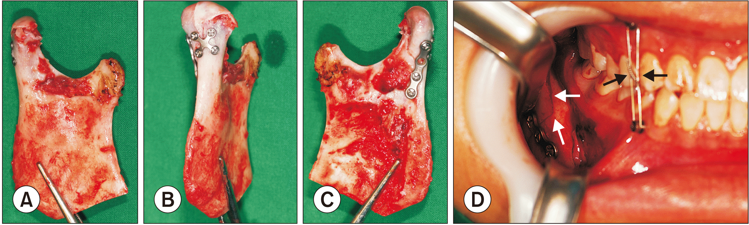
Intraoral vestibular and buccal gingival incision is needed to expose the mandibular oblique ridge and ascending ramus by dissection of the angle and internal ramus together. After identification of the fracture lines above the sigmoid notch, coronoidectomy could be considered for the better visualization. If the fractured condylar segment is visible and the sagittal split proximal portion can be easily detached from the surrounding masticatory muscles, the coronoid process could be preserved without resection. In most cases, a small triangular bony edge of the coronoid process can be removed for better visualization and safe repositioning of the extracorporeal fixated proximal segment.
Sagittal split osteotomy with a reciprocating saw, chisel, and mallet can be performed in the same way as for orthognathic surgery. The proximal segment is removed extracorporeally and kept in a warm saline bath in a metal bowl to maintain body temperature. After removal of the proximal segment, the remaining fractured condylar portion should be carefully dissected and removed as quickly as possible. Likewise, extracorporeal removal of the fractured segment should be carried out quickly and with as little trauma as possible. A retraction elevator or instruments should be readied by the surgical assistants during extracorporeal fixation by the main operator. This procedure is especially important to maintain the position of the condylar head for reinsertion to the original temporal fossa position. The removed fractured segment can be relocated to the proximal segment anatomically and should be fixed with miniplates and/or microplates as quickly as possible.(Fig. 1. A-1. C) Warm saline irrigation is recommended for the drilling procedure to maintain body temperature.
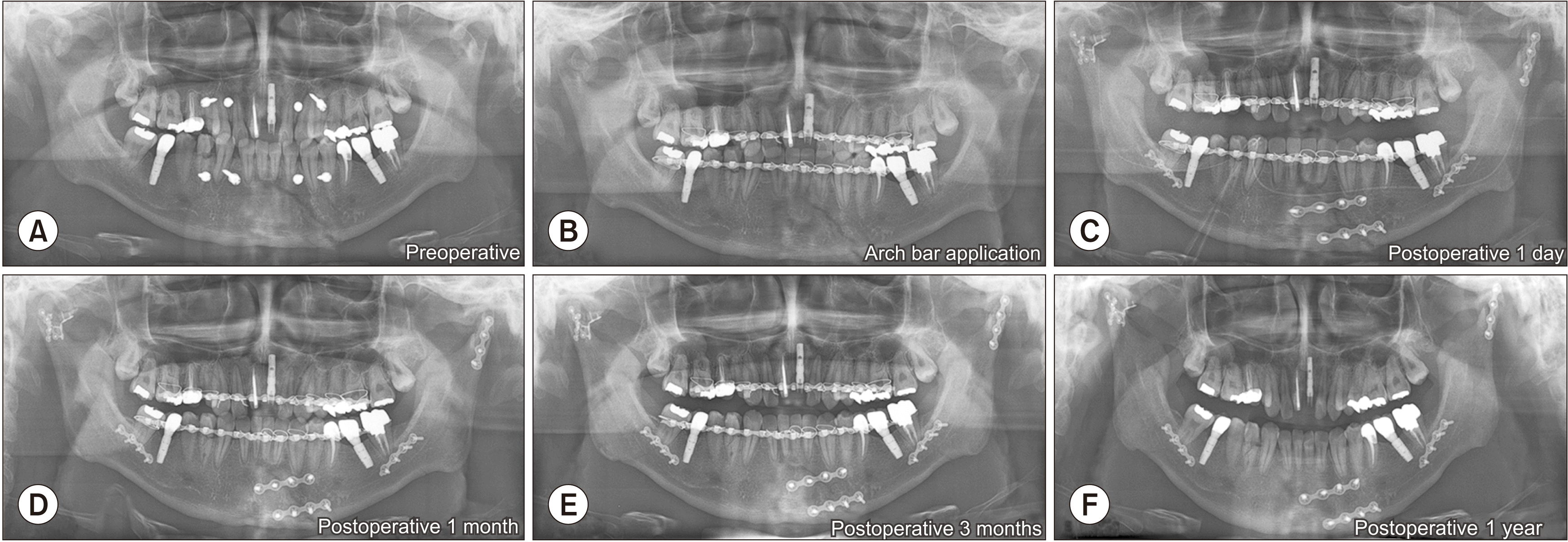
On the basis of our experiences treating more than 147 cases of MCF in 120 patients, we suggest that procedure time for removal and reinsertion of the fixated condylar segment to its original TMJ space should not exceed 30 minutes. Limiting this operative time helps to prevent postoperative complications, such as bone necrosis or cellular damage. After reinsertion, intermaxillary fixation through anchored screws or arch bars should be performed after confirmation of the original position of the sagittal split osteotomy line. This segment can be fixed with the same type of miniplate used in the orthognathic surgical procedure.(Fig. 1. D) Stable occlusion without lateral interference or deviation should be confirmed after boxing wire removal, and condylar translation with smooth rotation is also recommended.
The final surgical procedure involves insertion of Hemovac, an intraoral vacuum containing drain, along the inferior border of the mandible until the coronoid space adjacent to the fixated condylar head.(Fig. 2. C) This drain can also be anchored through the intraoral mucosa to avoid an unnecessary extraoral wound. Immediately after the procedure, the patient must be taught exercises for opening and closing the mouth to maintain occlusion of the upper and lower teeth. Sometimes, a rubber chain between the upper and lower anchorage screws or the arch bar can help to guide stable occlusion and rapid muscular reattachment to the reinserted bony segment.
Frequent buccal cheek massage with both hands can help reduce swelling and hematoma, and continuous cheek massage could facilitate Hemovac drainage until 2 to 5 days postoperatively. The patient can be discharged after drain removal within 5 to 7 days. The postoperative exercises should be performed for at least 4 weeks. We could achieve a successful final outcome as no pain or discomfort, stable occlusion without deviation, mouth opening greater than 35 mm, and symmetrical facial contour during 1-year follow-up check.(Fig. 2)
III. Discussion
Suggested surgical technical note is an intraoral approach basically; thus, we invoked the terminology of IREF rather than extraoral fixation. The ‘extracorporeal’ terminology has been used in cardiac or respiratory medicine, such as extracorporeal life support, extracorporeal membrane oxygenation, extracorporeal life or lung support, extracorporeal cardiopulmonary resuscitation, and extracorporeal shock wave therapy. For salvage of hypoxemic status in patients with complicated chest trauma, extracorporeal membranous oxygenation to achieve respiratory oxygenation has been widely used in the medical field7. Thus, our use of ‘extracorporeal fixation’ can be interpreted as ‘extraoral direct fixation of fractured segments’ in patients with MCF8-11.
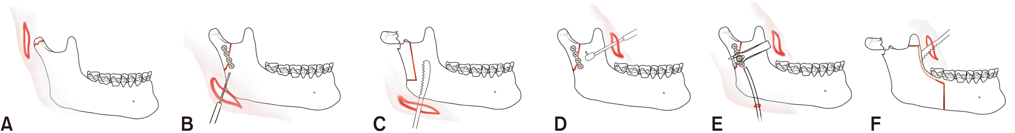
Various surgical approaches have been reported to treat MCF, and the advantages of intraoral reduction have been described. However, controversies have been continuing to surround the surgical treatment of condylar fracture12,13. In the position paper from the IBRA Symposium on Surgery of the Head, Condylar Fracture Osteosynthesis, Marseille, France 201214, the extraoral approach was subdivided into submandibular15, retromandibular, preauricular, and transparotid16 approaches. Authors have classified the extraoral approach as direct reduction with fixation via a submandibular or endaural approach, extracorporeal fixation with vertical ramus osteotomy, intraoral direct reduction and fixation with an angled driver or transbuccal set with endoscopic visualization7. Fig. 3 summarized various approaches to MCF including our suggested IREF.
From our experiences, the clinical and radiographic findings suggest that IREF could be a better choice than other recommended techniques because it leads to anatomically accurate reduction with a low risk of complications. The greatest advantages of IREF are that the intraoral approach prevents facial scars and facilitates anatomical and accurate repositioning through direct visualization. However, complications of IREF can occur because the procedure involves non-vascularized bone graft to the inside of the TMJ. Thus, only the surrounding blood supply from adjacent muscles and fascia should be recirculated to the grafted segment. During this process, a short ischemic time for the proximal segment and appropriate functioning of the Hemovac drain are the most important factors for successful outcomes. An intraoral Hemovac drain should be maintained for several days during the patient’s active mouth opening exercises. If the Hemovac is malfunctioned or removed too quickly, the whole proximal segment or the fractured segment may become necrotic17.
This study reported on the use of IREF for MCF with an intraoral approach that included sagittal split osteotomy, direct fixation with or without the use of a rectangular driver, and intraoral Hemovac insertion. In all of our cases, these advantages prevented an unattractive extraoral scar. The IREF procedure also involved large bony contact to the detached muscle fibers through intraoral sagittal split osteotomy, compared to a small bony segment in the case of extraoral vertical ramus osteotomy, which could have a narrow bony contact surface that could be addressed with another ostectomized bony margin and surrounding muscle fibers17.
In conclusion, this technical note showed that IREF is a reliable treatment option for condylar fracture management. If clinicians apply this minimally invasive method with a clean intraoral sagittal osteotomy, fast and accurate location of the fractured fragment and extracorporeal reduction, anatomical repositioning to the original condylar position, and active mouth opening exercises with extraoral muscular massage, the outcomes will be successful for patients with MCF.
 XML Download