Journal List > J Korean Assoc Oral Maxillofac Surg > v.47(6) > 1148684

Frimpong, Nguyen, Sodnom-Ish, Nimatu, Dampare, Rockson, Awuah, Amponsah, Newton, and Kim: Incidence and management of mandibular fractures in a low-resource health facility in Ghana

Abstract

Objectives

The mandible and other parts of the maxillofacial region suffer significant morbid injuries following road traffic accidents. Our study gives epidemiological description of mandibular fractures in Ghana and also evaluates the relevance of closed reduction and indirect fixation for managing mandibular fractures in low-resource health facilities in low-income countries like Ghana.
Patients and Methods This is a retrospective study involving 268 patients who reported to the Department of Oral and Maxillofacial Surgery of the Sunyani Regional Hospital with mandibular fractures from January 2010 to December 2019. Patient medical records were assessed for information on age, sex, fracture etiology, anatomic location of fracture, time of day of road traffic accident, and other associated injuries.

Results

A total of 268 patients were included in this study (males, 216 [80.6%]; females, 52 [19.4%]). Motor vehicular accident (MVA) was the leading cause of mandibular fractures (202 injuries, 75.4%). Other etiologies included assault (39, 14.6%), gunshot (13, 4.9%), falls (12, 4.5%), and industrial accidents (2, 0.7%). Of the 161 male cases caused by MVA, 121 (75.2%) occurred at night and in the evening while the remaining 40 (24.8%) occurred in the morning and afternoon. Among all managed 222 patients, 212 (79.1%) were treated with closed reduction and indirect fixation technique while 10 (3.7%) were treated with open reduction and direct fixation.

Conclusion

Closed reduction with indirect fixation could successfully be used to manage mandibular fractures in low resourced health facilities, especially in low-income countries. The poor lightening system on roads in Ghana is a major contributory factor to motor vehicular accidents.

I. Introduction

The mandible is a prominent bone in the maxillofacial region; thus, injuries to this bone tend to occur at higher rates than other areas of the body1-3. Motor vehicular accident (MVA) is the leading cause of mandibular fractures in low-income countries, while interpersonal violence is the leading cause in high-income countries2. Reckless driving, non-adherence to road safety regulation and poor road conditions are some of the major factors leading to MVA in developing countries like Ghana. Mandibular fracture, which can occur solitarily or in combination with other facial and skeletal injuries is also frequently caused by assault, industrial accidents, contact sports, falls, and firearm injuries.
The Department of Oral and Maxillofacial Surgery at the Sunyani Regional Hospital, which has a bed capacity of 450, supports the medical needs of the middle, northwestern, and northern belts of Ghana. This is the only accredited center in the middle and northern regions of Ghana. It thus receives referrals from five regions of Ghana as well as from eastern Cote d’Ivoire.
The objectives of this study were to describe the epidemiology of the mandibular fractures in Ghana and to evaluate the efficacy of closed reduction and indirect fixation techniques to successfully manage mandibular fractures in a low-resource health facility. Additionally, we tested the prediction that poor lightening systems for roads at night in Ghana contribute disproportionately to MVAs.

II. Patients and Methods

1. Patient data

This retrospective study included data collected from 268 patients who reported to the Department of Oral and Maxillofacial Surgery at the Sunyani Regional Hospital with mandibular fractures from January 2010 to December 2019. Patients’ medical records were assessed for information on age, sex, fracture etiology, anatomical location of fracture, time of day of road traffic accident, and other associated injuries. Patients who had incomplete records, suffered severe systemic or multi-organ injuries, were discharged against medical advice, or were referred to a tertiary center for further management were excluded from this study. The study protocol and access to patient records were approved by the Institutional Review Board of Seoul National University (S-D20200021), and the informed consent was waived by the IRB.

2. Statistical analyses

The data were entered into Microsoft Excel 2010 (Microsoft, Redmond, WA, USA) and analyzed with simple descriptive statistics using SPSS (ver. 15; SPSS, Chicago, IL, USA).

III. Results

A total of 268 patients were included in this study (males, 216 [80.6%], females, 52 [19.4%]). The male-to-female ratio was 4.2:1, and ages ranged from 2 to 59 years with a mean age of 28.6 years.(Table 1) MVA was the leading cause of mandibular fractures, accounting for 202 (75.4%) of all injuries. Other etiologies were assault (n=39, 14.6%), gunshot (n=13, 4.9%), falls (n=12, 4.5%), and industrial accident (n=2, 0.7%).(Table 2)
Ninety of the 161 male MVA cases (55.9%) occurred at night (9 p.m. to 6 a.m.) and 31 (19.3%) occurred in the evening (4 p.m. to 9 p.m.). The remaining MVAs occurred in the morning (n=24, 14.9%) and afternoon (n=16, 9.9%).(Table 3)
A total of 325 mandibular fractures were recorded in 268 patients with an average of 1.2 fractures per patient: 168 patients (62.7%) had a single fracture line, 89 (33.2%) had double fracture lines, and 11 (4.1%) had more than two fracture lines. Mandibular fractures involving the parasymphyseal region (n=121) were the most common fracture line recorded in this study, followed by fractures in the body of mandible (n=87) and the condyle (n=56). Other fracture types recorded in this study involved the angle (n=26), symphysis (n=22), ramus (n=11), and coronoid (n=2).(Table 4)
The most common multiple mandibular fracture recorded in this study involved the parasymphysis and the body, followed by the parasymphysis and the condyle. Associated injuries recorded in our study were distributed as follows: head injury (n=76), depressed skull fracture (n=13), Le Fort I (n=23), Le Fort II (n=14), Le Fort III (n=3), zygomatic fracture (n=17), frontal bone fracture (n=4), nasal bone fracture (n=7), ulna fracture (n=5), radius fracture (n=4), fibula fracture (n=10), cervical injury (n=13), and rib fracture (n=2).(Table 5)
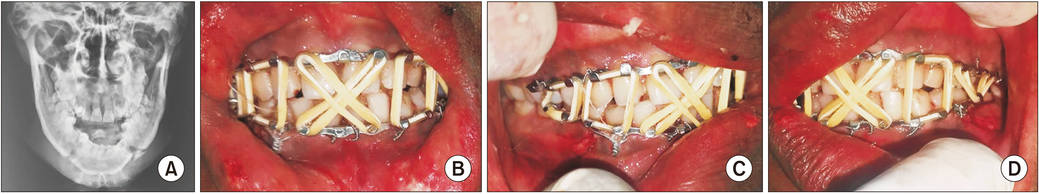
Total 222 cases were managed, and 212 cases (79.1%) were treated using closed reduction and indirect fixation techniques. Of these, 176 (65.7%) were treated with intermaxillary fixation using Erich arch bars, 28 (10.4%) were treated with direct dental wiring eyelet, and 8 (3.0%) were treated using acrylic cap splint with circummandibular wiring. Fig. 1 shows a pictorial description of a 36 year old with bilateral mandibular fracture treated using closed reduction and indirect fixation using Erich arch bars.
Few patients (n=10, 3.7%) were treated with open reduction and direct fixation, but of these, 3 received monocortical miniplates and 7 received transosseous wiring. Fifteen (5.6%) patients who presented with undisplaced condylar fracture with acceptable occlusion were managed conservatively and kept on a soft diet with periodic monitoring. Twenty-five (9.3%) patients were referred to a tertiary health facility due to other serious associated injuries that required urgent care that could not be provided at our facility. Six patients were discharged against medical advice.

IV. Discussion

Maxillofacial injuries continue to generate discussion among researchers globally. The mandible is the second most commonly fractured part of the maxillofacial skeleton because of its position and prominence. Fracture location and pattern are determined by injury mechanism and direction of the force vector4. Additionally, the patient’s age, presence of teeth, and the physical properties of the causing agent also directly affect the characteristics of the resulting injury. The mandible is the heaviest and strongest facial bone but is prone to fractures because it is an open arch, is located in the lower portion of the face, and atrophies during the aging process4,5. This study retrospectively evaluated 268 patients with mandibular fractures over an 8-year period from January 2010 to December 2019. In many countries, mandible fractures are reported significantly more often than middle-third facial fractures6-9.
In this study, people of all ages and gender were affected by mandibular fracture, but patients in their 20s had the highest fracture rates. About 48.9% of fracture patients in this study were 21-30 years old. This incidence is similar to estimates by Ragupathy and Pasupathy10. This could be because people in this age range are in the most active period of life and may be exposed to more dangerous situations that are more likely to result in accidents. The incidence was higher in males (4.2:1), which agrees with previous studies that estimated a worldwide male-to-female ratio of 4.43:11. This may be because males are more likely than females to be involved in violence, reckless driving, sports, drugs, and alcohol11.
In this study, the most common etiological factor was MVA, representing 75.4% of the total number of injuries, followed by assault, gunshot, falls, and industrial accidents. There is also a strong difference in the etiology of maxillofacial trauma between low-income and high-income nations. MVAs are more common in low-income nations, while interpersonal violence is more common in high-income countries12. Our study supports this conclusion and is consistent with findings from other economically similar countries; e.g., Fasola et al.13 and Olayemi et al.14 found that MVA was the most common cause of maxillofacial trauma in Nigeria. Further, according to Parkins et al.15, MVA was the most common cause of maxillofacial injuries in Ghana. The government of Ghana will therefore need to equip the Motor Traffic and Transport Department of the Ghana Police service to ensure proper enforcement of all road traffic regulations in the country. Furthermore, broader stakeholder consultation needs to be done on the legalization and regulation on the use of motorcycle as means of transportation for commercial activities. Our study found that 55.9% of cases caused by MVAs occurred at night. Although, no statistical correlation could be drawn between MVAs and the time of the day for its occurrence, further studies need to be con-ducted to determine if there is any scientific correlation for this observation made in our study. Assault was the second most common cause in this study, but this contrasts with some studies of high-income countries that found assault to be the leading cause of maxillofacial injuries, including mandibular fractures16-19. On the other hand, a Turkish study found that falls were the main cause of mandible fracture20.
In our study, 325 mandibular fractures were recorded in 268 patients with an average of 1.2 fractures per patient: 62.7% of patients had a single fracture line, 33.2% had double fracture lines, and 4.1% had more than two fracture lines. The most frequent location of mandibular fracture lines was in the parasymphyseal region (121/325 fracture lines), which is similar to findings from other studies21-25. Data from different countries show large variations in fracture location site. Adebayo et al.26 reported the body of the mandible as the most prominent site, while Ragupathy and Pasupathy10 and Țenţ et al.27 reported the condyle as the most frequent fracture site. In our study, the mandible body was the second most frequent fracture site. Differences in regional and patient factors, etiology, and injury mechanism may contribute to this variation28. The most common multiple mandibular fracture recorded in this study involved the parasymphysis and body, followed by the parasymphysis and condyle. This contrasts with a Turkish study that found a combination of parasymphyseal and condylar fractures to be most common29, and another study that reported the body and the angle as the most frequent mandibular fracture combination30.
The most common associated injury in this study was head injury (39.8%), followed by maxillary fractures (20.9%), which is consistent with findings by Fridrich et al.31 and Subhashraj et al.32, but differs from Sakr et al.33 and Elgehani and Orafi34, who found that maxilla fractures were the most commonly associated injuries due to the bone’s proximity to the mandible.
The main goal of mandibular fracture treatment is to achieve anatomical apposition and to restore function. There are many treatment options for maxillofacial fractures, and the choice may differ depending on many factors such as cost, feasibility, doctor’s preference and skill, and patient’s acceptance of the treatment. Minority of our patients (8.6%) who were treated with open reduction and internal fixation has severe unfavorable fracture which could not be reduced by closed reduction. The majority of patients treated in our study (79.1%) received closed reduction with arch bar fixation and few were treated with open reduction and internal fixation, which is consistent with another study5. Hill et al.35 and Olson et al.36 concluded that majority of mandibular fractures are capable of being managed by closed reduction with tolerable clinical result. It is also worth noting that treatment of mandibular fracture can be improved with open reduction and direct fixation to ensure early resumption of jaw function and also reduce long treatment duration which has huge impact on the socio-economic activities of patients.

V. Conclusion

Closed reduction with indirect fixation could be used to successfully manage mandibular fractures in low-resource health facilities, especially in low-income countries. Poor lightening systems on roads in Ghana are a major contributory factor to MVAs.

Acknowledgements

This study was supported by Basic Science Research Program funded by the Ministry of Education (2017R1D1A1B04029339) and by grant no 03-2019-0043 from the SNUDH Research Fund.

Notes

Authors’ Contributions

P.F. participated in data collection and wrote the manuscript. T.T.H.N., B.S-I., E.S.N., N.Y.A.D., R.R., and S.B.A. participated in the study design and performed the statistical analysis. E.K.A. and C.N. participated in the study design and helped to draft the manuscript. S.M.K. coordinated, read and approved the final manuscript.

Ethics Approval and Consent to Participate

The study protocol and access to patient records were approved by the Institutional Review Board of Seoul National University (S-D20200021).

Conflict of Interest

No potential conflict of interest relevant to this article was reported.

References

1. Jin KS, Lee H, Sohn JB, Han YS, Jung DU, Sim HY, et al. 2018; Fracture patterns and causes in the craniofacial region: an 8-year review of 2076 patients. Maxillofac Plast Reconstr Surg. 40:29. https://doi.org/10.1186/s40902-018-0168-y. DOI: 10.1186/s40902-018-0168-y. PMID: 30370262. PMCID: PMC6186527.
crossref
2. Abadi M, Pour OB. 2015; Genioplasty. Facial Plast Surg. 31:513–22. https://doi.org/10.1055/s-0035-1567882. DOI: 10.1055/s-0035-1567882. PMID: 26579866.
crossref
3. Anyanechi CE, Saheeb BD. 2011; Mandibular sites prone to fracture: analysis of 174 cases in a Nigerian tertiary hospital. Ghana Med J. 45:111–4.
4. Kim SM. 2021; Intraoral reinsertion after extracorporeal fixation in condylar fracture. J Korean Assoc Oral Maxillofac Surg. 47:476–9. https://doi.org/10.5125/jkaoms.2021.47.6.476. DOI: 10.5125/jkaoms.2021.47.6.476. PMID: 34969022.
crossref
5. Shah A, Shah AA, Salam A. 2007; Pattern and management of mandibular fractures: a study conducted on 264 patients. Pak Oral Dent J. 27:103–6.
6. World Health Organization (WHO). 2013. WHO global status report on road safety 2013: supporting a decade of action. WHO;Geneva:
7. Down KE, Boot DA, Gorman DF. 1995; Maxillofacial and associated injuries in severely traumatized patients: implications of a regional survey. Int J Oral Maxillofac Surg. 24:409–12. https://doi.org/10.1016/s0901-5027(05)80469-2. DOI: 10.1016/S0901-5027(05)80469-2.
crossref
8. Ellis E 3rd, Moos KF. el-Attar A. 1985; Ten years of mandibular fractures: an analysis of 2,137 cases. Oral Surg Oral Med Oral Pathol. 59:120–9. https://doi.org/10.1016/0030-4220(85)90002-7. DOI: 10.1016/0030-4220(85)90002-7.
crossref
9. Zhou H, Lv K, Yang R, Li Z, Li Z. 2016; Mechanics in the production of mandibular fractures: a clinical, retrospective case-control study. PLoS One. 11:e0149553. https://doi.org/10.1371/journal.pone.0149553. DOI: 10.1371/journal.pone.0149553. PMID: 26900699. PMCID: PMC4763280.
crossref
10. Ragupathy K, Pasupathy S. 2015; Incidence, aetiology and pattern of mandibular fractures in Pondicherry. J Evol Med Dent Sci. 4:16946. DOI: 10.14260/jemds/2015/2555.
11. Amin MR, Chowdhury MAP, Sarwar MG, Moula SM, Kabir MH, Ahmed TI, et al. 2016; A retrospective study of mandibular fracture in a 2- year period. Bangladesh Dent J. 32:1–3.
12. Barde D, Mudhol A, Madan R. 2014; Prevalence and pattern of mandibular fracture in Central India. Natl J Maxillofac Surg. 5:153–6. https://doi.org/10.4103/0975-5950.154818. DOI: 10.4103/0975-5950.154818. PMID: 25937725. PMCID: PMC4405956.
crossref
13. Fasola AO, Nyako EA, Obiechina AE, Arotiba JT. 2003; Trends in the characteristics of maxillofacial fractures in Nigeria. J Oral Maxillofac Surg. 61:1140–3. https://doi.org/10.1016/s0278-2391(03)00671-2. DOI: 10.1016/S0278-2391(03)00671-2.
crossref
14. Olayemi AB, Adeniyi AO, Samuel U, Emeka OA. 2013; Pattern, severity, and management of cranio-maxillofacial soft-tissue injuries in Port Harcourt, Nigeria. J Emerg Trauma Shock. 6:235–40. https://doi.org/10.4103/0974-2700.120362. DOI: 10.4103/0974-2700.120362. PMID: 24339654. PMCID: PMC3841528.
crossref
15. Parkins G, Boamah MO, Avogo D, Ndanu T, Nuamah IK. 2014; Maxillofacial and concomitant injuries in multiple injured patients at Korle Bu Teaching Hospital, Ghana. West Afr J Med. 33:51–5.
16. Ström C, Nordenram A, Fischer K. 1991; Jaw fractures in the County of Kopparberg and Stockholm 1979-1988. A retrospective comparative study of frequency and cause with special reference to assault. Swed Dent J. 15:285–9.
17. Layton S, Dickenson AJ, Norris S. 1994; Maxillofacial fractures: a study of recurrent victims. Injury. 25:523–5. https://doi.org/10.1016/0020-1383(94)90094-9. DOI: 10.1016/0020-1383(94)90094-9.
crossref
18. Kheirallah M, Mateńko D. 1994; [The epidemiological analysis of mandibular fractures in the material of I Department of Maxillofacial Surgery of Warsaw University in the years 1988-1992]. Czas Stomatol. 2:123–7. Polish.
19. Ansari S, Akhdar F, Mandoorah M, Moutaery K. 2000; Causes and effects of road traffic accidents in Saudi Arabia. Public Health. 114:37–9. https://doi.org/10.1038/sj.ph.1900610. DOI: 10.1038/sj.ph.1900610. PMID: 10787024.
crossref
20. Bereket C, Şener İ, Şenel E, Özkan N, Yilmaz N. 2015; Incidence of mandibular fractures in black sea region of Turkey. J Clin Exp Dent. 7:e410–3. https://doi.org/10.4317/jced.52169. DOI: 10.4317/jced.52169. PMID: 26330940. PMCID: PMC4554243.
crossref
21. Ravindran V, Ravindran Nair KS. 2011; Metaanalysis of maxillofacial trauma in the northern districts of Kerala: one year prospective study. J Maxillofac Oral Surg. 10:321–7. https://doi.org/10.1007/s12663-011-0264-3. DOI: 10.1007/s12663-011-0264-3. PMID: 23204748. PMCID: PMC3267926.
crossref
22. Natu SS, Pradhan H, Gupta H, Alam S, Gupta S, Pradhan R, et al. 2012; An epidemiological study on pattern and incidence of mandibular fractures. Plast Surg Int. 2012:834364. https://doi.org/10.1155/2012/834364. DOI: 10.1155/2012/834364. PMID: 23227327. PMCID: PMC3503282.
crossref
23. King RE, Scianna JM, Petruzzelli GJ. 2004; Mandible fracture patterns: a suburban trauma center experience. Am J Otolaryngol. 25:301–7. https://doi.org/10.1016/j.amjoto.2004.03.001. DOI: 10.1016/j.amjoto.2004.03.001. PMID: 15334392.
crossref
24. Atanasov DT. 2003; A retrospective study of 3326 mandibular fractures in 2252 patients. Folia Med (Plovdiv). 45:38–42.
25. Wong KH. 2000; Mandible fractures: a 3-year retrospective study of cases seen in an oral surgical unit in Singapore. Singapore Dent J. 23(1 Suppl):6–10.
26. Adebayo ET, Ajike OS, Adekeye EO. 2003; Analysis of the pattern of maxillofacial fractures in Kaduna, Nigeria. Br J Oral Maxillofac Surg. 41:396–400. https://doi.org/10.1016/s0266-4356(03)00165-7. DOI: 10.1016/S0266-4356(03)00165-7.
crossref
27. Țenţ PA, Popa D, Juncar RI, Haranguş A, Lung T, Juncar M, et al. 2017; The pattern and characteristics of mandibular fractures - a 3-year prospective clinical study. ASRJETS. 32:168–80.
28. Motamedi MH, Dadgar E, Ebrahimi A, Shirani G, Haghighat A, Jamalpour MR. 2014; Pattern of maxillofacial fractures: a 5-year analysis of 8,818 patients. J Trauma Acute Care Surg. 77:630–4. https://doi.org/10.1097/TA.0000000000000369. DOI: 10.1097/TA.0000000000000369. PMID: 25250606.
crossref
29. Rix L, Stevenson AR, Punnia-Moorthy A. 1991; An analysis of 80 cases of mandibular fractures treated with miniplate osteosynthesis. Int J Oral Maxillofac Surg. 20:337–41. https://doi.org/10.1016/s0901-5027(05)80261-9. DOI: 10.1016/S0901-5027(05)80261-9.
crossref
30. Ogundare BO, Bonnick A, Bayley N. 2003; Pattern of mandibular fractures in an urban major trauma center. J Oral Maxillofac Surg. 61:713–8. https://doi.org/10.1053/joms.2003.50118. DOI: 10.1053/joms.2003.50118. PMID: 12796884.
crossref
31. Fridrich KL, Pena-Velasco G, Olson RA. 1992; Changing trends with mandibular fractures: a review of 1,067 cases. J Oral Maxillofac Surg. 50:586–9. https://doi.org/10.1016/0278-2391(92)90438-6. DOI: 10.1016/0278-2391(92)90438-6.
crossref
32. Subhashraj K, Nandakumar N, Ravindran C. 2007; Review of maxillofacial injuries in Chennai, India: a study of 2748 cases. Br J Oral Maxillofac Surg. 45:637–9. https://doi.org/10.1016/j.bjoms.2007.03.012. DOI: 10.1016/j.bjoms.2007.03.012. PMID: 17524534.
crossref
33. Sakr K, Farag IA, Zeitoun IM. 2006; Review of 509 mandibular fractures treated at the University Hospital, Alexandria, Egypt. Br J Oral Maxillofac Surg. 44:107–11. https://doi.org/10.1016/j.bjoms.2005.03.014. DOI: 10.1016/j.bjoms.2005.03.014. PMID: 15896887.
crossref
34. Elgehani RA, Orafi MI. 2009; Incidence of mandibular fractures in Eastern part of Libya. Med Oral Patol Oral Cir Bucal. 14:e529–32. https://doi.org/10.4317/medoral.14.e529. DOI: 10.4317/medoral.14.e529. PMID: 19680213.
crossref
35. Hill CM, Crosher RF, Carroll MJ, Mason DA. 1984; Facial fractures--the results of a prospective four-year-study. J Maxillofac Surg. 12:267–70. https://doi.org/10.1016/s0301-0503(84)80257-x. DOI: 10.1016/S0301-0503(84)80257-X.
crossref
36. Olson RA, Fonseca RJ, Zeitler DL, Osbon DB. 1982; Fractures of the mandible: a review of 580 cases. J Oral Maxillofac Surg. 40:23–8. https://doi.org/10.1016/s0278-2391(82)80011-6. DOI: 10.1016/S0278-2391(82)80011-6.
crossref

Fig. 1
A pictorial description of a 36-year-old male patient with bilateral mandibular fracture treated using closed reduction and indirect fixation using Erich arch bars, modified Town’s view (A), closed reduction state with rubber boxing including frontal (B), right (C), and left (D) appearance.
jkaoms-47-6-432-f1.tif
Table 1
Age and sex distribution in our study population
Age group Male Female Total
1-10 6 (2.8) 2 (3.8) 8 (3.0)
11-20 11 (5.1) 3 (5.8) 14 (5.2)
21-30 99 (45.8) 32 (61.5) 131 (48.9)
31-40 65 (30.1) 4 (7.7) 69 (25.7)
41-50 19 (8.8) 9 (17.3) 28 (10.4)
51-60 16 (7.4) 2 (3.8) 18 (6.7)
Total 216 52 268

Values are presented as number (%).

Table 2
Distribution of subjects according to etiology
Etiology Male Female Total
Motor vehicular accident 161 (74.5) 41 (78.8) 202 (75.4)
Assault 36 (16.7) 3 (5.8) 39 (14.6)
Gunshot 11 (5.1) 2 (3.8) 13 (4.9)
Falls 6 (2.8) 6 (11.5) 12 (4.5)
Industrial 2 (0.9) 0 2 (0.7)
Total 216 52 268

Values are presented as number (%).

Table 3
Distribution of motor vehicular accident cases based on the time of the day
Time of the day No. of cases (%)
Morning (6 a.m. to 12 p.m.) 24 (14.9)
Afternoon (12 p.m. to 4 p.m.) 16 (9.9)
Evening (4 p.m. to 9 p.m.) 31 (19.3)
Night (9 p.m. to 6 a.m.) 90 (55.9)
Table 4
Distribution of fractures based on anatomic location
Site No. of cases (%)
Parasymphysis 121 (37.2)
Body 87 (26.8)
Condyle 56 (17.2)
Angle 26 (8.0)
Symphysis 22 (6.8)
Ramus 11 (3.4)
Coronoid 2 (0.6)
Table 5
Associated injuries recorded in our study population
Injury type No. of cases (%)
Head injury 76 (39.8)
Depressed skull fracture 13 (6.8)
Le Fort I 23 (12.0)
Le Fort II 14 (7.3)
Le Fort III 3 (1.6)
Zygomatic fracture 17 (8.9)
Frontal bone fracture 4 (2.1)
Nasal bone fracture 7 (3.7)
Ulna fracture 5 (2.6)
Radius fracture 4 (2.1)
Fibula fracture 10 (5.2)
Cervical injury 13 (6.8)
Rib fracture 2 (1.0)
Total 191 (100)
TOOLS
Similar articles