

 PDF
PDF Citation
 Print
Print


서 론
전립선암(prostate cancer, PCa)은 전세계적으로 남성에서 두번째로 흔한 암이며 암환자 사망의 주요 원인 중 8번째이다[1]. 전립선암의 선별검사, 진단 및 예후 예측을 위해서 전립선특이항원(prostate specific antigen, PSA)을 종양 표지자로 사용하는데[2], PSA는 정액 액화를 위해 전립선에서 특이적으로 방출되는 세린 단백분해효소이며[3, 4] 전립선암뿐만 아니라 전립선 비대증(benign prostate hyperplasia, BPH)이나 전립선염(prostatitis)과 같은 양성 전립선 질환에서도 증가한다. 또한 비만군에서는 혈액 희석에 의해 PSA가 감소하고 일부 전립선암군에서는 혈청에서 검출되지 않는 것으로 보고되었다[5, 6].
PSA를 이용한 전립선암 선별검사는 유익성과 위해성에 대한 논란이 있지만[7], 유럽의 무작위 임상연구 및 기타 연구에서는 PSA 선별검사를 통해 전립선암 사망률이 크게 감소했다는 보고가 있으며[8-10], 일본에서도 PSA에 기반한 선별검사를 통해 PSA 수치가 높은 암 비율이 감소하고 전립선암의 예후 향상에 기여했다는 보고가 있다[11, 12]. 미국의 USPSTF (US Preventive Services Task Force)는 55세에서 69세의 남성에 대해서 C등급(최소한 중간 정도의 확신으로 전립선암 선별검사를 통한 이득이 적다고 보며, 전문적인 판단 및 개인의 선호도에 따라 권장함), 70세 이상에서는 D등급(전립선암 선별검사에 대해 중간에서 높은 정도의 확신으로 선별검사를 통한 이득이 없거나 오히려 손해가 클 것으로 보아 권장하지 않음)으로 보고하였다[13]. 한편, 전립선암 치료 후 모니터링에는 PSA가 유용하고 중요한 검사인 것으로 알려져 있으며[14] 특히, 수술 후 8주 이상이 경과한 시점에서 두 번 연속 측정한 PSA 수치가 0.2 ng/mL 이상인 경우 생화학적 재발을 의심할 수 있다[15]. 이와 같이 PSA 결과는 환자들의 임상적 상황에 대한 정보를 토대로 해석해야 하지만 PSA의 임상적 유용성 및 필요성은 크다는 것을 알 수 있다.
이에 본 연구에서는 새로이 개발된 HISCL-5000 장비(Sysmex, Kobe, Japan)의 PSA 전용 시약을 이용하여 PSA 검사의 분석능을 평가했다. 또한 PSA의 전립선암 및 전립선비대증 진단능을 수신기작동특성곡선하면적(area under the receiver operating characteristic curve, AUROC)을 사용하여 분석하였다.
재료 및 방법
1. 연구 대상군
본 연구는 가톨릭대학교 인천성모병원의 기관심의위원회의 승인을 받아서 진행되었으며(OC170ISE0020), 2017년 3월부터 2018년 2월까지 PSA 검사가 진행된 환자의 결과 및 각 환자의 혈청 510개를 수집하였다. 510개의 검체는 각각 건강검진을 시행한 사람 중 의무기록 상 확인된 정상인 검체 147개, 전립선암 환자 검체 79개, 전립선비대증 환자 검체 187개, 전립선염 환자 9명, 다른 악성 질환 12개, 그리고 다른 양성 질환(요로 감염 등) 검체 76개이며(Supplementary Table 1), 각 검체의 PSA 결과는 Beckman Coulter UniCel DxI 800 (Beckman Coulter, Miami, FL, USA) 장비로 측정된 것이다.
2. 방법
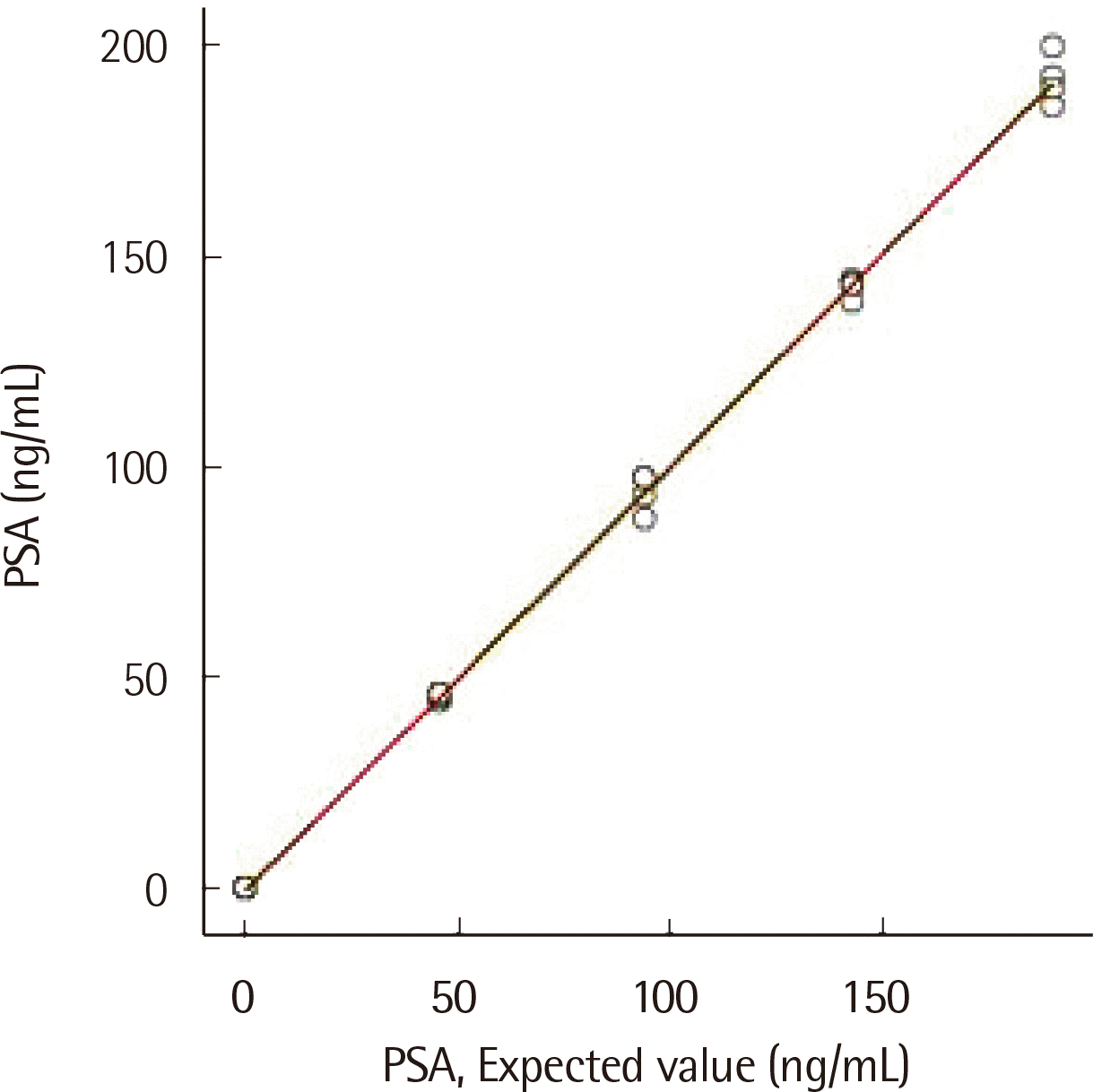
2) 직선성(Linearity)
직선성은 저농도(0.002 ng/mL) 및 고농도(189.900 ng/mL) 환자 검체를 혼합하여 5단계 농도를 사용하여 측정했다: 저농도(L), 0.25*L+0.75*H (저농도와 고농도 1:3 혼합), 0.5*H+0.5*L (저농도와 고농도 1:1 혼합), 0.75*L+0.25H (저농도와 고농도 3:1 혼합), 고농도(H) [18]. 각 검체를 4회 측정하고 평균값과 기대값을 비교했으며 1차, 2차, 3차 다항식 회귀 분석을 수행했다. 허용 가능한 비직선성(non-linearity)은 PSA의 바이어스(bias) 기준인 18.70%로 하였으며[17], 제조사에서 제시한 PSA의 직선성의 범위는 0.003-200 ng/mL이다.
3) 검출한계
두 개의 공시료 검체(P1, P2) 및 4단계로 희석된 검체(P3-P6)가 사용되었다. 모든 검체는 6일 동안 10회 측정되었다[19]. P3-P6는 제조사에서 제공하는 희석액을 사용하였다. 제조사가 제시한 공시료 검출한계(limit of blank, LOB) 및 검출한계(limit of detection, LOD)는 각각 0.00037, 0.08000 ng/mL이며, LOB와 LOD는 95% 신뢰수준에서 다음과 같이 계산하였다:
LOB= μB + 1.645σB (μB, 공시료 검체의 평균, σB; 공시료 검체의 표준편차)
LOD=LOB + 1.645σs (σs; 최저 농도의 표준편차).
5) 장비 간 비교평가(Method comparison)
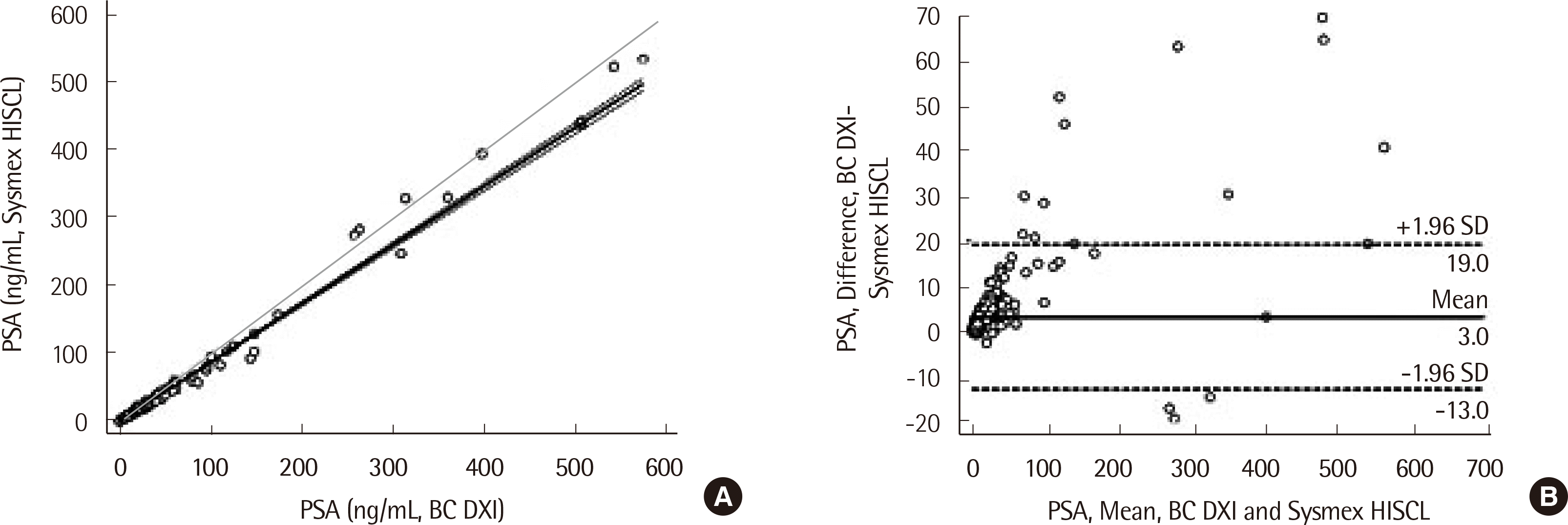
장비 간 비교를 위해 전립선암 환자 검체 79개, 전립선비대증 환자 검체 187개, 전립선염 환자 9명, 다른 악성 질환 환자 검체 12개, 그리고 다른 양성 질환(요로 감염 등) 환자 검체 76개를 사용했다. PSA 검체는 직선성 설정을 위해 사용되었다(0.003-200 ng/mL). HISCL-5000 장비 및 UniCel DxI 800 장비를 사용하여 비교하였으며, PSA는 0.01에서 541.56 ng/mL까지 측정되었고 UniCel DxI 800 장비의 변이계수 값은 2.16-3.85%였으며, 회귀분석을 통해 선형적합(linear fit)직선을 구하였다[21].
6) PSA의 임상적 평가
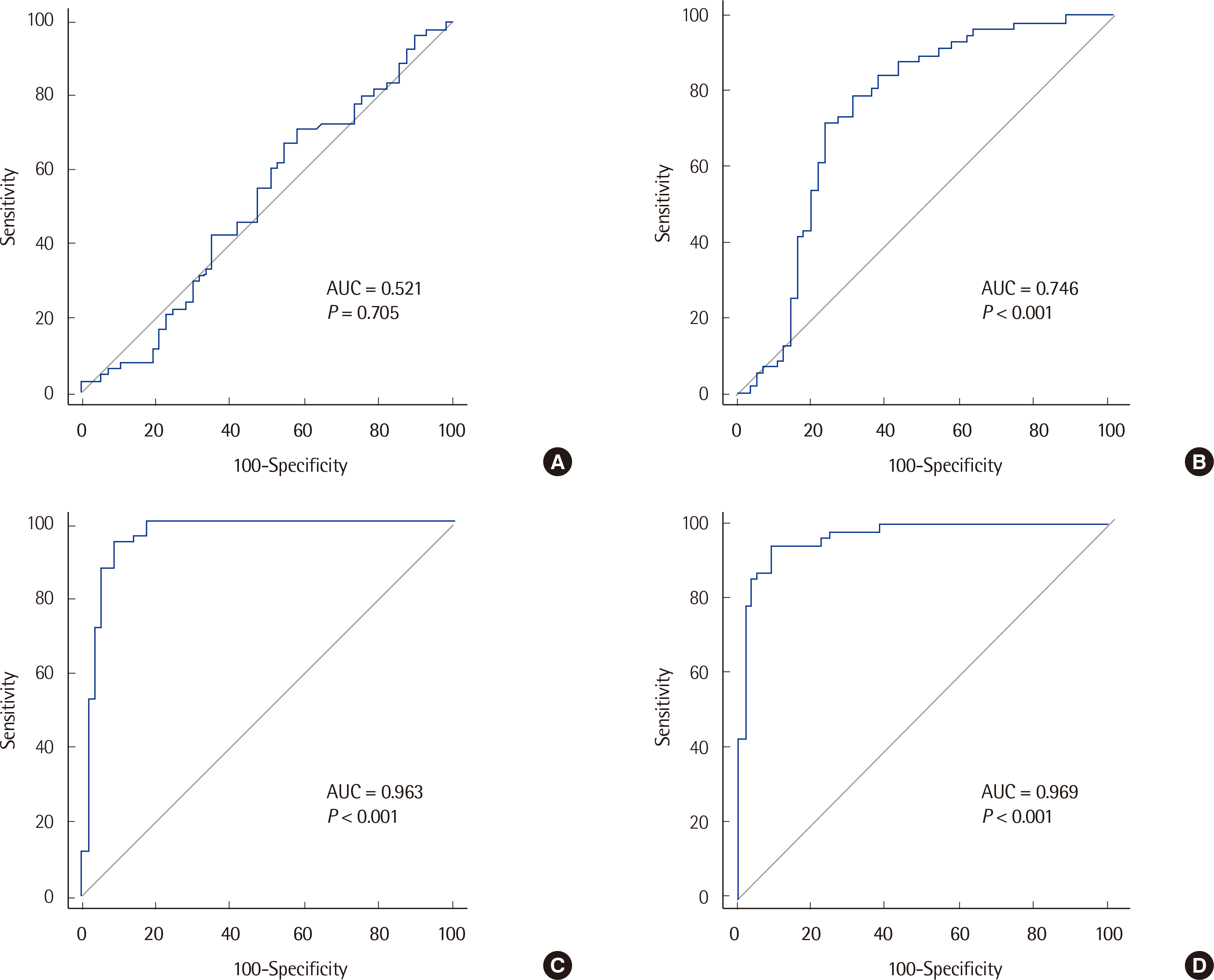
정상, 양성 질환, 악성 질환 그룹에서 각각 무작위로 57개의 검체를 선택하여[22] 각 그룹의 PSA 농도를 곡선하면적(AUROC) 분석으로 비교했다. 전립선암 및 전립선비대증에 대한 PSA의 진단 시 적용 가능성을 평가하였다.
7) 통계분석 프로그램
분석능 계산에 R 소프트웨어, 버전 3.4.4(Free Software Foundation, Inc., Boston, MA, USA)가 사용되었으며, 메드칼크, 버전 18.2.1 (MedCalc Software, Mariakerke, Belgium)을 사용하여 장비 간 비교 및 곡선하면적을 계산했다. 곡선하면적 분석을 위해 표본 크기는 다음과 같은 가정을 기반으로 계산되었다: 제1종 오류, 0.05; 제2종 오류; 음성/양성 그룹의 검체 크기 비율이 1인 경우 57개의 대조군 및 질환 검체가 필요. 대조군 및 질환 검체는 무작위로 선택되었다[22]. 범주형 데이터에 대해 카이제곱검정(Chi-square test) 혹은 피셔정확검정(Fischer’s exact test)을 수행했다.
결 과
1. 분석능
정밀 분석 결과 검사차례내, 검사차례간, 날짜간 및 검사실내변이계수 범위는 1.47-8.50%으로 확인되었다(Table 1). 분석된 모든 변이계수는 9.10% 이하로 이상적인 범위 내에 있었다. 직선성은 저농도와 고농도 정도관리 물질을 이용하여 확인하였다(Fig. 1). 기대값(x)과 측정값의 회귀 분석은 y=0.002+0.9999x (R2=0.99)였다. 바이어스 범위는 -9.70에서 11.16%까지였으며 생물학적 변이(18.70%) 내에 해당되었다. 공시료 검출한계 및 검출한계는 다음과 같이 각각 0.007 ng/mL, 0.020 ng/mL로 산출하였다. 정확성 평가에서 측정값은 기대값의 검증 간격 내에 있었고 바이어스 또한 허용 범위 안에 있었으며 가장 큰 바이어스(11.16%)는 최저 농도에서 확인되었다(Table 2). 장비 간 비교를 위해 질환군(363개)의 PSA 검체를 사용하였고, 상관계수는 0.99 (P<0.001), 회귀 방정식은 y= −1.0605 + 0.9223x (R2=0.99)이었는데 농도에 따른 계통오차는 없는 것으로 판단하였다(Fig. 2).
고 찰
본 연구에서는 HISCL-5000 장비의 분석능을 평가하고 PSA의 전립선암 및 전립선비대증 진단능을 곡선하면적을 사용하여 분석했다. 총 변이계수는 1.47-8.50%로서, 비정밀도 기준인 9.10% 내에 있었으며[23] 이전 보고들과도 일치하였다[24, 25]. 직선성은 저농도(0.002 ng/mL)에서 고농도(189.900 ng/mL)까지 관찰하였고, 이는 제조사에서 제시한 직선성 구간 내에 포함되었다. 정확성 검증을 위한 기대값은 검증 간격 내에 있었다. 일치도 평가를 위해 383개의 비정상 PSA 검체가 사용되었으며, PSA의 상관계수는 이전의 연구와도 유사한 수준(0.99, P<0.001)으로 확인되었다[26]. 이와 같은 높은 수준의 검출 한계와 낮은 농도에서의 정밀도는 다른 보고의 결과와 비교해도 임상에서 필요로 하는 PSA 보고에 적합하다[27, 28].
전립선암 선별 검사에서 PSA의 임상적 유용성은 여전히 불분명하고 논란의 여지가 있으며[11], 본 연구의 결과에서도 확인된다. PSA는 기관 특이적인 것으로 알려져 있지만, 본 연구에서는 PSA가 전립선염, 요로 질환 등 양성 질환의 영향을 받는 것으로 확인되었다. 곡선하면적 분석에 의한 전립선암과 전립선비대증의 곡선하면적은 각각 0.75, 0.52였다. 정상인과 비교했을 때는 전립선암과 전립선비대증의 곡선하면적이 0.97과 0.96이었다. 실제 임상 환경에서는 전립선염, 요로 질환 등의 양성 질환자가 있을 수 있기 때문에 PSA의 전립선암 진단 유용성은 떨어질 수 있다.
본 연구의 한계점으로는 PSA의 임상적 검증이 완전히 평가되지 않았다는 것이다. 현재로서는 표준화된 치료를 받는 환자의 치료 효과 평가, 수술 혹은 항암 후의 전립선암 환자의 예후를 확인하는데 유용하다.
결론적으로, HISCL-5000 장비에 의한 PSA 측정은 신뢰할 수 있는 성능을 보였으며 결과값은 평가 기준 내에 있었다. PSA의 임상적인 적용은 신중하게 고려해야 하며 추가 연구가 필요할 것이다.
 XML Download