

 PDF
PDF Citation
 Print
Print


INTRODUCTION
Carbon monoxide (CO) is a harmful substance that is a common cause of morbidity and mortality from poisoning [1]. However, CO is also endogenously synthesized upon the heme degradation by heme oxygenases (HOs). Atrial and ventricular cardiac myocytes constitutively express HO-2, and inducible HO-1 is increased by various stress factors [2], including myocardial infarction [3]. In addition, HOs prevent myocardial infarction [4], heart failure [5], and ischemia-reperfusion injury [4,6].
CO is an important cellular messenger that is considered to be both a physiological signaling molecule like nitric oxide (NO) and a potential therapeutic [7]. CO is being studied as an inhalation therapy with numerous potential clinical applications [7]. CO shows positive effects on the cardiovascular system, including vasodilatory effects [8,9], anti-apoptotic activity [10,11], and immune modulation effects [12]. These properties may be executed in conjunction with NO [13].
Recently, ion channels have been recognized as important effectors in CO activities. CO has a wide range of ion channel targets, including K+ channels, that play key roles in its beneficial effects. K+ channels play a critical role in cardiac electrophysiology, and their dysfunction is linked to intracellular signaling, metabolism, remodeling, and arrhythmogenesis in many cardiovascular diseases [14]. Voltage-dependent K+ channels (VDKCs) regulate resting membrane potential, proliferation, and contractile responses in the heart. There are two types of VDKCs: delayed rectifier K+ (Kv) channels and large-conductance Ca2+-activated K+ (BK) channels. Each K+ channel type has distinct kinetics and regulation.
Cardiac Kv channels conduct K+ currents during the plateau phase of action potentials and play pivotal roles in cardiac repolarization, cardiac physiology, and pathophysiology. Disruption of normal Kv channel functions renders the heart susceptible to abnormal electrical activity and predisposes to arrhythmia. Inherited mutations or drug blockage of Kv channels can cause cardiac arrhythmias [15].
CO activates the BK channel [16] and TREK1 channel [17] and inhibits the Kv2.1 channel [11] and the inward rectifier K+ channel [18]. CO also induces both activation and inhibition of the epithelial Na+ channel [19,20] and L-type Ca2+ channel [21-23]. CO modulates these proteins through a variety of different mechanisms, although the precise mechanism by which CO differentially regulates each of these ion channels remains controversial.
Human cardiac fibroblasts (HCFs) are the most abundant cell type in the heart, making up about two-thirds of the cardiac cellular population [24,25]. Although cardiac myocytes occupy approximately 75% of normal myocardial tissue volume, they only account for 30%–40% of cell numbers [26]. Therefore, HCFs are crucial for maintaining the cardiac extracellular matrix [27] and play a relevant role in myocardial structuring, cell signaling, and electro-mechanical function in healthy and diseased myocardium [24,28,29]. These cells are capable of synchronizing the electrical activity of multicellular cardiac tissue over extended distances through electrotonic interactions. This synchronization is accompanied by extremely large local conduction delays, which might contribute to arrhythmia generation in fibrotic hearts [30].
HCFs are non-excitable cells, however, they have multiple ion currents whose distribution and properties are distinct from cardiomyocytes [31-33]. They can affect cardiomyocyte electrical activity [30] and induce arrhythmogenesis [34]. The direct electrical coupling between fibroblasts and ventricular cardiomyocytes has been demonstrated in co-culture conditions and the whole heart [17,24,30] via connexin-based gap junctions [35] and indirectly via the extracellular matrix and the release of soluble mediators [24].
Kv and BK channels are the main K+ channels in HCFs [32,36]. CO activates the BK channels of HCFs through protein kinase G (PKG), protein kinase A (PKA), and S-nitrosylation [37], but the CO effect and related mechanism for the Kv channels of HCFs are still unclear. We identified the effect of CO on Kv channels of HCFs and the involved signaling pathways using the whole-cell mode patch clamp technique.
Go to : 
METHODS
Cell culture and reagents
We used commercial human adult ventricular cardiac fibroblasts (HCF-av, Cat #6310; ScienCell, San Diego, CA, USA). Cells were cultured in Dulbecco’s Modified Eagle’s Medium (DMEM; Welgene, Gyeongsan, Korea) with fetal bovine serum (10%; Welgene) and penicillin-streptomycin solution (100×; GenDEPOT, Barker, TX, USA) in an incubator with a humidified atmosphere of 5% CO2 and 95% air at 37°C. Confluent fibroblasts were detached by incubation with trypsin (0.25%; Welgene) and ethylene diamine tetraacetic acid (0.02%) in DMEM for several minutes. The detached cells were pelleted by centrifugation and then the supernatant was removed. The pellet was suspended in 1 mL of bath solution and the cells used in this study. Only cells in early passages (P4 to P7) were used to limit possible culture variation. Passage (P) is the number of times the cells are processed with trypsin and transferred to another flask.
Electrophysiological recordings
The Axopatch 200B Patch Clamp Amplifier (Axon Instruments, Foster City, CA, USA) was used for whole-cell mode patch clamping to record K+ currents from single HCFs. The K+ currents were filtered at 2 kHz and digitized at 10 kHz. pCLAMP 9.0 software (Axon Instruments) was used for data acquisition and analysis of the whole-cell currents. The recording patch pipettes were prepared from filament-containing borosilicate tubes (TW150F-4; World Precision Instruments, Sarasota, FL, USA) using a two-stage microelectrode puller (PC-10; Narishige, Tokyo, Japan) and were fire-polished using a microforge (MF-830; Narishige). The pipetted material exhibited a resistance of 2–3 MΩ. All electrophysiological experiments were carried out at room temperature. The bath solution to record delayed rectifier K+ currents (IK) contained 150 mM NaCl, 5.4 mM KCl, 1 mM CaCl2, 2 mM MgCl2, 10 mM glucose, and 5 mM HEPES (pH adjusted to 7.35 with NaOH). The pipette solution contained 130 mM KCl, 1 mM CaCl2, 2 mM MgCl2, 10 mM HEPES, 10 mM EGTA, and 2 mM Mg-ATP (pH adjusted to 7.3 with KOH). All chemicals were purchased from Sigma-Aldrich (St. Louis, MO, USA). To record only the IK of the cells, 100 nM iberiotoxin (a specific large-conductance calcium-activated K+ channel blocker) was added to the bath solution and 10 mM EGTA was added to the pipette solution during the experiments.
Statistical analysis
SPSS version 22.0 software (IBM Corp., Armonk, NY, USA) was used for statistical analysis. The paired Student’s t-test was used to evaluate differences between the means of two groups, whereas one-way analysis of variance was used for multiple groups. p-values < 0.05 were considered statistically significant. The results are presented as the mean ± standard error of the mean (SEM).
Go to :
RESULTS
CORM3 activated IK in HCFs
We recorded macroscopic outward K+ currents of HCFs using whole-cell mode patch clamping with a voltage protocol that consisted of depolarizing steps (from −80 mV to +50 mV) in 10 mV increments for 400 ms with −80 mV of holding potential. The recorded K+ currents of the HCFs showed behavior typical of IK: voltage dependency, fast activating but not inactivating properties to depolarization, and maintenance throughout the test pulse (Fig. 1). The K+ currents were blocked by 1 µM diphenyl phosphine oxide-1 (DPO-1), a specific blocker of the Kv1.5 channel and ultra-rapid type IK (–94.5 ± 0.2% of control, at +30 mV, n = 7, Fig. 1A, B).
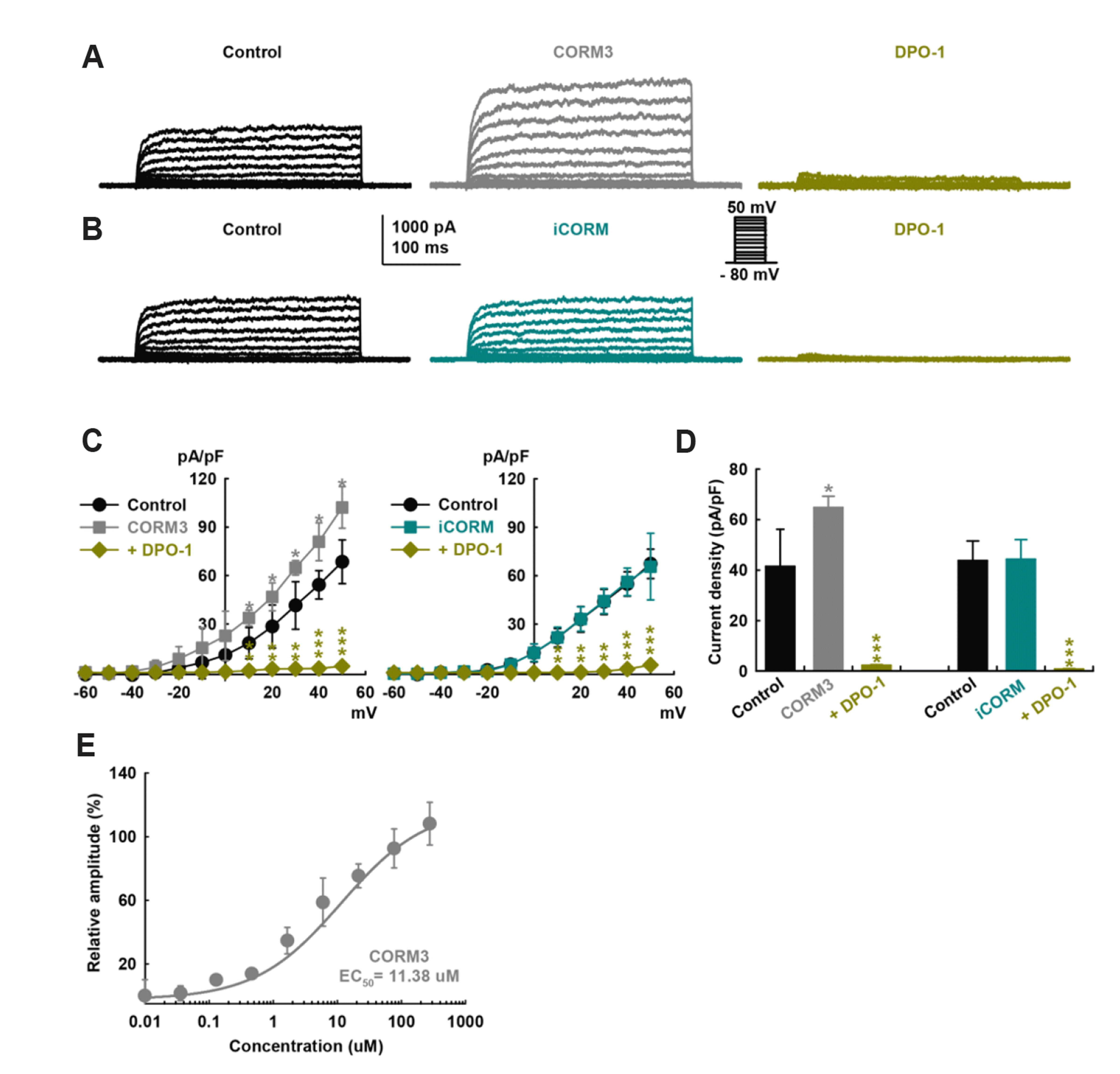 | Fig. 1Carbon monoxide (CO) increases delayed rectifier K+ currents (IK) of human cardiac fibroblasts (HCFs).Original recordings of the K+ currents were obtained by repeated voltage step depolarization from −80 to +50 mV for 400 ms duration (holding potential was –80 mV) by whole-cell mode patch clamp recordings. (A) Representative outward K+ currents show the changes before (control) and after application of (A) carbon monoxide-releasing molecule-3 (CORM3, a CO donor, 10 μM) or (B) iCORM3 (inactive CORM3, 10 μM). The currents were blocked by diphenyl phosphine oxide-1 (DPO-1, a blocker of the delayed rectifier K+ channel and the Kv1.5 channel, 1 μM). (C) Summarized current–voltage (I–V) curves of steady-state currents for the effects of CORM3, iCORM3, and DPO-1. (D) Bar graphs showing the current density changes of the K+ currents at +30 mV regarding the effects of CORM3, iCORM3, and DPO-1 (n = 7, each). (E) Concentration-dependent activation curve of IK by CORM3. The solid line shows the fit based on a standard dose-response relationship, which yielded an estimated half maximal effective concentration (EC50) of 11.38 μM for CORM3. Values are mean ± SEM, *p < 0.05, **p < 0.01, and ***p < 0.001 compared to control (n = 7, each).
|
CORM3 (10 μM, a CO donor) significantly increased the IK amplitude (+56.2 ± 3.1% of control, at +30 mV, n = 7, Fig. 1A). iCORM3 (the inactive form of CORM3) had no significant effect on the currents (10 µM, +1.2 ± 9.9% of control, at +30 mV, n = 7, Fig. 1B). In current–voltage (I–V) curves (Fig. 1C), the currents were activated from –20 mV and also did not show strong outward rectification, a characteristic of large-conductance Ca2+-activated K+ currents (IBK). IBK is the other main K+ current of HCFs. We added iberiotoxin (100 nM, the specific BK channel blocker) to the bath solution and 10 mM EGTA to the pipette solution to block IBK (*p < 0.05, **p < 0.01, ***p < 0.001). CORM3 increased IK current densities (41.5 ± 14.5 pA/pF at control, 64.8 ± 4.4 pA/pF at CORM3, 2.28 ± 0.27 pA/pF at DPO-1, +30 mV, n = 7, *p < 0.05, ***p < 0.001, Fig. 1D). CORM3’s concentration-response relationship showed that steady-state currents normalized by control data were fitted with the Hill equation, producing an estimated half-maximal excitation concentration (EC50) value of 11.38 µM and a Hill coefficient of 0.7 (n = 7, Fig. 1E).
Nitric oxide synthase (NOS)/ NO involvement in CO–induced IK activation of HCFs
To investigate the regulation of IK by CO, we first explored the involvement of NO because CO is known to activate NOS and soluble guanylate cyclase (sGC) [22]. The CORM3-induced IK activation was significantly suppressed by pretreatment with a NOS inhibitor, L-NG-monomethyl arginine citrate (L-NMMA, 100 µM) for 20 min (–10.2 ± 9.3% of control, n = 7, Fig. 2A, B). Pretreatment of cells with other NOS blocker, L-NG-nitroarginine methyl ester (L-NAME, 100 µM) also attenuated the CORM3-induced IK effect (+4.0 ± 11.7% of control, n = 7, Fig. 2D, E). L-NMMA or L-NAME itself did not affect IK amplitude. The current densities at +30 mV of the CORM3 effects after L-NMMA or L-NAME pretreatment did not change (n = 7 each, Fig. 2C, F), suggesting that CO could activate IK through NO formation by NOS.
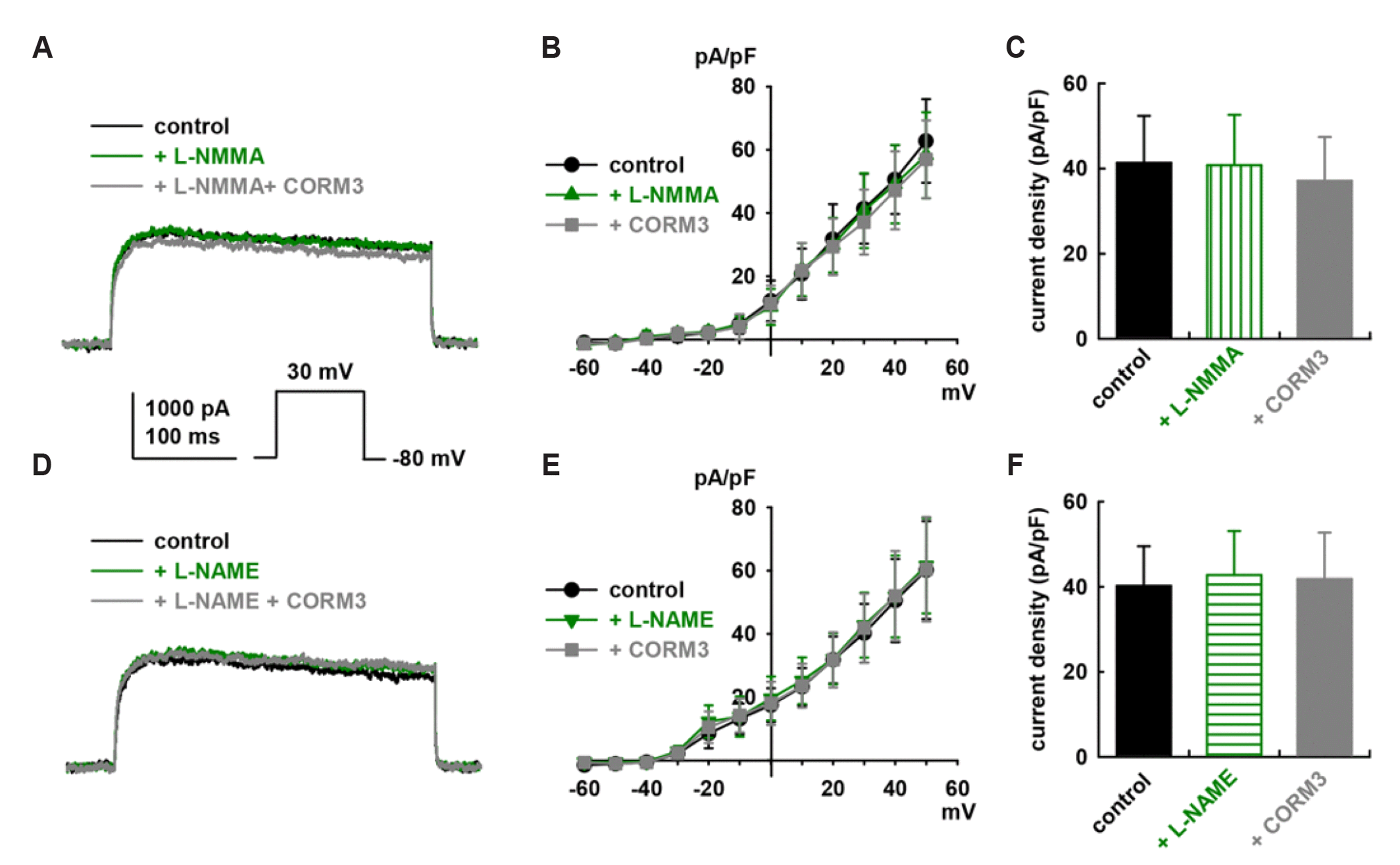 | Fig. 2Nitric oxide synthase (NOS) blockers inhibits carbon monoxide (CO)–induced activation of delayed rectifier K+ currents (IK) in human cardiac fibroblasts (HCFs).Currents are obtained in which cells were repeatedly depolarized from −80 mV to +50 mV (400 ms duration). carbon monoxide-releasing molecule-3 (CORM3) was applied either to untreated cells or to cells pretreated for 20 min with NOS blockers. (A) Representative original recordings of IK show the CORM3 (10 μM) effect after pretreatment of L-NG-monomethyl arginine citrate (L-NMMA, 100 μM) for 20 min. (B) Summarized current–voltage (I–V) relationship curves for IK and (C) bar graphs for the summary of current density changes of IK (at +30 mV) also show the effect of CORM3 (10 μM) on IK after pretreatment of L-NMMA (100 μM). (D) Representative original recordings of IK. (E) Summarized I–V relationship curves of IK, and (F) bar graphs for the summary of current density changes of IK (at +30 mV) also show the CORM3 (10 μM) effect on IK after L-NG-nitroarginine methyl ester (L-NAME, 100 μM) pretreatment (n = 7 each). Values are mean ± SEM (n = 7 each).
|
cGMP and cAMP signaling pathway involvment in CO-induced IK activation of HCFs
We tested the stimulating effect of CORM3 on IK was mediated by the cGMP signaling pathway. Under pretreatment by KT5823 (a PKG inhibitor, 1 µM), 10 µM CORM3 failed to increase IK (+1.3 ± 11.3% of the control, n = 7, Fig. 3A). With ODQ (a soluble guanylate cyclase inhibitor, 10 µM) pretreatment, CORM3 could not increase IK (–4.9 ± 8.8% of the control, n = 7, Fig. 3B). The current densities did not change significantly (n = 7, each, Fig. 3C).
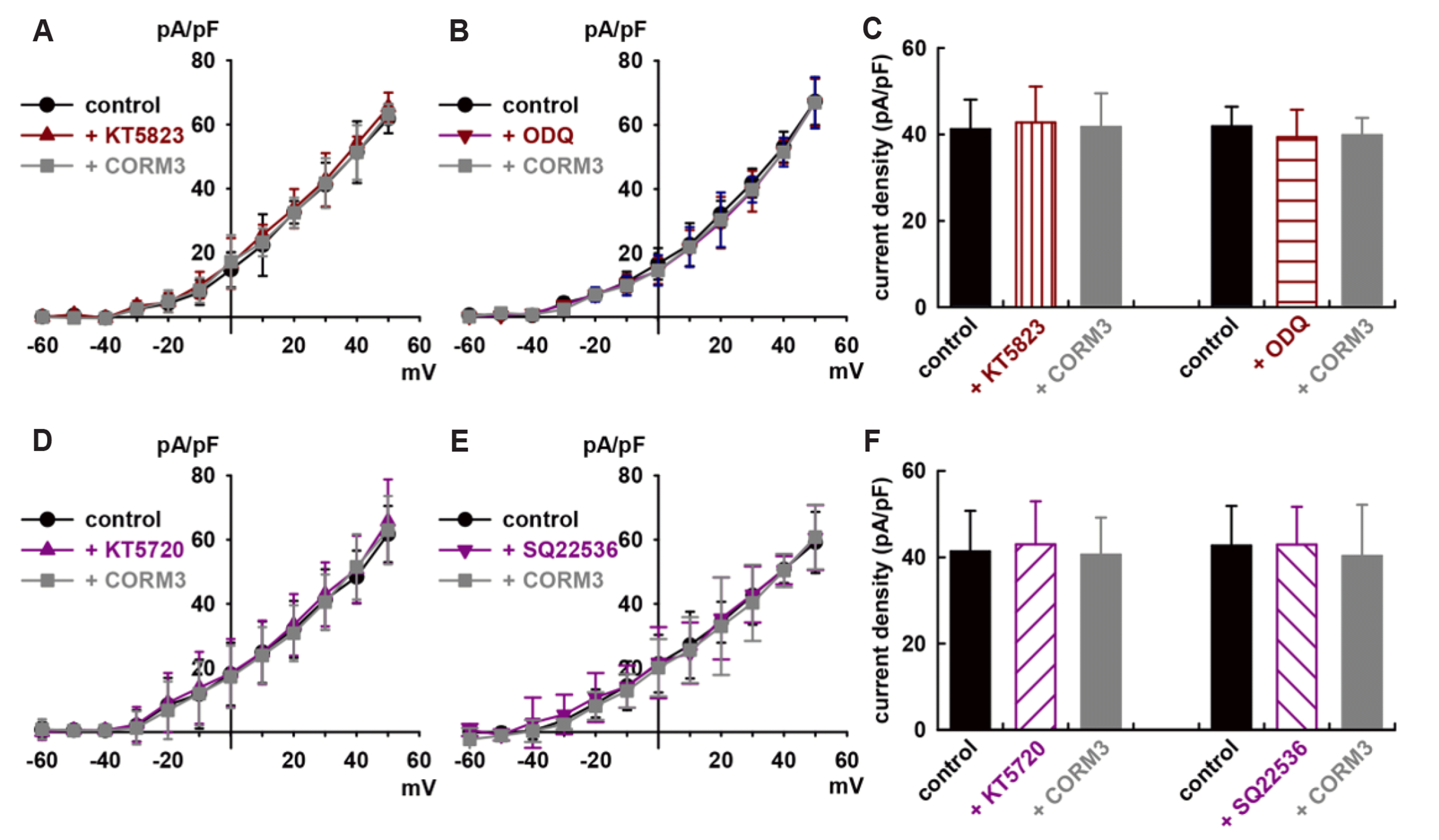 | Fig. 3cGMP and cAMP signaling pathways involved in carbon monoxide (CO)–induced activation of delayed rectifier K+ currents (IK) in human cardiac fibroblasts (HCFs).Summarized current–voltage (I–V) curves from –60 mV to +50 mV show the effects of carbon monoxide-releasing molecule-3 (CORM3, 10 μM) on IK after pretreatments with (A) KT5823 (a PKG blocker, 1 μM) or (B) ODQ (a soluble guanylate cyclase blocker, 1 μM) (n = 7 each). (C) Bar graphs show summarizing current density changes for the CORM3 effects on IK after pretreatments with KT5823 or ODQ (at +30 mV). Summarized I–V curves for the effects of CORM3 on IK after pre-incubation with (D) KT5720 (a PKA blocker, 1 μM) or (E) SQ22536 (an adenylate cyclase blocker, 1 μM). (F) Bar graphs show current density changes at +30 mV for the effects of CORM3 on IK after pre-incubation with KT5720 or SQ22536 (n = 7 each). Values are mean ± SEM.
|
We also tested whether the cAMP signaling pathway is involved in CO’s effect of CO on IK in cells. A KT5720 (a PKA inhibitor, 1 µM) pretreatment for 20 min inhibited CORM3’s stimulation effect of on IK (–1.8 ± 9.1% of the control, n = 7, Fig. 3D). A SQ22536 (an adenylate cyclase inhibitor, 100 µM) pretreatment also inhibited the CORM3 effect (–5.6 ± 12.9% of the control, n = 7, Fig. 3E). There were no significant changes in current density by CORM3 after KT5720 or SQ22536 pretreatment (Fig. 3F). These results suggest that both cGMP and cAMP signaling pathways are involved in CO’s effects on IK in HCFs.
S-nitrosylation involvement in CO-induced IK activation of HCFs
We examined whether CO activated IK via direct S-nitrosylation of the target proteins’ thiol residues. An alternative pathway for NO’s biological effects because CO activates IBK through this mechanism [37]. With N-ethylmaleimide (NEM, a thiol-alkylating reagent, 0.5 mM) pretreatment for 20 min, CORM3 failed to increase IK (–1.28 ± 10.1% of the control, n = 7, Fig. 4A, B). The current densities of the IK were unchanged by CORM3 after NEM treatment (Fig. 4C), which suggests that the ultimate target of CO is a thiol residue. NEM alone did not increase the currents. DL-dithiothreitol (DTT, a reducing agent, 5 mM) reversed CORM3-induced enhancement of IK (CORM3, +68.6 ± 25.6% of the control; *p < 0.05 vs. control; DTT, +14.7 ± 13.9% of control, n = 7, #p < 0.05 vs. CORM3, Fig. 4D, E). The current densities of the IK were significantly increased by CORM3 (42.5 ± 3.5 pA/pF at control, 71.6 ± 8.9 pA/pF at CORM3, n = 7, *p < 0.05) and reversed by DTT significantly (48.7 ± 4.8 pA/pF, n = 7, #p < 0.05 vs. CORM3, Fig. 4F). We conclude that CO also activates IK of HCFs through S-nitrosylation.
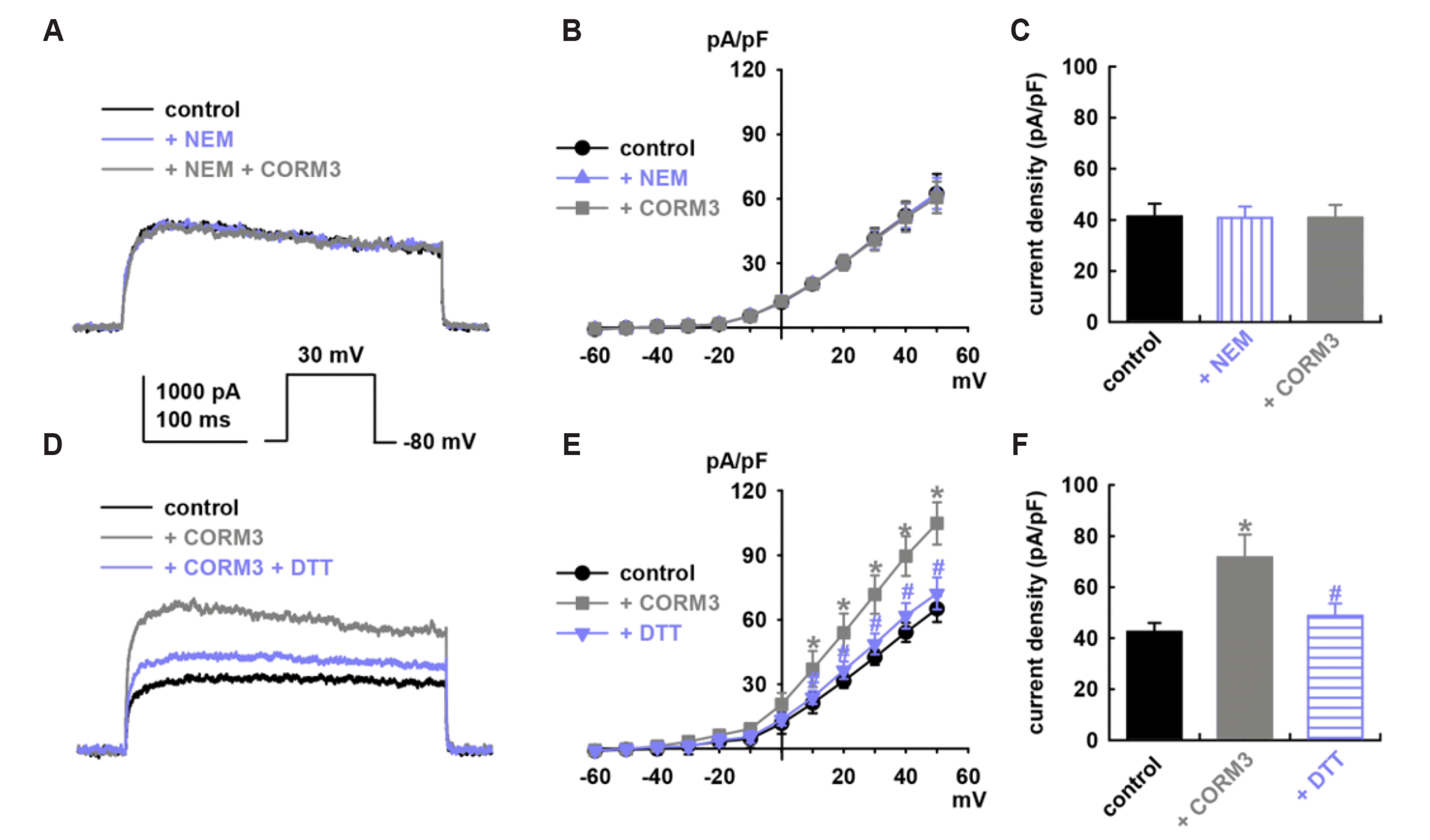 | Fig. 4S-nitrosylation involves carbon monoxide (CO)–induced activation of delayed rectifier K+ currents (IK) in human cardiac fibroblasts (HCFs).(A) Original recordings for the effect of carbon monoxide-releasing molecule-3 (CORM3, 10 μM) on IK after pretreatment with N-ethylmaleimide (NEM, a thiol-alkylating reagent, 0.5 mM) at +30 mV stimulation. (B) Summarized current–voltage (I–V) curves for the effect of CORM3 (10 μM) on IK after pretreatment with NEM (0.5 mM). (C) Bar graphs showing the current density changes for the CORM3 effects on IK after pretreatment with NEM (at +30 mV, n = 7 each). (D–F) The reversing effect of DL-dithiothreitol (DTT, a reducing agent, 5 mM) for CORM3-induced activation on IK (*p < 0.05 compared to control, #p < 0.05 compared to CORM3, n = 7 each).
|
MAPK signaling pathways involvement in CO-induced IK activation of HCFs
We also tested the mitogen-activated protein kinase (MAPK) signaling pathway for the CO-induced activation of IK because MAPKs are believed to be involved in the regulation of Kv channels [38]. Pretreatment with SB239063 (a p38 MAPK inhibitor,10 µM) inhibited the CORM3 effect on IK (–11.9 ± 7.7% of the control, n = 7, Fig. 5A). Pretreatment with PD98059 (a p44/42 MAPK inhibitor, 10 µM) also inhibited CO-induced IK activation (–3.7 ± 4.8% of the control, n = 7, Fig. 5B). SB239063 (10 µM) or PD98059 (10 µM) did not increase the current density of IK of HCFs (Fig. 5C). These results suggest that the MAPK signaling pathway is also involved in the CO-induced activation of the IK effect.
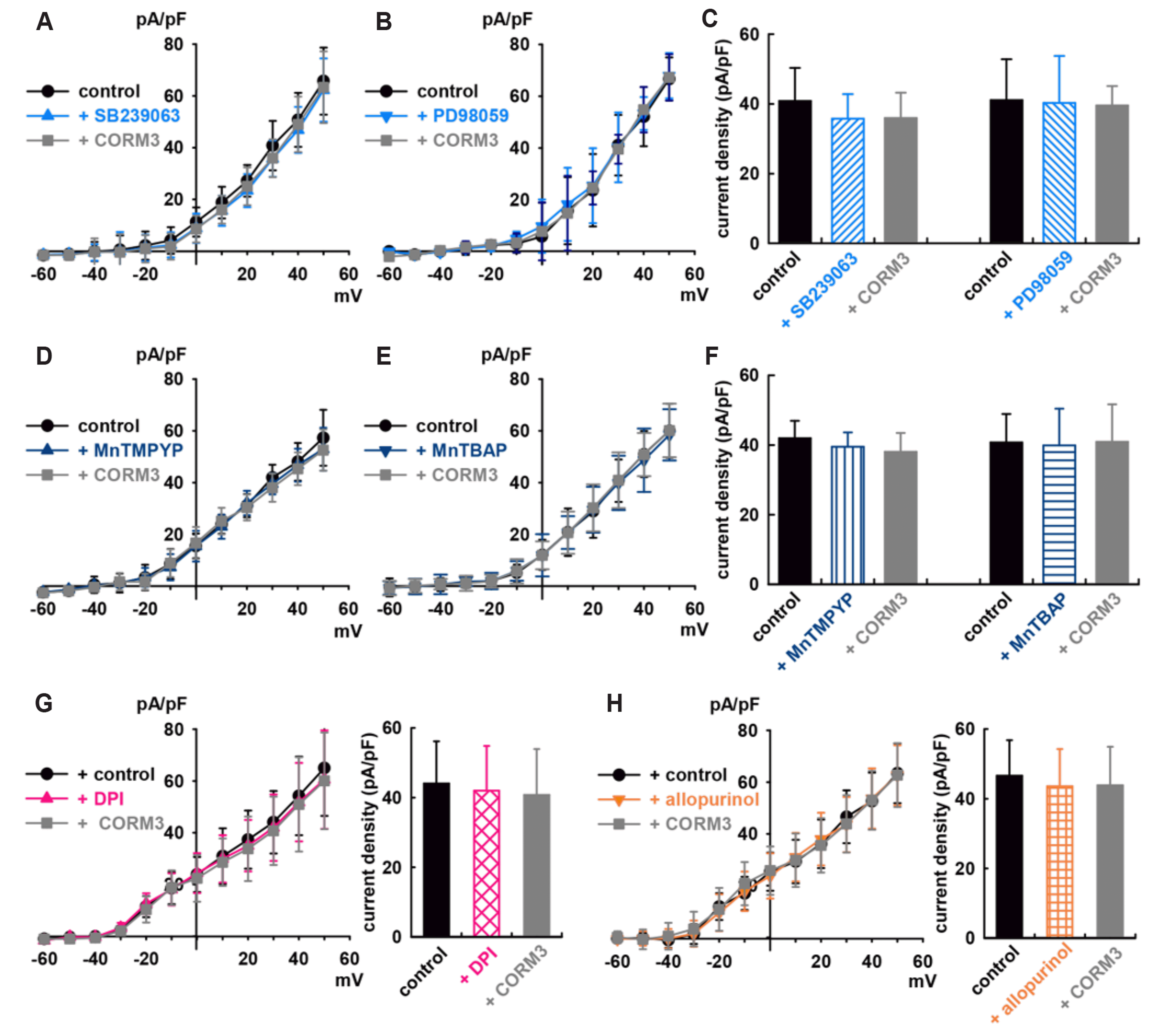 | Fig. 5Effect of mitogen-activated protein kinase (MAPK) and redox signaling pathways on carbon monoxide (CO)-induced activation of delayed rectifier K+ currents (IK) in human cardiac fibroblasts (HCFs).(A, B) Summarized current–voltage (I–V) curves and (C) bar graphs of current density changes for the carbon monoxide-releasing molecule-3 (CORM3, 10 μM) effects on IK after pretreatment with SB239063 (a p38 MAPK inhibitor, 10 μM) or PD98059 (a p44/42 MAPK inhibitor, 10 μM), n = 7 (each). (D, E) Summarized I–V curves and (F) bar graphs for the effects of 10 μM CORM3 on IK after pretreatments with superoxide dismutase mimetics; MnTMPYP (50 μM) or MnTBAP (10 μM), n = 7 (each). Summarized I–V curves and bar graphs of current density changes for the effects of CORM3 on IK after (G) diphenylene iodonium (DPI, a NADPH oxidase inhibitor, 3 μM, n = 7) or (H) allopurinol (a xanthine oxidase inhibitor, 1 μM, n = 7).
|
Redox signaling pathway involvement in CO-induced IK activation of HCFs
To explore the source of reactive oxygen species (ROS) involved in CO-mediated activation of IK of HCFs, cells were pretreated with two superoxide dismutase (SOD) mimetics, 5,10,15,20-tetrakis(1-methylpyridinium-4-yl)-21H,23H porphyrin manganese (III) pentachloride (MnTMPyP, 50 µM) and manganese (III) tetrakis (4-benzoic acid) porphyrin chloride (MnTBAP, 10 µM). Both inhibited CO-induced IK activation (–9.3 ± 10.9% of the control, n = 7, Fig. 5D; +0.4 ± 13.1% of the control, n = 7, Fig. 5E). MnTMPyP (50 µM) and MnTBAP (10 µM) did not increase IK of HCFs (Fig. 5F). Diphenylene iodonium (DPI; 3 µM, an inhibitor of NADPH oxidases) or allopurinol (1 µM, a xanthine oxidase inhibitor) inhibited the CORM3-induced activation on IK (–7.5 ± 10.9% of the control, n = 7, Fig. 5G; –5.9 ± 10.8% of the control, n = 7, Fig. 5H). These results strongly suggest that ROS contributes to CO-mediated activation of IK of HCFs.
Go to :
DISCUSSION
The major findings of the present study are CO activates IK of cultured HCFs. NO through NOS, phosphorylation by PKG, PKA, and MAPKs, S-nitrosylation and reduction/oxidation (redox) signaling are involved in the CO-induced activation of IK.
Identification of Kv channels in HCFs
In our experiments, the recorded outward K+ currents of HCFs in 100 nM iberiotoxin (a specific BK channel blocker) in the bath solution and 10 mM EGTA in the pipette solution showed typical characteristics of the IK (Fig. 1): rapid activation and no inactivation in kinetics, voltage dependency, and significant inhibition by DPO-1, a specific blocker for ultra-rapid type of IK (IKur) and hKv1.5 currents [39].
Kv channels include the slow, rapid, and ultra-rapid delayed rectifiers (IKs, IKr, and IKur) [14]. A functionally significant Kv channel with components corresponding to IKr and IKs is present in human ventricular cells, whereas IKur is only present in atrial myocytes [40,41]. These findings explain the molecular, physiological, and pharmacological determinants of human atrial and ventricular repolarization and arrhythmias [42].
Kv1.5 is a rapidly activating, voltage-gated K+ channel encoded by KCNA5 that inactivates slowly and incompletely [43,44]. Kv1.5 also contributes to repolarization of vascular smooth muscle cell membrane potential, limiting Ca2+ entry and vascular tone. It is essential for balancing coronary blood flow with the metabolic demands of the working myocardium [45]. This channel is thought to be the major contributor to the IK in the human heart [46,47]. Its expression is largely confined to the atria and generates the IKur, the major repolarizing current that is active throughout phases 1–3 of the atrial action potential [48,49], and determines the duration and membrane potential of the plateau phase [41]. Because IKur is atrium specific, Kv1.5 channel is promising target as new, safer antiarrhythmic drugs to prevent atrial fibrillation without the risk of inducing QT prolongation and ventricular arrhythmias. DPO-1 also increases action potential duration in atrial but not ventricular myocytes and prevents atrial arrhythmias [50].
However, the strong mRNA expression of Kv1.5 for the Kv channels is also in HCFs [32,51] and ventricular cardiomyocytes [52]. These findings may indicate a functional role of these ion channel subunits in action potential formation in the human atrium and ventricle. This channel is also highly expressed in most vascular smooth muscle cells and is important for regulating cell excitability and maintaining basal tone.
HCFs make up the largest cell population in the heart and have cell–cell communication with cardiomyocytes and other cells [27]. The cardiomyocyte–cardiac fibroblast interactions are important in normal heart function and the development of diseases such as cardiac arrhythmia and fibrosis [35]. Kv channels may be useful therapeutic targets for cardiovascular disease.
Effect of CO on IK in HCFs
To test the CO effect on IK, CORM3 was used as a CO donor because suitable delivery systems were required. CORMs are low-molecular weight compounds that release one or more CO molecules upon decomposition [9]. We achieved a reliable result for CO with CORM3 on IBK of HCFs [37].
In this experiment, CO stimulated IK in HCFs concentration-dependently (Fig. 1). This is the first report that CO induces activation for IK, to the best of our knowledge. In previous reports, CO inhibited IK in ventricular cardiomyocytes, native Kv1.5 recorded in HL-1 murine atrial cells [53], and recombinant Kv2.1 (KCNB1) expressed in HEK293 cells [11]. These differences could be due to differences in the cell type or expressed by other Kv channel mRNA. The native IK differ dramatically in terms of kinetics, amplitude, and drug response. Tissue and species–specific Kv gene expression contributes to the current heterogeneity [45]. HCFs also showed strong gene expression of Kv1.1, Kv1.2, and Kv3.1 in the Kv channels of these cells [51].
Effect of CO on IK through variable signaling pathways
NOS/NO pathway: To investigate the regulation of the IK channel by CO, the involvement of NO was first explored because CO and NO can cross–talk [54,55] and NO increases the expression of HO-1 [56,57].
We demonstrated that the activation of IK by CO in HCFs was abolished by pretreatment with NOS blockers (L-NMMA or L-NAME), which indicates that NO formation by CO–induced NOS stimulation has an important role in IK activation in HCFs by CO. These NO-dependent CO effects are also reported in the BK channels of HCFs [37] and L-type Ca2+ channels of intestinal smooth muscle cells [22]. However, CO inhibits recombinant Kv1.5 expressed in HEK293 cells through NO [53].
NO also showed a similar difference for IK. NO inhibited the hKv1.5 channel current, which activates IK in transfected Chinese hamster ovary cells and mouse ventricular myocytes [47] but also activated the current in HCFs [51], cardiomyocytes [58] and the sino-atrial node cells of guinea-pigs [59]. Considering the differing CO effects for IK, the importance of HCFs in the heart cell population, and the connection between other cell types of the myocardium, more research is required on CO as a therapeutic agent for cardiovascular disease.
sGC/cGMP/PKG pathway: Previously, we demonstrated that CO-mediated augmentation of the BK channel is NO-dependent and involves channel S-nitrosylation and the PKG and PKA pathways [37].
The present study shows that CO activates the IK of HCFs through the cGMP-dependent pathway (Fig. 3A–C). These results are consistent with previous reports that CO binding can result in sGC activation, leading to the cellular generation of cGMP [60,61] and the CO effect for Kv1.5 expressed in HEK293 cells through sGC [53].
In case of NO, NO regulates diverse target proteins through different modes of post-transcriptional modification sGC/cGMP/PKG-dependent phosphorylation [62]. NO also blocks hKv1.5 channels by cyclic GMP-dependent mechanism [47].
AC/cAMP/PKA pathway: We have also shown that the cAMP-dependent pathway is involved in CO-mediated activation of IK in HCFs (Fig. 3D–F). Our results indicate that both the PKG and PKA pathways are involved in the CO-induced activation of IK. NO also can regulate both adenylate cyclase and guanylate cyclase in cardiac myocytes and increase cAMP by activation of adenylate cyclase in cardiomyocytes [63]. CO also activates BK currents in HCFs through the cAMP-dependent pathway [37]. cAMP modulates significant alterations of cardiac electrical activity via a cGMP-dependent mechanism [64] and should be considered a novel regulator of cardiac electrophysiology.
S-nitrosylation: NO exerts ubiquitous signaling via post-translational modification of cysteine residues, a reaction termed S-nitrosylation. Specifically, S-nitrosylation modulates the major currents involved in the generation of the action potential and many cardiac ion channels such as the voltage-gated sodium channels, L-type Ca2+ channels, and voltage-gated potassium channels [58].
NO regulates diverse target proteins through S-nitrosylation [62] and blocks hKv1.5 channels by S-nitrosylation [47]. CO also caused S-nitrosylation of Kv1.5 of HL-1 murine atrial cells [53], and elevated NO levels in myocytes, and S-nitrosylation of the Nav1.5 channel [65] and BK channel of HCFs [66]. However, S-nitrosylation is not involved in the NO-dependent activation of IK in HCFs [51]. Therefore, this pathway suggests that a direct reaction of CO or a consequence of some interaction of CO with other mechanisms (e.g., MAPK or the redox signaling pathways) is required to activate IK channels.
MAPK pathway: Although the underlying mechanism and specific molecular targets involved are unknown, there is a significant body of evidence that indicates that CO can also interfere with MAPK-dependent pathway signaling [67,68]. MAPKs are involved in Kv channel modulation in VSMCs [69] and rat coronary arterial myocytes [38].
In our results, MAPK pathway inhibition with SB239063 (p38 MAPK inhibitor) or PD98059 (p44/42 MAPK inhibitor) depressed CO-induced IK stimulation. This suggests that MAPK pathway plays an important role in the CO-induced activation of IK channels in HCFs.
Activation of p38 MAPK by CO may involve upstream MAP kinase kinase-3 [12] or may involve the regulation of phosphatases or sGC activation (reviewed by [70]).
Redox signaling pathways: We also tested the redox signaling pathway for the CO effect because exposure to high amounts of CO inhibits mitochondrial respiration, generates ROS, and enhances ventricular arrhythmia after oxidative stress [71].
In our results, CO-induced IK activation was blocked by pretreatments with two superoxide dismutase mimetics: a nicotinamide adenine dinucleotide (NADPH) oxidase inhibitor, and a xanthine oxidase inhibitor (Fig. 5). CO-mediated activation of IK was partly attenuated by the SOD mimetics (MnTMPyP and MnTBAP). These agents presumably increase H2O2 levels through superoxide dismutation, and H2O2 has an augmenting effect on the Kv1.5 channel [53].
CO increases ROS and enhances ventricular arrhythmia after oxidative stress [71]. In the heart, redox signaling regulates several physiological processes (e.g., excitation–contraction coupling) and is involved in a wide variety of pathophysiological and homoeostatic or stress response pathways. ROS can be produced by a variety of enzyme systems associated with heart failure and is involved in cardiac redox signaling, derived from many sources, including xanthine oxidase [72], NOSs [73], and NADPH oxidases (NOXs) [74]. Among the ROS sources in the heart, NOXs are particularly important in redox signaling. NOX isoforms are expressed in multiple cell types, including cardiomyocytes and fibroblasts [75].
Modulation of the NOX1/NADPH oxidase signaling pathway may be a novel therapeutic strategy for preventing heart failure after myocardial injury [76]. NOX4-derived increase in ROS induces inhibition of the hKv1.5 channel [77]. HO-1 expression is increased in atrial fibrillation (AF) and appears to provide protection against the oxidative stress associated with this condition [78,79]. Given the important role of Kv1.5 in normal atrial function, its redox sensitivity and the likely involvement of HO-1 are protective in AF.
Data continues to establish CO as an important gasotransmitter alongside NO, which provides a range of beneficial cardiovascular effects. CO dilates coronary and other vessels [80] and HO-1 induction, which produces CO, protects against myocardial infarction, hypertension and vascular injury [68]. CO accounts for many of the effects of HO-1 induction [81-83] and CO inhalation, as well as CORMs, are being developed for cardiovascular therapy [7].
While several studies have shown the cardioprotective effect of CO or CORMs, their application has not yet reached clinical practice and their responses depend on the cell types [84,85].
Moreover, these variable responses to treatment in different tissues and organs could be accompanied by unexpected side effects. Therefore, it is still early to consider CO as a therapeutic, since it has only been shown to have a positive effect on specific individual organs. Our findings add to the growing understanding of the complexity of CO signaling in cardiac tissues by describing a new ion channel target for regulation.
In our experiments, CO significantly activated the IK of HCFs. Since the IK of HCFs was significantly inhibited by DPO-1, a specific Kv1.5 channel blocker, Kv1.5 channels appears to be the main component of IK. Effects of CO on Kv1.5 was reported by Al-Owais et al. [53]. However, different from our results, the voltage-gated Kv1.5 channels of HEK293 and HL-1 cells are inhibited by CO [53]. Even though the effects of CO in experimental studies differ depending on the cell or tissue in question, further evaluation is required. We also found multiple redundant inhibitory effects of various pharmacological inhibitors on the CO effects on the IK of HCFs. It was very difficult to suggest a single line of signaling cascade to explain the results obtained by the inhibitors of signaling pathways. Our results may demonstrate the possible involvement of multiple mechanisms for the effect of CO on the IK of HCFs. Further studies are necessary to describe how CO modulates the currents in HCFs.
Go to :
 XML Download