TO THE EDITOR: Minor
BCR-ABL1-positive chronic myeloid leukemia (CML) is a very rare subtype of CML, with only 1–2% of patients with CML exhibiting this fusion gene as a sole rearrangement [
1]. Amongst the several reported cases in literature, and to the best of our knowledge, only one case has been reported in Korea so far [
2]. In this letter, we report a Korean patient diagnosed with minor
BCR-ABL1-positive CML.
An 81-year-old male with a history of hypertension and dyslipidemia was referred to our hospital because of marked leukocytosis. The complete blood count (CBC) at referral indicated anemia, leukocytosis, and thrombocytopenia, with a hemoglobin (Hb) level of 7.5 g/dL, white blood cell (WBC) count of 37.59×10
9/L, and a platelet count of 89×10
9/L. The differential counts of WBC were myelocytes 1%, metamyelocytes 2%, band neutrophils 13%, neutrophils 45%, eosinophils 2%, basophils 6%, lymphocytes 12%, and monocytes 19% (
Fig. 1). Bone marrow (BM) biopsy and related cytogenetic studies were performed; bone marrow biopsy showed hypercellularity, with an estimated cellularity of 70–90% (
Fig. 2) with granulocytic proliferation. Additionally, the number of megakaryocytes and dwarf megakaryocytes had increased (
Fig. 3). Approximately 5.3% of all nucleated cells (ANCs) were counted as blasts, whereas the population co-expressing CD34(+), CD117(+), and myeloperoxidase corresponding to myeloblasts, was 2.28% of the total cells observed using flow cytometry (FCM). Pseudo-Gaucher cells were not observed. Monocytes accounted for 17.1% of the ANCs, consistent with the FCM results of 17.4%. Conventional chromosome analysis using BM cells exhibited 46,XY,t(9;22)(q34;q11.2)[20] (
Fig. 4). Reverse transcriptase-polymerase chain reaction (RT-PCR) showed a minor (P190)
BCR-ABL1 transcript of the e1a2 type (
Fig. 5). No major (P210) or micro (P230)
BCR-ABL1 transcripts were detected. Therefore, the patient was diagnosed with minor
BCR-ABL1-positive CML in chronic phase.
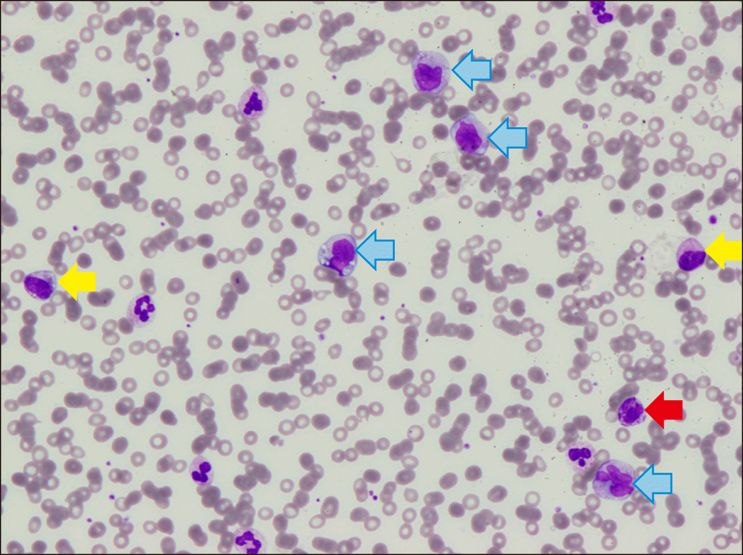 | Fig. 1A peripheral blood smear showing leukocytosis with monocytosis (blue arrows), basophilia (red arrow) and left shift (yellow arrow) (white blood cell count 37.59×10⁹/L with 0.19 monocytes and 0.04 basophils, Wright-Giemsa stain, ×400). 
|
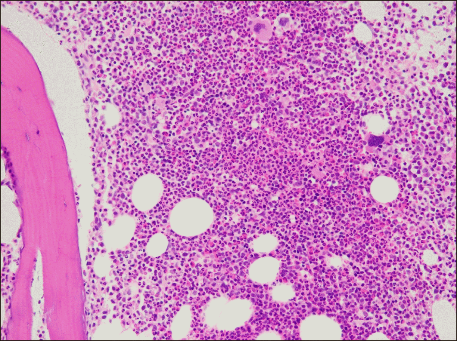 | Fig. 2Bone marrow biopsy showing hypercellularity (estimated cellularity of 70–90%, Hematoxylin & Eosin stain, ×200). 
|
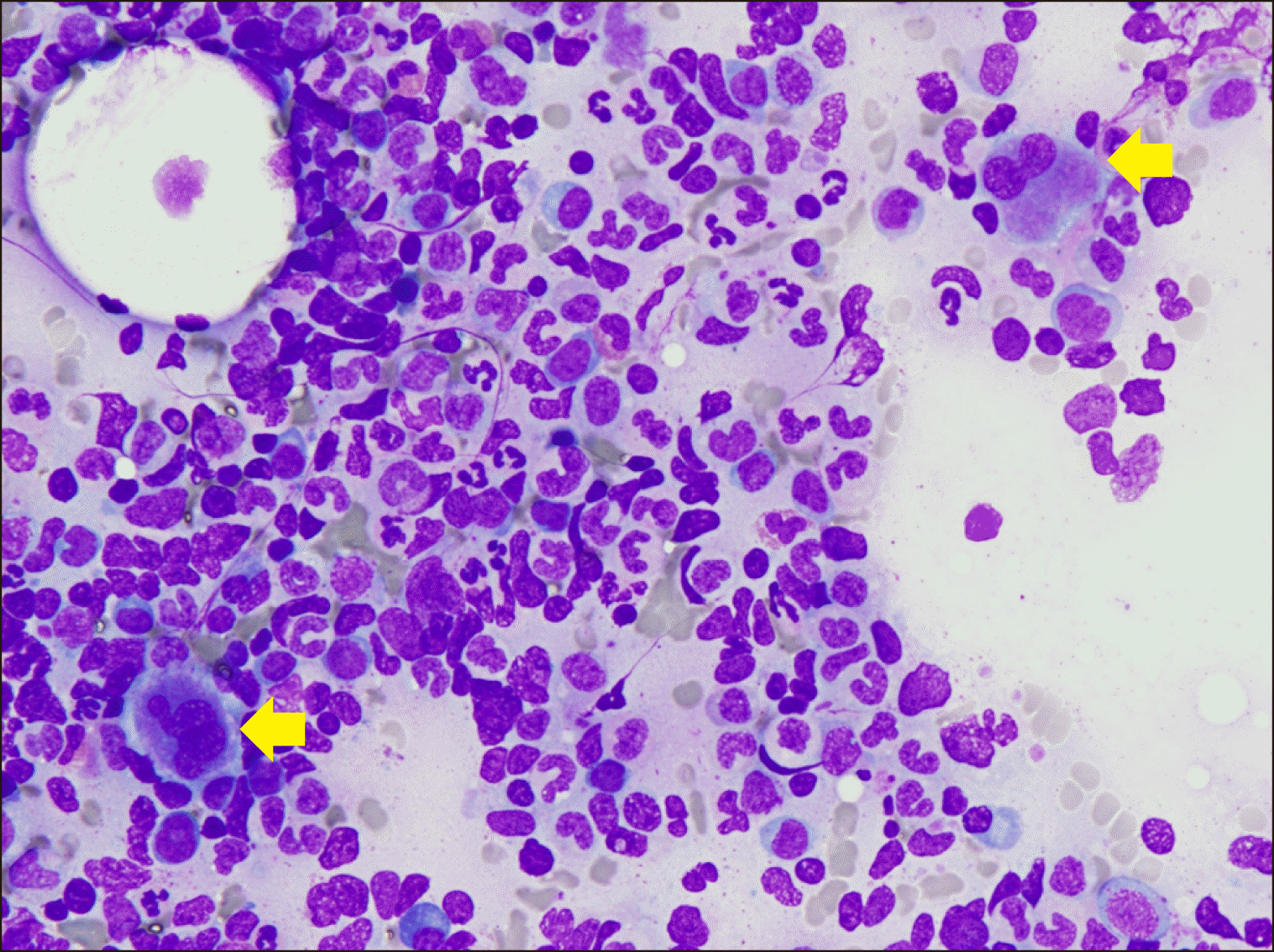 | Fig. 3Dwarf megakaryocytes (yellow arrow) with hypolobated nuclei were seen in the bone marrow (Wright-Giemsa stain, ×400). 
|
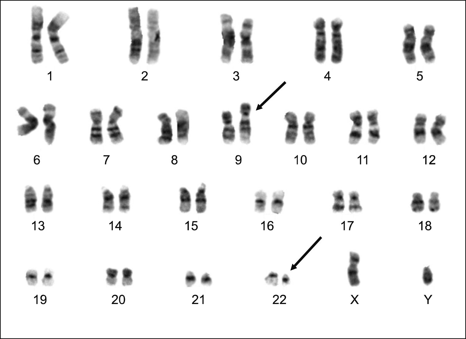 | Fig. 4Karyotyping results exhibiting 46, XY, t(9;22)(q34;q11.2)[20]. 
|
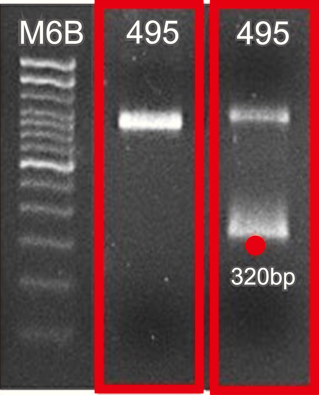 | Fig. 5The minor BCR-ABL1 fusion gene was found as a sole rearrangement in the case presented here. Nested reverse transcriptase polymerase chain reaction revealed the minor BCR-ABL1 fusion transcript (P190) with an approximately 320 bp length, indicating it to be the e1a2 type transcript. No amplicons corresponding to the 320 bp position were detected for the major BCR-ABL1 transcripts. 
|
The Philadelphia chromosome, resulting from the chromosomal translocation t(9;22), contains the
BCR-ABL1 fusion gene, which is a hallmark of CML. Over 95% of CML patients are
BCR-ABL1-positive [
3]. The fusion gene product varies in size depending on the breakpoint in the
BCR gene. The fusion products thus formed are major p210, minor p190, and micro p230. The most common form—the major
BCR-ABL1 rearrangement—is mostly observed in patients with CML, whereas the minor
BCR-ABL1 rearrangement is more common among patients with acute lymphoblastic leukemia (ALL) [
4]. Although our patient was positive for the minor
BCR-ABL1 rearrangement (
Fig. 5), ALL was ruled out based on morphological and FCM results.
The case we have described here constitutes the second case of minor
BCR-ABL1-positive CML reported in Korea [
2]. Several similar reports have been made globally [
5,
6]. Our case is in line with these prior reports with respect to minor
BCR-ABL1 CML being associated with significant monocytosis. The patient did not undergo tyrosine kinase inhibitor (TKI) therapy immediately after diagnosis owing to the presence of a ureter stone complicated by infection that required surgery. On November 26, the patient began treatment with dasatinib, a second-generation TKI. Thus, it was too early (<1 mo) to predict treatment response. Nevertheless, minor
BCR-ABL1-positive CML has been correlated with a poor prognosis. CML patients with a minor
BCR-ABL1 fusion gene respond more slowly to TKI treatment and are less likely to achieve a major molecular response [
7]. Accordingly, a less favorable outcome can be expected in our patient compared with that of a patient with a typical major
BCR-ABL1-positive CML.
Go to :


 PDF
PDF Citation
 Print
Print


 XML Download