Journal List > Korean J Gastroenterol > v.78(6) > 1148613

내시경적 절제술로 치료된 직장 Mucosa-associated Lymphoid Tissue 림프종 1예

Abstract

A primary extranodal B-cell lymphoma of mucosa-associated lymphoid tissue (MALT) is a distinct clinical-pathological entity that develops in diverse anatomic locations. However, colorectal involvement is rare. The authors encountered a case of a MALT lymphoma of the rectum in a 69-year-old woman who complained of up to 3 kg weight loss during 3 months. A colonoscopy demonstrated a 1.0×1.0 cm sized subepithelial tumor (SET) at the lower rectum. The patient underwent an endoscopic mucosal resection with ligation for the SET. Pathological analysis revealed marked small to medium-sized lymphocytic infiltration. Immunohistochemistry revealed neoplastic cells positive for CD20 and bcl-2. A polymerase chain reaction detected immunoglobulin H gene rearrangement. Finally, rectal MALT lymphoma was diagnosed. Computed tomography and positron emission tomography scans showed that there was no lymph node metastasis or other organ involvement. A bone marrow biopsy was found to be negative for any neoplastic process. Therefore, the Ann Arbor stage was IeB. Helicobacter pylori (H. pylori) was not detected in the gastric biopsy specimens. Thus far, she has had no relapse since the endoscopic resection. The incidence of rectal MALT is very rare, and treatment is unclear. This paper reports a case of rectal MALT lymphoma treated successfully with only an endoscopic resection.

서 론

위장관 mucosa-associated lymphoid tissue (MALT) 림프종은 전체 위장관 림프종 중 두 번째로 빈도수가 높은 림프종으로 알려져 있다. 위에서 가장 높은 빈도수를 보이며 결장직장에서는 상대적으로 드물게 발견되는 것으로 보고되고 있다.1 내시경 진단 기술이 발전함에 따라 위장관 림프종의 진단율은 증가하고 있으나 결장직장의 MALT 림프종은 여전히 드문 질환으로 알려져 있다.2 또한 내시경적 형태 및 특징이 정확하게 알려져 있지 않아 진단과 치료의 명확한 기준이 아직까지 정립되어 있지 않은 상황이다.3 본 증례는 직장 MALT 림프종을 내시경적으로 진단 및 치료를 실시한 증례로 직장 MALT 림프종에 대한 이해와 진단 및 치료 기준 정립에 도움이 될 수 있을 것이다.

증 례

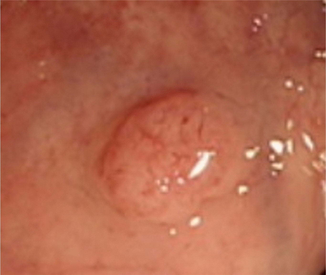
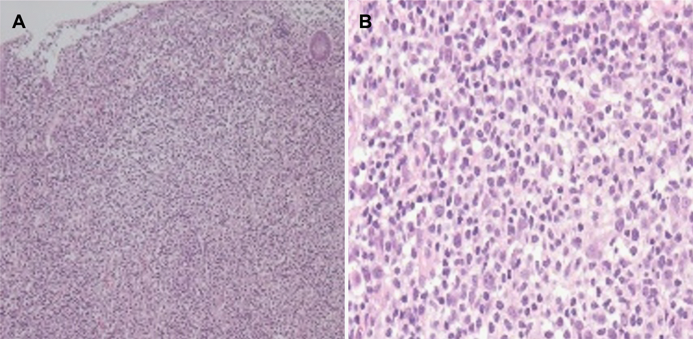
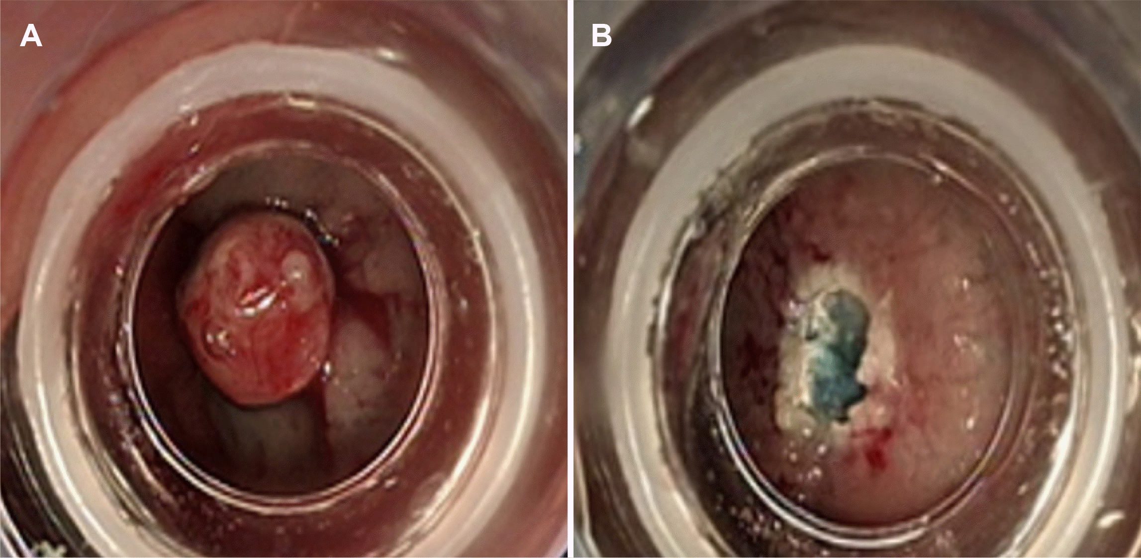
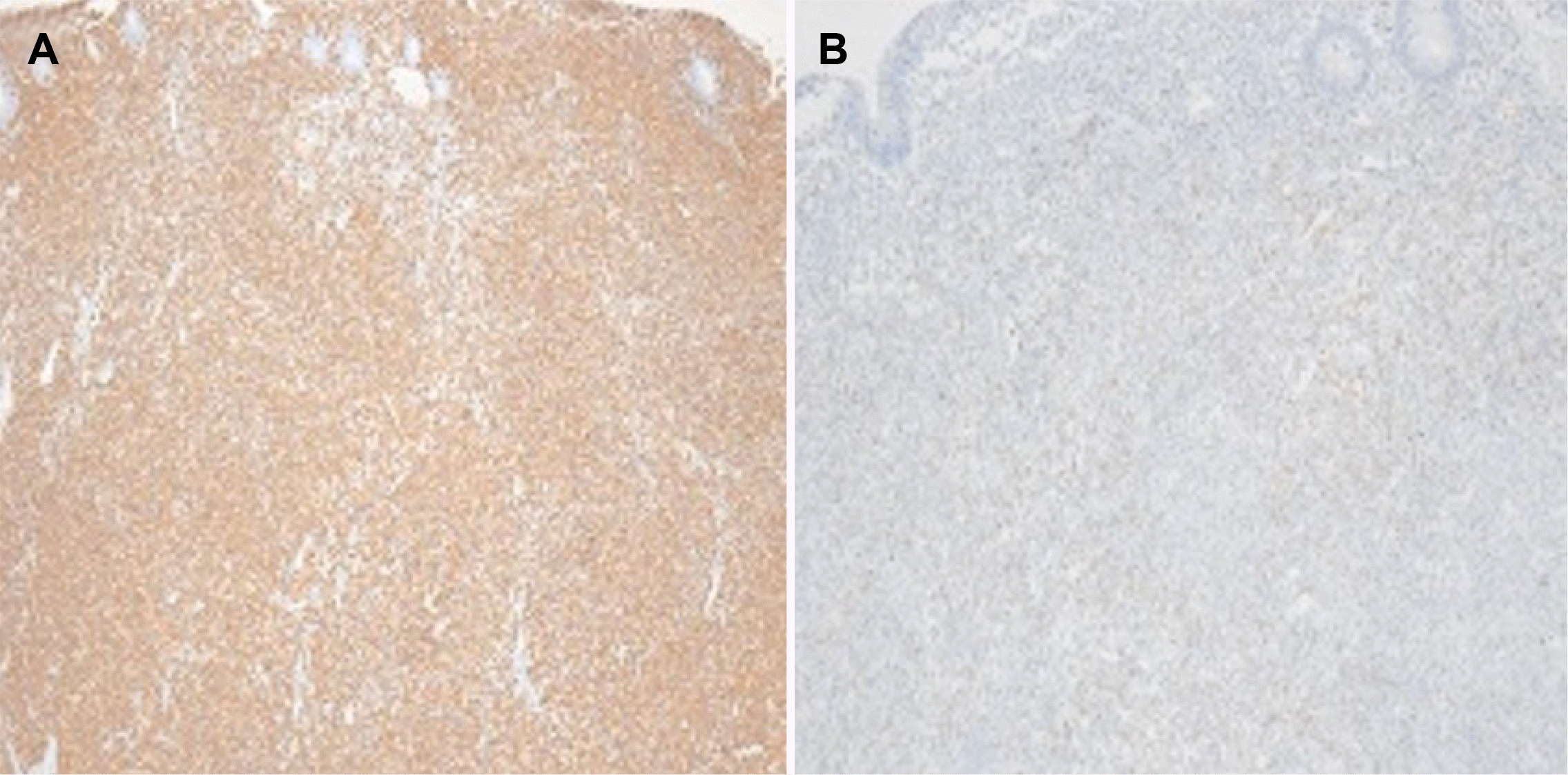
69세 여자 환자가 식욕부진, 체중감소로 소화기내과 외래로 내원하였다. 환자는 3개월 전부터 약 3 kg가량의 체중감소가 있었으나 전신상태는 양호하였다. 환자는 고혈압, 당뇨, 결핵, 간염 등의 병력은 없었고, 특이병력으로 하시모토 갑상선염이 있어 갑상선 호르몬 복용 중이었다. 활력징후는 혈압 110/60 mmHg, 맥박수 72회/분, 체온 36℃로 측정되었다. 키 153.2 cm,체중 55.5 kg, 체질량지수 23.6이었으며, 음주력과 흡연력은 없었다. 신체 검사에서 복부는 평탄하고 부드러웠고, 압통과 반발압통이 없었으며, 촉지되는 종괴는 없었다. 서혜부, 경부, 액와부위 등에 촉지되는 림프절 종대는 없었다. 대장내시경에서 하부 직장 내 약 0.7 cm 크기의 둥근 모양의 점막 융기 병변이 발견되었다. 경계가 명확한 점막하종양 형태로 보였고 표면의 명확한 색조 변화는 없었으나 미란이 일부 관찰되어 조직 검사를 시행하였다(Fig. 1). 조직 병리 검사에서 작은 크기의 비정형 림프구 침윤이 관찰되었다(Fig. 2). Wotherspoon score 3로 mucosa-associated lymphoid tissue lyphoma (MALT 림프종) 의심 하에 확진을 위한 추가적인 진단 및 치료 목적으로 내시경적 절제술(endoscopic mucosal resection with ligation, EMR-L)을 시행하였다(Fig. 3). 1 cm 미만의 점막하 병변의 절제술에 있어서 band ligation을 이용한 절제술은 치료 성공률이 높고 비교적 안전하기 때문에 band ligation에 의한 절제술을 선택하였다. 절제조직으로 시행한 면역 화학 검사에서 CD 20에는 양성을, CD 3에는 음성을 보였고 bcl-2에는 양성을 보였다(Fig. 4). 중합효소연쇄반응(PCR)을 이용한 면역글로불린 중쇄유전자 재배열(immunoglobulin heavy chain gene rearrangements)을 분석한 결과 B세포 단클론성(monoclonality)이 증명되어 MALT 림프종(MALT lymphoma)으로 최종 진단하였다.
말초혈액 검사에서 백혈구 5,860/mm3, 혈색소 12.7 g/dL, 혈소판 156,000/mm3로 정상이었고, 혈청 생화학 검사에서도 LDH 314 U/L, AST 32 IU/L, ALT 21 IU/L, CRP 0.73 mg/L로 특이 소견을 보이지 않았다. 종양표지자 검사에서 암배아 항원(carcinoembryonic antigen, CEA)은 상승되지 않았다. 단순 흉부 및 복부 X선 사진은 정상이었다. 복부 전산화단층촬영(CT) 검사에서 직장 주변의 다른 장기 침범은 없었으며, 직장 주변 영역, 대혈관 및 장간막 주위 등의 림프절 종대는 관찰되지 않았다. 흉부 CT와 경부 CT 모두 정상 소견이었고, 골수 검사 및 위내시경 검사에서도 특이 소견이 없었으며 조직 검사를 통한 Giemsa 염색 및 요소분해 검사(rapid urease test) 상 Helicobacter pylori (H. pylori) 감염은 증명되지 않았다. 전신 전자방출단층촬영(fluorine-18-fluoro-2-deoxy-D-glucose PET CT)에서 이상 소견을 보이지 않았다. 골수 검사도 정상 소견을 보였다. 병기는 Ann arbor 분류에 따라 Ib였으며, 내시경 절제술에 의한 조직 결과에서 변연부 음성을 보여 근치적 절제가 확인되었다. 추후 경과에 따라 방사선 치료가 필요할 수 있음을 설명하였으나 환자가 거부하였다. 환자는 시술 후 현재 37개월까지 재발 없이 외래 추적 관찰 중에 있다.

고 찰

MALT 림프종은 1983년 Isaacson과 Wright4가 위에 발생한 MALT 림프종을 기술함으로써 최초로 개념이 제시되었으며, 조직학적으로 림프형질세포의 점막 침윤에 의한 림프상피성 병변이 특징적으로 나타난다. MALT 림프종은 원발성 위 림프종의 40%를 차지하여 45-50%를 차지하는 미만성 거대 B세포 림프종과 함께 가장 흔한 원발성 위장관 림프종으로 알려져 있다.5 조직학적 소견뿐만 아니라 저등급 위 MALT 림프종의 경우 면역글로불린중쇄 유전자 재배열 분석을 하여 B세포 단클론성을 확인하는 것이 진단에 도움이 되는데, 양성률은 연구에 따라 다양하며 국내 보고에 의하면 약 55%에서 양성을 보인다고 알려져 있다.6 H. pylori 감염은 위 MALT 림프종 발생의 대표적 원인으로 알려져 있고, H. pylori 양성인 환자에서 제균 치료를 통해 위 MALT 림프종의 관해를 유도할 수 있다.7 하지만 위를 제외한 림프절 외 기관에서 발생한 MALT 림프종의 경우 H. pylori 양성률은 45%에 그쳐 이러한 경우에는 H. pylori가 발병 원인 인자인지 불분명하고 일차 치료로서 제균 치료의 유용성은 떨어진다고 알려져 있다.8 대장에 발생하는 원발성 악성 림프종은 전체 대장 악성 종양의 0.20-0.65%로 드문 질환으로 알려져 있다.9,10 Ahlawat 등11은 2006년까지 직장 MALT 림프종이 전 세계적으로 30예가 보고 되었다고 정리한 바 있고, 국내에서는 1999년 이후 2012년까지 9예가 보고되었고 2018년 Jeon 등이 20예를 보고한 바 있다.3,12,13 Jeon 등3이 발표한 연구에서는 총 51예의대장 MALT 림프종 중 20예(39%)가 직장 MALT 림프종으로대장에서는 직장 MALT 림프종이 가장 흔하게 발생하는MALT 림프종으로 보고하였다.
매우 드문 유병률과 비특이적인 임상 양상으로 인해 원발성 직장 MALT 림프종의 정립된 치료 방법은 없는 상태이다. 대부분의 예에서 화학요법과 수술을 1차 치료로 시행하였고,3,13-16 드물게 제균 치료로 관해된 보고도 있다.17 전이가 없는 국소적인 직장 MALT 림프종에 대해서는 방사선 치료 또는 수술적 절제술이 고려되고 있다.18 일부 증례에서는 직장 MALT 림프종 환자에서 방사선 치료를 시행하여 완전 관해를 유도할 수 있었다고 보고하였다.19 본 증례와 비슷하게 Mathew 등은 직장 MALT 림프종이 내시경적 절제술 단독 치료로 완치된 예를 보고한 바 있다.20-22 Jeon 등3은 51예의 대장 MALT 림프종 중 17예(33%)의 국소병변에 대해서는 내시경 절제술 단독으로 치료를 실시하였고 좋은 예후를 보였다. 점막하종양의 형태를 보이는 직장 MALT 림프종에서 어떤 내시경 절제술을 선택할 것인지에 대한 근거는 아직까지 부족한 실정으로 점막하종양의 형태를 비슷하게 보이는 직장 신경내분비종양의 연구를 참고할 수가 있겠다. 직장 신경내분비종양의 경우 여러 내시경적 절제술에 대한 연구가 진행이 되었고 10 mm 미만의 경우 Lim 등23은 점막하절제술에 비해 ligation 보조 점막절제술의 치료 성공률은 비슷하게 우수하면서 치료 시간은 단축시킬 수 있다고 보고하였고, Lee 등24은 cap 보조 점막절제술과 비교하였을 때 ligation 보조 점막절제술이 근치적절제율과 조직학적 완전 절제율에서 더 뛰어난 결과를 보고하였다. 이번 증례 또한 신경내분비종양의 치료 결과를 참고하여 ligation 보조 점막절제술을 실시하였고 좋은 예후를 보였다.
대장의 점막하 종양은 흔하게 발견되는 병변으로 악성 종양의 가능성이 있어 감별 진단이 중요하다. 특히 직장은 과증식 용종이 많이 발견되는 부위로 본 증례와 같은 크지 않은 점막하종양은 흔히 놓치기 쉽다. 선암 및 신경내분비종양 뿐만 아니라 점막하 종양의 형태를 보이는 림프종 또한 직장에서 발견될 수 있는 악성 병변임을 인지하고 내시경 검사에 임하는 것이 필요하며 점막 표면이 발적이나 미란, 궤양을 보이거나 색조 및 경도의 변화를 보이면 적극적으로 조직 검사를 시행하는 것이 필요하다. 조직 검사에서 MALT 림프종이 의심되는 상황이라면 확진 및 치료를 위해 내시경 절제술을 고려해 볼 수 있다.
이번 증례는 우연히 발견된 직장의 점막하 종양에 대해 MALT 림프종으로 진단하고 치료 목적으로 내시경적 절제술을 시행한 예이다. 다른 장기의 전이 없이 직장에만 국한되어 있었고 완전 절제가 이루어져 추가 치료 없이 경과 관찰 중에 있으며 현재까지 재발은 없었다. 아직까지는 H. pylori 감염이 확인되지 않은 직장의 국소 MALT 림프종에 대한 표준치료는 정립되지 않은 상황이나 여러 문헌에서 전이가 없는국소 병기의 치료법으로 내시경 절제술에 대한 좋은 결과를보고하였다. 이번 증례 또한 타 장기 전이가 없고 국소적으로분포하는 경우 내시경적으로 완전 절제가 이루어졌다면 추가치료 없이 주의 깊은 경과 관찰만으로 좋은 예후를 보일 수있는 예가 될 것이다.

Notes

Financial support

None.

Conflict of interest

None.

REFERENCES

1. Stanojevic GZ, Nestorovic MD, Brankovic BR, Stojanovic MP, Jovanovic MM, Radojkovic MD. 2011; Primary colorectal lymphoma: An overview. World J Gastrointest Oncol. 3:14–18. DOI: 10.4251/wjgo.v3.i1.14. PMID: 21267399. PMCID: PMC3026053.
crossref
2. Howell JM, Auer-Grzesiak I, Zhang J, Andrews CN, Stewart D, Urbanski SJ. 2012; Increasing incidence rates, distribution and histological characteristics of primary gastrointestinal non-Hodgkin lymphoma in a North American population. Can J Gastroenterol. 26:452–456. DOI: 10.1155/2012/480160. PMID: 22803021. PMCID: PMC3395447.
crossref
3. Jeon MK, So H, Huh J, et al. 2018; Endoscopic features and clinical outcomes of colorectal mucosa-associated lymphoid tissue lymphoma. Gastrointest Endosc. 87:529–539. DOI: 10.1016/j.gie.2017.08.027. PMID: 28882576.
crossref
4. Isaacson P, Wright DH. 1983; Malignant lymphoma of mucosa-associated lymphoid tissue. A distinctive type of B-cell lymphoma. Cancer. 52:1410–1416. DOI: 10.1002/1097-0142(19831015)52:8<1410::AID-CNCR2820520813>3.0.CO;2-3. PMID: 6193858.
crossref
5. Alkan S, Karcher DS, Newman MA, Cohen P. 1996; Regression of salivary gland MALT lymphoma after treatment for Helicobacter pylori. Lancet. 348:268–269. DOI: 10.1016/S0140-6736(05)65578-X. PMID: 8684218.
crossref
6. Choi SM, Jung HY, Kang GH, et al. 2001; Early histologic response to eradication therapy in low-grade gastric mucosa-associated lymphoid tissue lymphoma according to polymerase chain reaction-based B-cell monoclonality. Korean J Gastroenterol. 37:247–253.
7. Wotherspoon AC, Doglioni C, Diss TC, et al. 1993; Regression of primary low-grade B-cell gastric lymphoma of mucosa-associated lymphoid tissue type after eradication of Helicobacter pylori. Lancet. 342:575–577. DOI: 10.1016/0140-6736(93)91409-F. PMID: 8102719.
crossref
8. Grünberger B, Wöhrer S, Streubel B, et al. 2006; Antibiotic treatment is not effective in patients infected with Helicobacter pylori suffering from extragastric MALT lymphoma. J Clin Oncol. 24:1370–1375. DOI: 10.1200/JCO.2005.02.9025. PMID: 16549831.
crossref
9. Shepherd NA, Hall PA, Coates PJ, Levison DA. 1988; Primary malignant lymphoma of the colon and rectum. A histopathological and immunohistochemical analysis of 45 cases with clinicopathological correlations. Histopathology. 12:235–252. DOI: 10.1111/j.1365-2559.1988.tb01939.x. PMID: 3366441.
crossref
10. Jinnai D, Iwasa Z, Watanuki T. 1983; Malignant lymphoma of the large intestine--operative results in Japan. Jpn J Surg. 13:331–336. DOI: 10.1007/BF02469515. PMID: 6645123.
11. Ahlawat S, Kanber Y, Charabaty-Pishvaian A, et al. 2006; Primary mucosa-associated lymphoid tissue (MALT) lymphoma occurring in the rectum: a case report and review of the literature. South Med J. 99:1378–1384. DOI: 10.1097/01.smj.0000215855.98512.9d. PMID: 17233195.
crossref
12. Kim JH, Moon YS, Lee SH, et al. 2008; A case of primary B cell mucosa-associated lymphoid tissue lymphoma presenting as a solitary rectal mass. Korean J Gastrointest Endosc. 36:102–106.
13. Park H, Chung JW, Kim AJ, et al. 2012; A case of rectal mucosa-associated lymphoid tissue lymphoma diagnosed by endoscopic unroofing technique. Korean J Gastroenterol. 59:428–432. DOI: 10.4166/kjg.2012.59.6.428. PMID: 22735876.
crossref
14. Matsuo S, Mizuta Y, Hayashi T, et al. 2006; Mucosa-associated lymphoid tissue lymphoma of the transverse colon: a case report. World J Gastroenterol. 12:5573–5576. DOI: 10.3748/wjg.v12.i34.5573. PMID: 17007004. PMCID: PMC4088249.
crossref
15. Nakase H, Okazaki K, Ohana M, et al. 2002; The possible involvement of micro-organisms other than Helicobacter pylori in the development of rectal MALT lymphoma in H. pylori-negative patients. Endoscopy. 34:343–346. DOI: 10.1055/s-2002-23643. PMID: 11932795.
crossref
16. Inoue F, Chiba T. 1999; Regression of MALT lymphoma of the rectum after anti-H. pylori therapy in a patient negative for H. pylori. Gastroenterology. 117:514–515. DOI: 10.1053/gast.1999.0029900514b. PMID: 10465640.
crossref
17. Matsumoto T, Iida M, Shimizu M. 1997; Regression of mucosa-associated lymphoid-tissue lymphoma of rectum after eradication of Helicobacter pylori. Lancet. 350:115–116. DOI: 10.1016/S0140-6736(05)61818-1. PMID: 9228971.
crossref
18. Li B, Shi YK, He XH, et al. 2008; Primary non-Hodgkin lymphomas in the small and large intestine: clinicopathological characteristics and management of 40 patients. Int J Hematol. 87:375–381. DOI: 10.1007/s12185-008-0068-5. PMID: 18409078.
crossref
19. Kobayashi T, Takahashi N, Hagiwara Y, et al. 2008; Successful radiotherapy in a patient with primary rectal mucosa-associated lymphoid tissue lymphoma without the API2-MALT1 fusion gene: a case report and review of the literature. Leuk Res. 32:173–175. DOI: 10.1016/j.leukres.2007.04.017. PMID: 17570523.
crossref
20. Mathew A, Humburg BC, Bayer MG. 2009; A case of rectal MALT lymphoma treated by endoscopic resection. Am J Gastroenterol. 104:255–256. DOI: 10.1038/ajg.2008.47. PMID: 19098890.
crossref
21. Shah RM, Kuo V, Schwartz A. 2020; Endoscopic mucosal resection and cure for rectal mucosa-associated lymphoid tissue lymphoma. Proc (Bayl Univ Med Cent). 34:305–306. DOI: 10.1080/08998280.2020.1836939. PMID: 33678972. PMCID: PMC7901432.
crossref
22. Wei YL, Min CC, Ren LL, et al. 2021; Laterally spreading tumor-like primary rectal mucosa-associated lymphoid tissue lymphoma: a case report. World J Clin Cases. 9:3988–3995. DOI: 10.12998/wjcc.v9.i16.3988. PMID: 34141757. PMCID: PMC8180215.
crossref
23. Lim HK, Lee SJ, Baek DH, et al. Resectability of rectal neuroendocrine tumors using endoscopic mucosal resection with a ligation band device and endoscopic submucosal dissection. Gastroenterol Res Pract. 2019; Sep. 16. [Epub ahead of print]. DOI: 10.1155/2019/8425157. PMID: 31687016. PMCID: PMC6811798.
crossref
24. Lee J, Park YE, Choi JH, et al. 2020; Comparison between cap-assisted and ligation-assisted endoscopic mucosal resection for rectal neuroendocrine tumors. Ann Gastroenterol. 33:385–390. DOI: 10.20524/aog.2020.0485. PMID: 32624659. PMCID: PMC7315720.
crossref

Fig. 1
Colonoscopy demonstrated a 0.7×0.5 cm sized well-demarcated subepithelial tumor at the lower rectum.
kjg-78-6-344-f1.tif
Fig. 2
Pathological analysis revealed marked small to medium-sized lymphocytic infiltration. (A) Hematoxylin and eosin stain (H&E) staining, ×100 and (B) H&E staining, ×400.
kjg-78-6-344-f2.tif
Fig. 3
The patient underwent endoscopic mucosal resection with ligation (endoscopic mucosal resection with ligation) for the rectal subepithelial tumor. (A) Colonoscopy finding after ligation and (B) after resection.
kjg-78-6-344-f3.tif
Fig. 4
Immunohistochemical staining. (A) CD20 immunohistochemical staining, ×40. (B) BCL-2 immunohistochemical staining, ×100.
kjg-78-6-344-f4.tif
TOOLS
Similar articles