This article has been
cited by other articles in ScienceCentral.
Abstract
Natural killer (NK) cell enteropathy is not well understood. Although NK-cell enteropathy is typically benign, aggressive NK-cell lymphoma is an important differential diagnosis. This paper reports a rare case of NK-cell enteropathy of the stomach presenting with dyspepsia and weight loss. In this case, upper endoscopy revealed an ulcer-like lesion (approximately 7 mm) surrounded by erythema, edema, and mucosal hemorrhage. The biopsy revealed atypical lymphocyte cells expressing CD56 without an Epstein-Barr virus infection, which is consistent with NK-cell enteropathy. Follow-up endoscopy 2 months later revealed lesion regression with mild erythema. The patient was observed closely for 6 months, with no evidence of lymphoma.
Go to :

Keywords: Killer cells, natural, Intestinal diseases, Lymphoma, Stomach
INTRODUCTION
Natural killer (NK) cell enteropathy is a poorly understood entity that was first described by Vega et al.
1 in 2006 as an indolent NK-cell lymphoproliferative disease of the gastro-intestinal (GI) tract. Subsequently, a case series from the USA reported NK-cell enteropathy,
2 whereas similar cases from Japan were reported as lymphomatoid gastropathy of the NK-cell type.
3 NK-cell enteropathy is typically a benign disease presenting with vague abdominal symptoms. It can be misdiagnosed as aggressive NK-cell lymphoma because of its indolent course. Therefore, this entity should be identified accurately to avoid unnecessary chemotherapy. In Korea, Koh et al.
4 reported the first case of NK-cell gastropathy in adolescents. To the best of the authors’ knowledge, previous Korean reports have not described NK-cell enteropathy of the stomach in adults. This paper reports a rare case of NK-cell enteropathy of the stomach in an elderly woman that presented with abdominal symptoms.
Go to :

CASE REPORT
An 81-year-old woman presented with recurrent dyspepsia and epigastric pain that began 2 months earlier. She also lost 4 kg of weight over the previous 2 months. She has been receiving medications for hypertension and underwent a hemithyroidectomy for thyroid cancer 3 years earlier. The physical examination and laboratory findings were unremarkable. The patient had stable vital signs, with a normal blood pressure and body temperature during the initial outpatient examination.
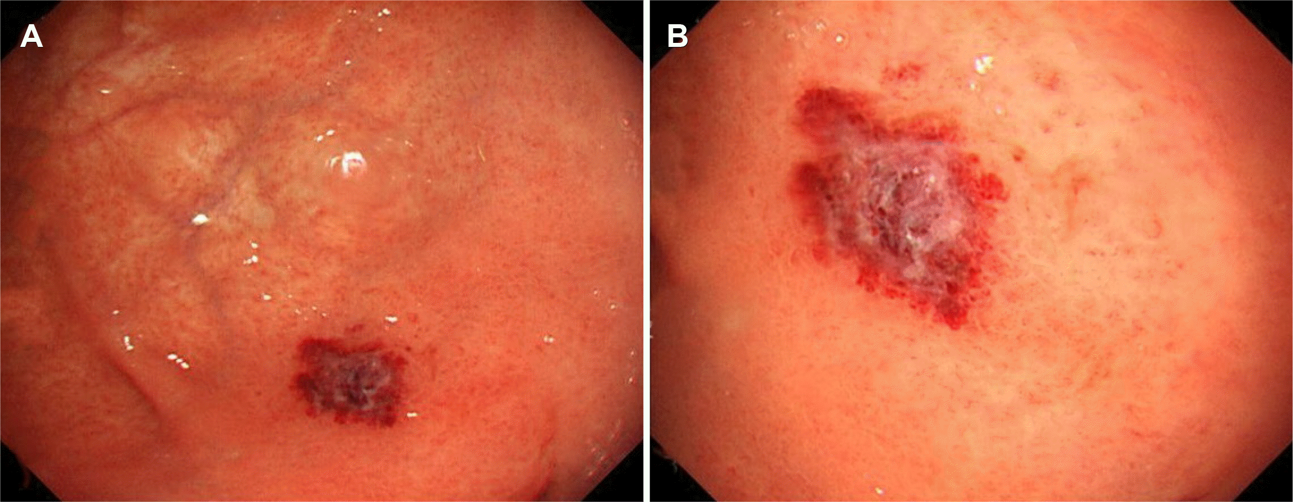
The patient had a medical history of thyroid cancer and presented with alarm symptoms, including recent weight loss and persistent dyspepsia. Hence, upper endoscopy, colonoscopy, and abdominal CT were performed to evaluate the malignancy. Upper endoscopy revealed an ulcer-like lesion (approximately 8 mm) surrounded by erythema, edema, and mild mucosal hemorrhage in the gastric fundus (
Fig. 1). A
Helicobacter pylori infection was not observed, and no other endoscopic abnormalities were noted. There were no abnormal findings on the previous upper endoscopy in 2015. Colonoscopy and abdominal CT revealed no specific findings. Based on the endoscopic findings, the lesion possibly caused dyspepsia and weight loss. Biopsy samples were taken from the ulcer surrounded by erythema and edema.
 | Fig. 1Initial endoscopic images. (A, B) Esophagogastroduodenoscopy shows an 8-mm, reddish ulcer-like lesion surrounded by erythema, edema, and mild mucosal hemorrhage on the gastric fundus. 
|
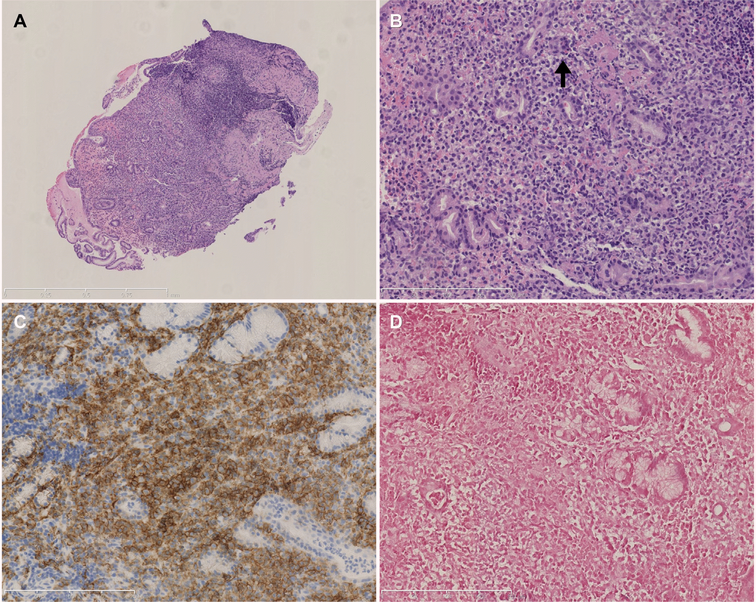
A histopathological evaluation revealed atypical lymphoid cell proliferation in the lamina propria on low-power magnification (
Fig. 2A). On high magnification, the cells had a clear cytoplasm and mildly enlarged nuclei, suggesting lymphoepi-thelial lesions (
Fig. 2B). On immunohistochemical staining, the tumor was positive for CD3, CD5, T-cell intracellular antigen 1, and CD56 (
Fig. 2C). In contrast, CD20 was negative. The MIB-1 labeling index was 60%. Ebstein Barr virus in situ hybridization was negative (
Fig. 2D). The pathology findings were consistent with those of NK-cell enteropathy. An A T-cell receptor rearrangement polymerase chain reaction was not performed.
 | Fig. 2Histopathological findings. (A) In low-power magnification, medium-sized lymphocytes proliferated in lamina propria (hematoxylin and eosin stain [H&E], ×50). (B) Lymphoid cells showed clear cytoplasm and slightly enlarged nuclei. Lymphoepithelial lesions were noted (black arrow). (H&E, ×200). (C) CD56 immunohistochemical staining was positive (CD56 stain, ×200). (D) EBER in situ hybridization was negative (EBV ISH, ×200). 
|
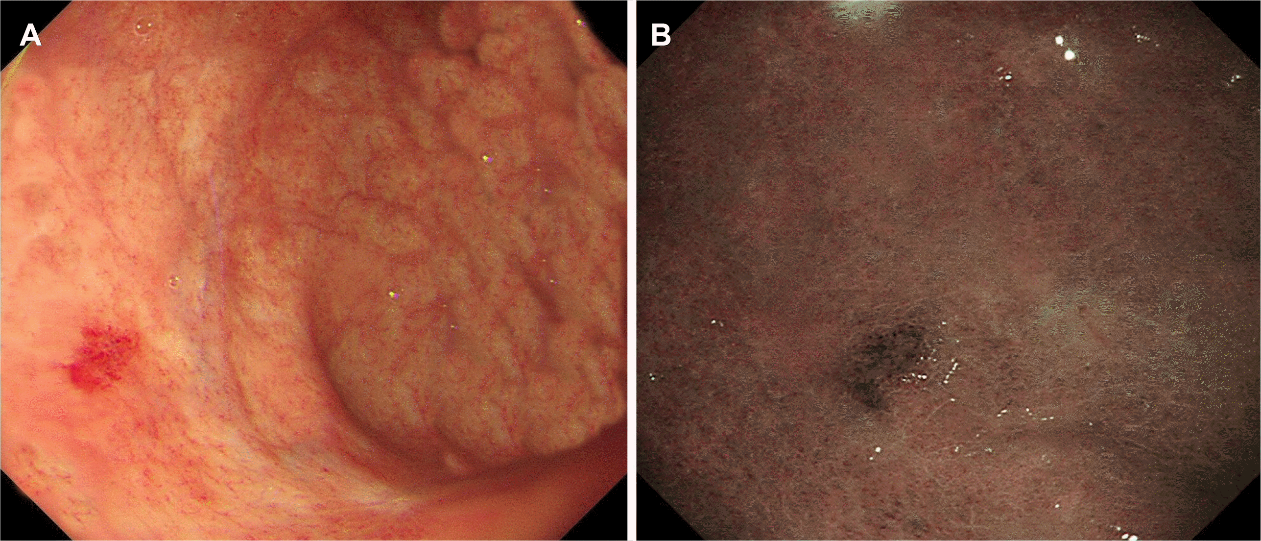
The abdominal symptoms improved with an oral proton pump inhibitor. The follow-up endoscopy performed 2 months later showed improvement of the lesion. The mucosal edema, inflammation, and bleeding regressed, but only mild erythematous lesions persisted (
Fig. 3).
 | Fig. 3Follow-up endoscopic images. (A) Two months later, repeated endoscopy shows regression of lesion with mild erythema. (B) Narrow-band image with magnification. 
|
Go to :

DISCUSSION
NK-cell enteropathy is a rare and clinically benign lymphoproliferative disease involving the gastrointestinal tract. This disease was recently described as “lymphoid gastropathy.” It is characterized by the proliferation of atypical lymphocyte cells expressing CD56 without an Epstein-Barr virus infection.
5 NK-cell enteropathy is a poorly understood disease and is not included in the current World Health Organization classification.
6 In 2006, Vega et al.
1 initially described it as an indolent NK-cell lymphoproliferative disease of the GI tract. Subsequently, Mansoor et al.
2 reported eight cases using the term NK-cell enteropathy. Takeuchi et al.
3 reported 10 similar cases of lymphomatoid gastropathy of the NK-cell type. Most cases from Japan were asymptomatic and detected incidentally by routine endoscopy. On the other hand, the cases from the United States of America exhibited abdominal symptoms, such as dyspepsia, epigastric pain, weight loss, or diarrhea. Two cases have been reported in Korea. Koh et al.
4 reported NK-cell enteropathy of the stomach in a 14-year-old adolescent, and Hwang et al.
7 reported an indolent NK cell proliferative lesion in the gallbladder. The present case report describes a rare case of NK-cell gastropathy in an elderly female patient presenting with a solitary, small gastric ulcer.
NK-cell enteropathy is found mainly in the stomach and colon. It rarely affects the extragastrointestinal organ, such as the gallbladder.
7 The endoscopic findings of NK-cell enteropathy of the stomach have not been clarified. In previously reported cases,
1-4,8 most lesions were less than 1 cm in size. The endoscopic findings varied, but most lesions manifested as an erosion or ulcer with marked erythema.
9 Both solitary and multiple lesions were reported in previous cases. These findings resembled benign inflammatory diseases, including erosive gastritis, gastric ulcer, or malignant lymphomas. Thus, NK-cell enteropathy is difficult to distinguish from these diseases. Hence, endoscopists and pathologists should consider this rare disorder in a differential diagnosis.
Differentiating between NK-cell enteropathy and NK/T-cell lymphoma is important. Because of their similarities, NK-cell enteropathy, an indolent and benign disease, can be misdiagnosed as an aggressive NK-cell lymphoma. Therefore, careful and accurate identification of this entity is important for avoiding unnecessary chemotherapy. In the case series by Mansoor et al.,
2 three patients were initially misdiagnosed with lymphoma and underwent chemotherapy. The main difference between these two entities is the Epstein-Barr virus (EBV) association. In malignant lymphoma, the tumor cells are infected with the EBV, whereas NK-cell enteropathy is not associated with the EBV. Clinically, NK/T-cell lymphoma presents with an aggressive, ulceroinfiltrative mass lesion, while NK-cell enteropathy appears as a superficial ulcer with an indolent and benign course.
10 This disease is characterized by the self-limited proliferation of atypical lymphocyte cells in the gastric mucosa. It has a good prognosis and can resolve spontaneously without treatment. An exacerbation of this disease or death has not been reported. In the present case, follow-up endoscopy performed after 2 months showed improvement of the lesion.
In conclusion, this paper described a rare case of NK-cell enteropathy of the stomach in an elderly patient who presented with abdominal symptoms. This condition should be distinguished from intestinal NK/T-cell lymphoma to avoid mis-diagnosis and overtreatment. Although NK-cell enteropathy is benign, close observation and follow-up endoscopy are needed because the cause of this entity remains unknown.
Go to :


 PDF
PDF Citation
 Print
Print


 XML Download