

 PDF
PDF Citation
 Print
Print


Introduction
The epiphora, mainly caused by primary acquired nasolacrimal duct obstruction (PANDO), occurs often in patients aged over 70 years old and at least 4 times more common in females [1]. Despite the obscure status of etiology of PANDO, some factors including previous conjunctival infections, nasal diseases, sinusitis, topical timolol or chloramid exposure are considered to be precipitating causes [2].
The dacryocystorhinostomy (DCR) is a standard surgery for blockage of the lacrimal outflow tract to connect the lacrimal sac directly to the nose by detaching the layers of bone and mucosa that keep these two structures apart. Since the 1890s the traditional approach has been the external (EX-DCR) or transcutaneous route which was replaced by an endonasal approach (EN-DCR) in last two decades only for either primary surgery or revising failed cases [3]. The intranasal pathology including a deviated nasal septum or sinusitis can be concurrently managed by EN-DCR to give effect on handling lacrimal symptoms [4].
The structures which are critical to the EN-DCR surgery are the middle turbinate (MT), the uncinate process (UP), and the agger nasi (AN) [5]. The postoperative examination of the nasal fossa is adversely affected by and the failure of DCR is caused by, amongst the most commonly reported ones, the synechiae occurring between the head of the MT and the lateral wall of the nasal fossa [5]. A planned unciformectomy was recently reported to address the vulnerability of endonasal anatomical landmarks [6]. In most Asian countries, it is concluded that the lacrimal sac fossa is preferably approached through an uncinectomy which is a process attached to the frontal process of the maxilla and to the lacrimal bone most frequently [7].
The guidelines for EN-DCR are substantially based on the studies which analyzed the lacrimal system of Caucasian race only. We have experienced that DCR procedures were not equally suitable for Asians and Caucasians and it can be explained by the anatomic distinctions in the intranasal structures between two races [7].
When we started doing an endoscopic EN-DCR surgery in Mongolia, no record of study on this surgery was found and there was lack of paper on anatomy of lacrimal fossa among Mongolians. This is one of the rationales behind our study.
Materials and Methods
Two hundred ninety two consecutive patients (over age of 16 years) who had a primary acquired NLDO cases’ orbital CT scans between January 31, 2020 and January 31, 2021 were studied at Department of Anatomy and Department of Ophthalmology, Mongolian National University of Medical Sciences University and University affiliated Oculoplastic eye clinic.
This study was approved by the Ethics Committee of Mongolian National University of Medical Sciences (approval number no. 2020/3-01).
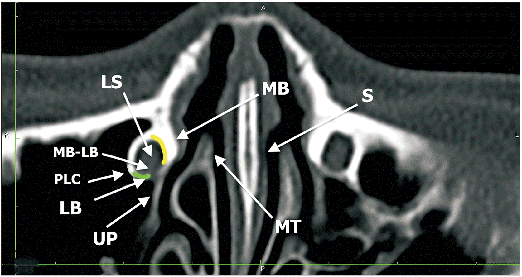
We measured morphometry of lacrimal sac fossa and anatomical variations around this structure (Fig. 1). The maximum thickness of lacrimal bone was measured. An axial-section image of the lacrimal sac fossa was used to measure lacrimal bone portion of the lacrimal sac fossa in millimeters. We picked the maximum thickness of the lacrimal bone. The maximum thickness of frontal process of the maxillary bone was studied. An axial-section image of the right lacrimal sac fossa was used to measure maxillary bone portion of the lacrimal sac fossa millimeters.
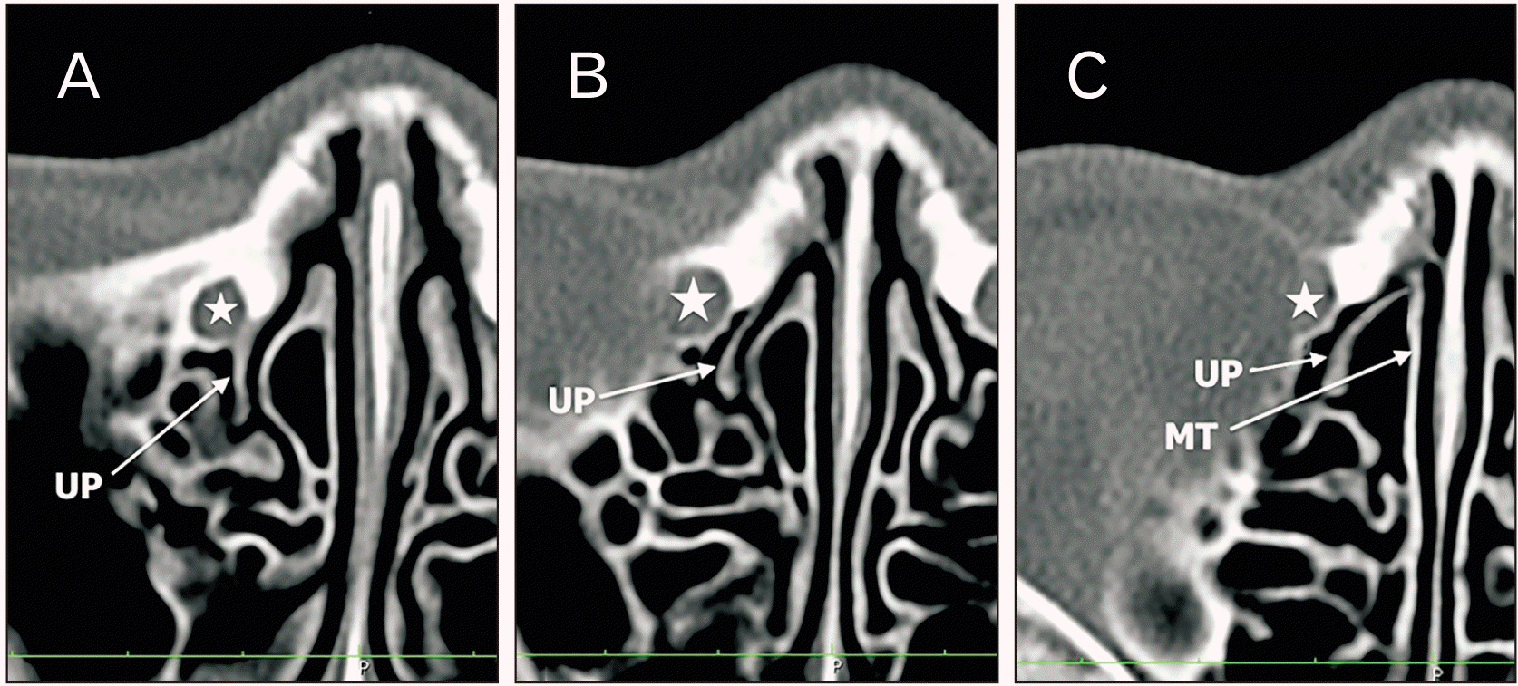
On the axial computed tomography (CT) image, the lacrimal sac fossa was divided into the following 3 levels: upper, middle, and lower. The first axial level showing the uppermost part of the lacrimal sac fossa was regarded as the upper level, the axial slice showing the lacrimal sac fossa just before joining the nasolacrimal duct was the lower level, and the mid-slice level between the 2 levels was determined as the middle level. On each level, insertion of the UP was categorized into 4 types as follows (Fig. 2):
Type-I: the UP insertion is posterior to the posterior lacrimal crest.
Type-II: the UP insertion is anterior to the posterior lacrimal crest and posterior to the junction between the lacrimal bone and the maxillary bone.
Type-III: the UP is inserted onto the maxillary bone.
Type-IV: the UP is inserted onto the lateral wall of the MT.
The relationship between operculum of the MT and lacrimal sac fossa was studied. There are 3 types of operculum of the MT insertion to the lateral wall of the nose:
Type-I: At the lacrimal sac fossa
Type-II: Behind the posterior lacrimal crest
Type-III: Above the lacrimal sac fossa
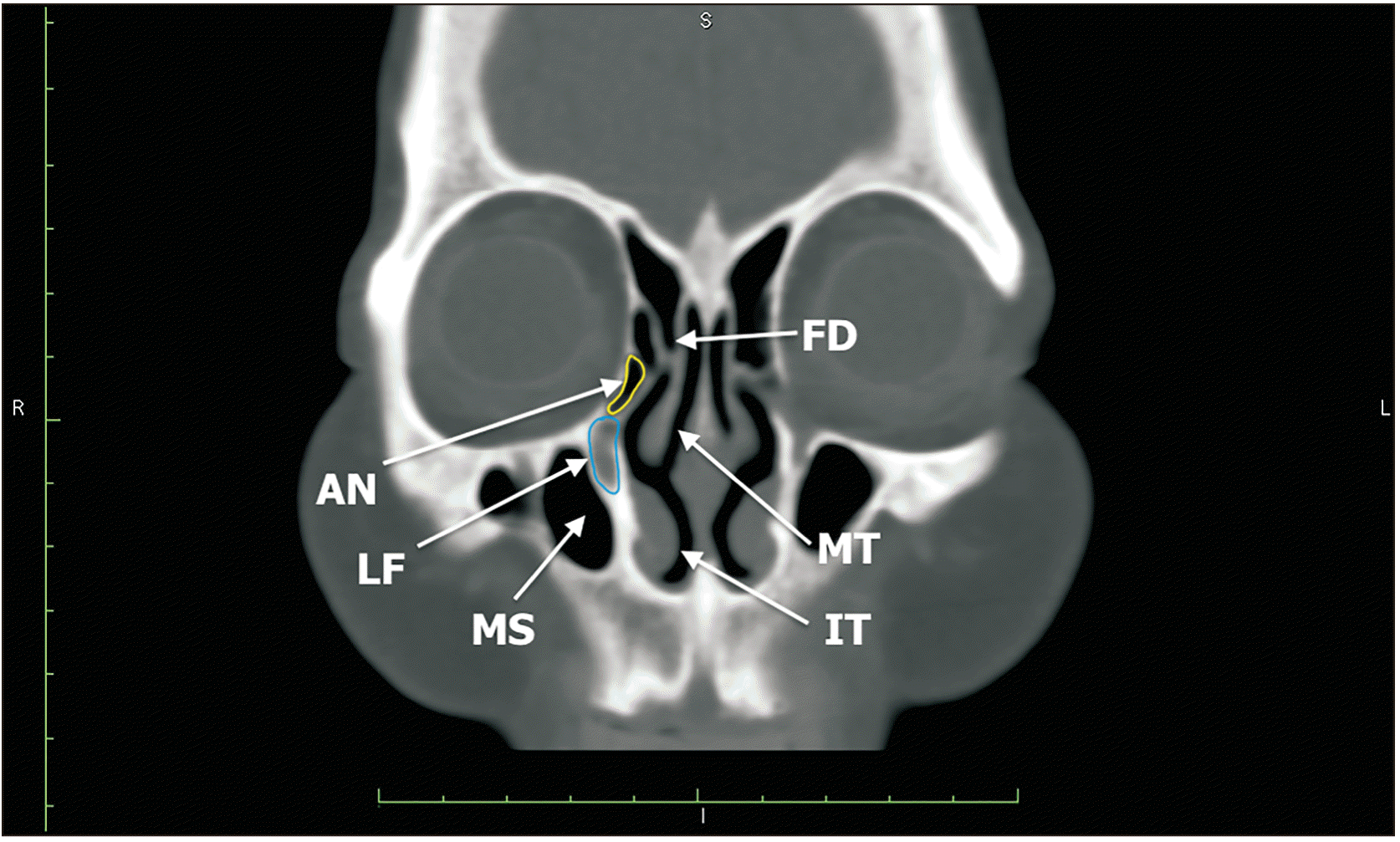
An AN cell is considered as the most anteriorly placed anterior ethmoid cell (Fig. 3). It can be either adjacent to the lacrimal sac fossa (type-I) or not adjacent to the lacrimal sac fossa (type-II).
The structure was termed “adjacent to the lacrimal fossa” when observed to contact the lacrimal sac on ≥1 section on CT scan and was termed “distant from the Lacrimal Fossa” in all other cases.
The sample size calculation indicated that 291 patients were required to undergo complete follow up in order to detect, with 80 percent power and 5 percent significance level. Thus, our study group is limited to 292 consecutive patients >16 years of age (146 male and 146 female; male-to-female ratio=1:1) who had a PANDO before DCR surgery done (146 external, 146 endonasal; external to endonasal ratio=1:1). The patients’ orbital CT scans were analyzed between January 31, 2020 and January 31, 2021.
We studied anatomy of lacrimal sac fossa and factors affecting success rate in DCR surgery by duration of surgery, Lac-Q questionnaire on patient’s post-operative satisfaction and irrigation test to check patency of created passage by DCR surgery. Criteria for successful DCR involved resolution of symptom (no tearing) and no reflux in the opposite canaliculus on lacrimal irrigation. All failed procedures had recurrent nasolacrimal duct obstruction which was confirmed by lacrimal irrigation tests after 1 month, 3 months, and 6 months.
Exclusion criteria
1. Pre-sac nasolacrimal duct obstruction
2. Eyelid malposition
3. History of lacrimal surgery
4. Lost follow up within 6 months
5. Facial bone fracture
6. History of nasal and paranasal sinus surgery
7. History of brain surgery
8. Facial palsy
9. Allergic rhinitis
All patients under study had thin section 2 mm CT sections obtained in the transverse, axial and coronal planes throughout the orbits and nasal/ paranasal structures (Somatom Sensation; Siemens firm, München, Germany). The images were analyzed with a digital image workstation (Materialise Mimics 10.01, Leuven, Belgium; Osirix, Bernex, Switzerland). Two ophthalmologists performed measurements separately in the first 40 patients. Interobserver reliability was determined with intraclass correlation coefficient (ICC); subsequently, one ophthalmologist performed the rest of the measurements in patients.
Continuous variables are presented as mean±standard deviation (SD) when normally distributed (assessed by the Kolmogorov-Smirnov test and distribution histograms) and as median (and interquartile range) when not normally distributed. Categorical variables are presented as frequencies and percentages. Differences in continuous variables across the study groups were evaluated using independent samples t-tests (Wilcoxon’s rank-sum test when indicated), while differences in categorical variables were compared by chi-square tests (Fisher’s exact test when indicated). We used chi-square goodness of fit test to compare our results with Fayet and associates’ study [5] since these are categorical variables. We used a logistic regression to assess whether anatomical variance of lacrimal sac fossa affects surgical success rate or not. All continuous variables were assessed per one unit change in each variable. All statistical tests were two-sided, and a P-value of <0.05 was considered to be statistically significant. All statistical analyses were performed using a standard software package (Stata, version.11.2; Stata Corp, College Station, TX, USA).
Result
In total, 292 participants were enrolled in DCR surgery group (146 EX-DCR, 146 EN-DCR group; ratio=1:1). Average age of the participants were 63.1 years (SD=11.3 years), no gender difference was observed between 2 groups (P<0.05).
Average thickness of frontal process of maxilla is 4.41±1.96 mm in successful surgery group, 4.97±1.04 mm in failed group and it was significantly different in 2 groups (P<0.05). The anterior insertion of UP is not posterior to the posterior lacrimal crest (retrolacrimal variation) in all levels in our study (Table 1). The lacrimal variation or the UP insertion is anterior to the posterior lacrimal crest and posterior to the junction between the lacrimal bone and the maxillary bone was observed 11.1% in middle level, 82.1% in lower level and there was no case with this variation in upper level. The maxillary variation or the UP inserted into the maxillary bone was identified 23.0% in upper level, 64.5% in middle level, 17.1% in lower level. Turbinal variation or the UP inserted onto the lateral wall of the MT was observed 77.0% in upper level, 24.3% in middle level and no case was identified at lower level. The UP was attached to the lacrimal sac fossa in 79.0% of successful surgery group but 100% in failed surgery group (P<0.05).
The AN cell was observed in 263 out of 292 cases (90.1%) group and 256 out of 292 control group (Table 2). The AN cell is adjacent to the lacrimal sac fossa in 79.1% of successful cases and 100% in failed group (Table 3).
The operculum of the MT was attached to the lacrimal sac fossa in 276 out of 292 cases (94.5%) and located behind the posterior lacrimal crest in 2 only 2 cases and above lacrimal sac fossa in 4 cases (Table 2). In both successful cases and failed group of cases, the operculum of the MT was positioned dominantly relative to the lacrimal sac fossa and it was not significantly different among failed and successful groups (Table 3).
Average duration of surgery was significantly shorter for EN-DCR 10.2 minutes (SD=6.1 minutes) compared to EX-DCR 28.5 minutes (SD=10.5 minutes) and it was significantly different between 2 groups (P<0.0001). Surgical success was achieved in 95.8% of the EN-DCR group and 94.5% of the EX-DCR group (P=0.85). Patients who underwent EX-DCR reported a median 6.0-point improvement (interquartile range, 3.0–10.0) in total Lac-Q scores (Table 4). An 11.0-point improvement (interquartile range, 9.0–16.5) was seen in the endonasal group (P<0.005).
Discussion
Anatomic differences of the lacrimal sac fossa and its surrounded structures shall be taken into account during DCR surgery. Different races result in variations in the lacrimal sac fossa region such as thickness of frontal process of maxillary bone, dissimilarity of operculum of the middle turbinate (OMT), AN and UP.
According to multiple studies, the anterior portion of the UP is located close to or contacted with the medial aspect of the lacrimal sac fossa [8-11].
Our study found that the UP was adjacent to the lacrimal sac fossa at the lower level in the majority of cases. This finding shows that UP can indicate the lower portion of the lacrimal sac fossa during endoscopic DCR surgery among Mongolians. Zhang et al. [9] found similar results during the cadavers and CT scan study on lacrimal systems of 42 Asians. According to the study, the UP was found attached laterally to the frontal process of the maxilla in all subjects and to the lacrimal bone in 79% of the total cases. This shows that an uncinectomy is essential procedure for DCR surgery among Mongolians.
Having a CT scan for Caucasian population, Fayet et al. [6] revealed unprecedented results about the anatomy of the UP relative to the lacrimal sac fossa. As per the study, the UP contacts the retrolacrimal portion in 32% of the patients in the lower level and 5.2% in the upper and middle level, respectively. When we did our study, the retrolacrimal position of the UP was not found. Therefore, the uncinectomy has to be preferred to Asian patients rather than in Caucasians in order to have a sufficient ostium [12].
As described in the most of anatomic drawings and descriptions, the operculum of the MT remains a landmark of the lacrimal sac fossa as not more than 20% of the sac is located on the top of the attachment of the operculum of the MT [11, 13]. Yet, a recent study suggested to remove a large part of the lacrimal sac fossa for a full exposure of the sac as it was situated above the operculum of the MT [6]. This study matched with the one conducted on Caucasian subjects to find the relative vertical position of the operculum of the MT to the lacrimal sac fossa.
The AN cell descent was found in 53.9% the lacrimal sac fossa by Woo et al. [7] and in 82.9% of our cases and it suggests to consider of a removal of the AN cell to allow the lacrimal sac fundus be fully exposed. During an interracial research using axial CT scans it was found that the ethmoid cell was located more anteriorly to the lacrimal sac in Asians compared to Caucasian population. Considering the above findings, we can prefer to remove the AN cell more frequently in Mongolians than in Caucasians.
The thicker frontal process of the maxilla and the thinner lacrimal bone form the lacrimal sac fossa. The study found that the significant portion of the lacrimal sac fossa of the Asian patients were dominated by the frontal process of the maxilla. As per the previous and current study results, the frontal process of the maxilla is found considerably thick in Asian patients [14-17]. When Woo et al. [7] measured 4.63±1.0 mm among Koreans, Fayet et al. [6] measured 4.09±2.06 mm among Caucasians. According to the result of the same study we performed, it was 4.85±1.94 mm which is thicker than both studies. The study on DCR reveals certain difficulties in osteotomy in Asian patients. Thus, during a surgery for Mongolian patients with a thick frontal process of the maxilla shall be treated with special surgical techniques and instruments including surgical drills to uncover the upper portion of the sac fossa. The anatomical relationship that describes the distance between the anterior lacrimal crest to the anterior ethmoidal foramen (AEF), the AEF to the posterior ethmoidal foramen (PEF), and the PEF to the optic canal, is known as the ‘24-12-6’ orbital surgical guideline or the “Rule of Halves” in western textbooks. Recent studies illustrate, there is significant variability in the medial orbital wall measurements between different populations and against the surgical guideline [18]. Our study result support this idea on the surgical anatomical difference of lacrimal sac fossa is existing in Mongolians.
In conclusion, although the literature study remains silent, most anatomic descriptions of the lacrimal system in the Western literature touch on Caucasian population as shown in surgical experiences in this region.
Mongolians have a thicker lacrimal fossa bone comparing to Caucasian population. During endoscopic DCR, in case of thick lacrimal fossa bone, it may cause a challenge to make adequately sized osteotomy. Thus, special surgical techniques and instruments, such as surgical drills, need to be equipped for the Mongolian patients due to thick frontal process of the maxilla to expose the upper portion of the sac fossa. The anterior insertion of UP is not posterior to the posterior lacrimal crest (retrolacrimal variation) in all levels in our study. The lacrimal variation or the UP insertion is anterior to the posterior lacrimal crest and posterior to the junction between the lacrimal bone and the maxillary bone was observed dominantly in lower level. Therefore, the UP can be a surgical landmark to define the lower part of the lacrimal fossa and uncinectomy is needed to be done in most of the Mongolians for adequately sized osteotomy during DCR surgery.
In our study, the OMT was positioned dominantly relative to the lacrimal sac fossa. OMT is mentioned as a vertical landmark, from an endonasal approach for predicting the position of the upper limits of the lacrimal fossa, in many studies that were done among Caucasion. The variation of OMT which is attached to the lacrimal sac was observed in majority of the cases of our study. Hence, OMT is preferred to be removed partially to secure a sufficient osteotomy in Mongolian patients during DCR.
The variation of AN cell that is adjacent to the lacrimal sac fossa was predominantly observed in Mongolians comparing to Caucasions. As a result, it can be preferred to remove the AN cell more frequently in Mongolians than in Caucasians.
 XML Download