

 PDF
PDF Citation
 Print
Print


Introduction
The atlas (C1) is known to show congenital anomalies in its anterior and posterior arches. Though posterior arch anomalies are well known, the anterior arch anomalies are seldom reported in the literature [1]. Currarino et al. [1] have classified posterior arch anomalies into five types: Type A, failure of fusion of two hemi arches; Type B, defect in one side of the arch, Type C, defects on both sides of arch, Type D, absence of arch except posterior tubercle, and Type E, absence of whole arch including tubercle. Type A is the frequent type and it occurs in 5.4% of the population and 97% of all posterior arch defects. When compared to posterior arch, the occurrence of anterior arch anomalies is rare [2]. So far only few cases of anterior arch of C1 including either absence or clefts have been reported [3].
Anomalies of C1 may cause atlantoaxial joint instability which necessitates the treatment with a cervical collar or surgery. Furthermore, these anomalies may increase the risk of cranio-vertebral and/or cervical injuries [4]. There are reported cases of C1 arch anomalies that were confused with fractures and resulted in misdiagnosis [5, 6]. Most of the congenital C1 arch anomalies are incidental and asymptomatic. However, they may increase the risk of developing myelopathy, anterior rachischisis and early cervical degenerative disc disease [7, 8]. The knowledge about the occurrence and various types of arch anomalies is clinically important in the current practice of surgery and radiology. To date, prevalence and existing variants of C1 arch anomalies among Omani population are not known. Hence, we sought to determine the prevalence and various existing variations of congenital anomalies of C1 arch in Omani patients who were referred for radiological investigation to a tertiary care hospital in Oman.
Materials and Methods
The present study was conducted by reviewing the cervical spine computed tomography (CT) scans of an indoor and outdoor patients aged ≥18 years, who were referred for head and neck CT scan to the Radiology Department of Sultan Qaboos University Hospital (SQUH) in Oman. In case of more than one cervical CT scan for a patient, the most recent one was included. All patients with the history of spinal fracture and non-Omani nationalities were excluded from the study.
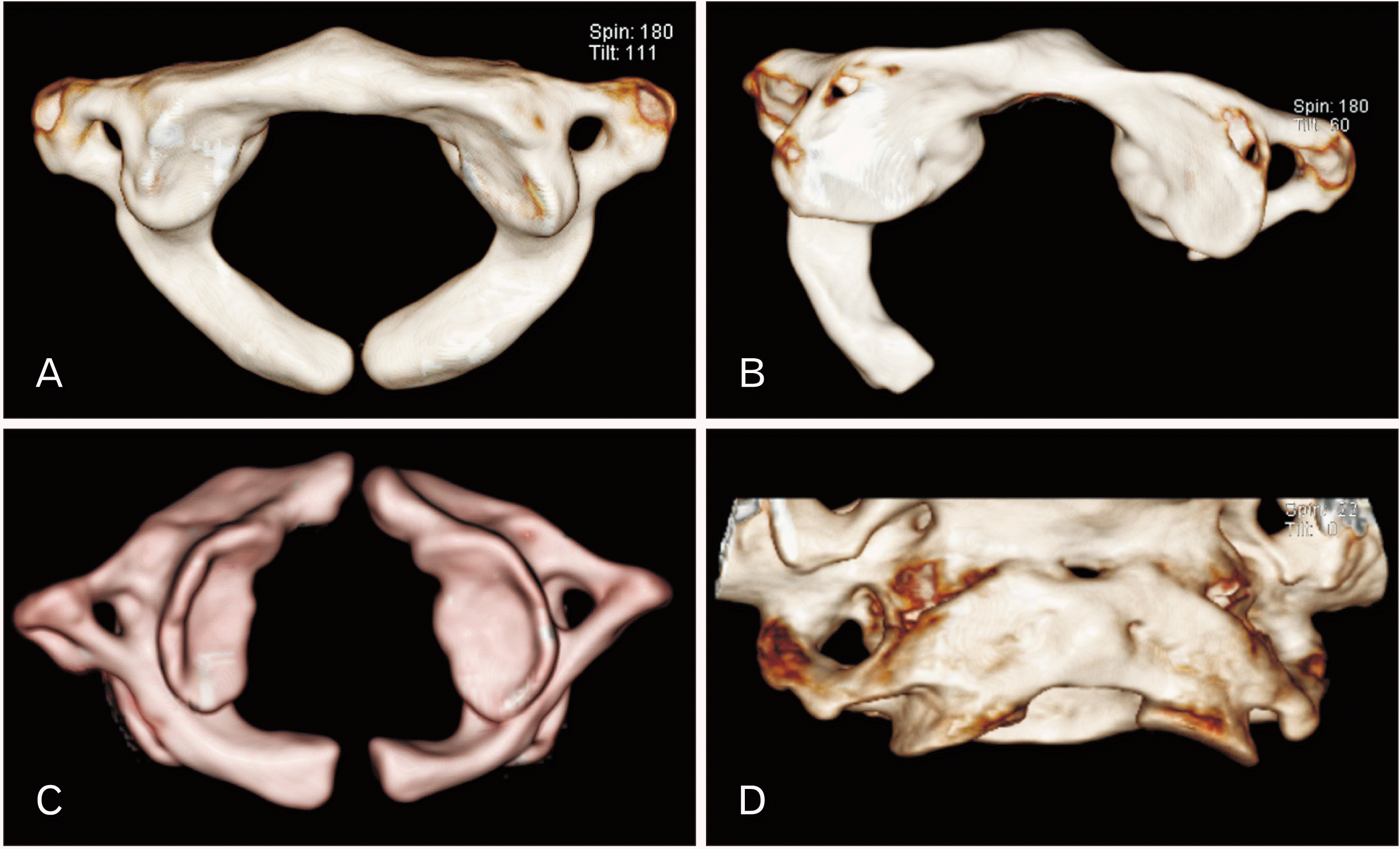
Patients data were collected retrospectively from the “TRACKCARE” system at SQUH from January 2016 till December 2017. The types of the arch anomalies of C1 were recorded based on Currarino’s classification (Fig. 1) [1]. The characteristic CT appearance of C1 was used to differentiate the anomalies from fractures. The anomalies are identified by smooth and well-corticated margins without any associated soft tissue swelling. Contrary to this, the fractures usually have irregular jagged borders with associated soft tissue swelling [6]. The prevalence of anterior and posterior arch anomalies was calculated. The study was approved by the Medical Research Ethics Committee, College of Medicine, SQUH (REF. NO. SQU-EC/181/18).
Statistical analysis
IBM SPSS v. 23 (IBM Corp., Armonk, NY, USA) software was used to analyze the data. Descriptive statistics were applied to evaluate the prevalence of C1 arch anomalies among different age groups and sex. Chi-square test was used to determine the influence of sex on occurrence of C1 arch anomalies. A P-value of <0.05 was considered of statistical significance.
Results
A total of 663 subjects were evaluated in the present study. Among these patients 65.3% (n=433) were males. Overall prevalence of C1 arch anomalies was 4.37% (29/663). Among the observed anomalies, there were 4.07% of isolated posterior (27/663) arch anomalies, 0.3% (2/663) of combined anterior and posterior arch anomalies. In males and females, the prevalence of C1 arch anomalies was found to be 3.69% (16/433) and 5.65% (13/230), respectively. There was no sex influence on prevalence of C1 anomalies (P=0.33). Among isolated posterior arch anomalies, the type A and type B posterior arch defects were found in 3.77% and 0.3% of cases, respectively while type C, D and E defects were not detected. The representative images of C1 arch anomalies were shown in the Fig. 1. The mean age of the patients was 37.2±15.9 years. Age distribution for all subjects and those with C1 arch defects were noted in Table 1. There was no statistically significant association between the age groups and prevalence of C1 arch anomalies (P>0.05). Atlanto-occipital assimilation was noted in only one case of total study subjects (Fig. 1). However, this is not considered as the C1 anomaly.
Discussion
To the best of our knowledge, this is the first study to report the prevalence of congenital arch anomalies of C1 through the CT examinations of the cervical spine in Omani population. In the existing literature, the reported prevalence of C1 arch anomalies varied between 0.95% and 5.65% [1, 9-13]. In a study by Geipel [9], congenital C1 defects were found in 4% of 1,613 adult specimens. Senoglu et al. [10] observed congenital anomalies of 2.95% in a cohort of 1354 cases from United States. In a cohort of 1153 mainly Asian patients from Korea, Kwon et al. [11]. have found C1 defects only in 0.95% of cases. Guenkel et al. [12]. reported defects in 3.8% of 1,069 cases from Switzerland. A recent study by Hyun et al. [13], have noted congenital C1 arch anomalies of 5.65% among 3,273 subjects from United States. Evidence from these studies suggests that prevalence of C1 anomalies is more common in Caucasians when compared to Asians [10-13]. In the present study, the reported prevalence of C1 arch anomalies was 4.37%. It is relatively high when compared to other reported studies [1, 9-12]. However, it is low when compared to a recent study by Hyun et al. [13].
Posterior arch anomalies are more common than anterior arch anomalies. This can be explained based on the fact that the posterior arch develops from two ossification centers and the possibility of non-fusion of these centers is very high [1]. Type A posterior arch anomaly occurs when there is failure of fusion in the bilateral ossification centers, hence, it is the most common. Other types of posterior arch anomalies, and anterior arch anomalies are less common, because they occur when there is a defect in any one of the three ossification centers [1]. In the present study, type A posterior arch anomaly was found to be the most common, followed by type B posterior arch anomaly, other types of posterior arch anomalies were not detected. The reported prevalence of various types of posterior arch anomalies were described in Table 2 [1, 9-13]. In all these studies, type A was the most common. Next to type A, type B anomaly was frequently reported while type C and type E were rarely observed and type D was not detected. In recent study by Hyun et al. [13], females had a significantly higher prevalence than males. Contrary to this study, in the present study, the prevalence rate of C1 arch anomalies was similar in both sex. Occipitalisation is reported to occur in 0.08% to 3% of the general population without any sex difference in prevalence [14]. In the present study, it was observed in only one case of the total study subjects.
Generally, the congenital anomalies of C1 are asymptomatic incidental findings [15]. Majority of the anomalies are detected during investigations of neck pain and stiffness or trauma of head and neck region. There are many clinical cases of symptomatic C1 arch anomalies particularly posterior arch anomalies. The reported symptoms of C1 arch defects include myelopathy, anterior rachischisis, cervical degenerative disc disease, occipital headache and weakness in upper and lower limbs [7, 8, 16, 17]. Most of the neurological symptoms are due to atlantoaxial instability caused by the defects in anterior arch or posterior arch or combined defects. The baseline data of congenital anomalies of C1 reported in the present study may be clinically important in identifying the specific type of anomaly in order to distinguish it from fractures, in the setting of cervical trauma and proper management.
The present study has following limitations. Since, our study being a single-centered, it is relatively difficult to estimate the rare types of posterior arch anomalies. A future prospective study involving the subjects from more health centers and exploring the correlation of anomalies with associated clinical symptoms and cervical spine degenerative diseases would be ideal.
In conclusion, The prevalence rate of C1 arch anomalies is relatively high in Omani subjects. The baseline data C1 anomalies reported in the present study has a great impact on clinical practice, due to the fact that studying and evaluating the types of congenital anomalies helps in their accurate diagnosis and early intervention.
 XML Download