Abstract
The aim of this study is to investigate the volumetric measurements of the maxillary sinus among Indonesian population through computed tomography (CT) scan semi-automated segmentation. This project collected 802 retrospective head CT scan archives from Department of Radiology, Hasan Sadikin Hospital, Bandung, Indonesia between 2019–2020. Patients with craniofacial anomalies/pathology fracture in proximity of the maxillary sinuses, and mediocre image quality were excluded from this study resulting only 97 CT scan archives (194 maxillary sinuses; 52 males; 45 females; age range 0–25 years old). Three-dimensional craniofacial structures were reconstructed and volumetric measurements of the maxillary sinus were computed through semi-automated segmentation using ITK-SNAP. This study recorded the initial phase of maxillary sinus pneumatization during infancy. The maxillary sinus developed until reaching the maximum of average maxillary sinus volume at 13,278.73 mm3 in 16 to 20 years old group in which afterwards fell to 12,325.21 mm3. There was no difference found between right and left maxillary sinus volume. This study revealed that the pneumatization of maxillary sinus begin during infancy and climb until reaching the second decade of life, in which after that slowly decrease. Moreover, no difference between right and left maxillary sinus volume was detected. The volumetric dimension of maxillary sinus presented in this study may serve as the basis knowledge surgical intervention of maxillary sinus and its related structures.
Go to : 
Paranasal sinuses are complex anatomical structured consist of aired area ethmoid, frontal, maxillary and sphenoid sinuses with significant variation of shape, morphology and size [1, 2]. Evaluation of paranasal sinus abnormalities using computed tomography (CT) radiograph can be based on segmentation of the volume of the paranasal sinus cavities, including mucosal thickening which considered as an objective diagnostic criterion for chronic sinusitis [3]. The volume of paranasal sinuses have been found in correlation with each other indicating harmonious growth [4].
Maxillary sinus volume (MSV) has been explored in otolaryngology, dentistry, oral maxillofacial surgery, and forensic fields. These attentions are due to the proximity of the maxillary sinus to the neighboring structures and the vital function in respiratory system. As a result, plethora of maxillary sinus quantitative research have been conducted using cadaveric dissection, conventional radiographs, CT, cone-beam computed tomography (CBCT), and magnetic resonance imaging (MRI) scans. Nowadays, three-dimensional (3D) rendering allowed comprehensive morphometric analysis of the maxillary sinus [5].
CT is gold standard in paranasal sinuses imaging as it can capture the morphology, dimensions and abnormality of paranasal sinuses [6-8]. Volumetric calculation of paranasal sinuses can be performed manually, semi-automated [9], or automated [10-12]. Previous studies performed sinus volumetric measurements using CT Scan using different methods: axial CT [13] and 3D reconstruction data CT with V-works 4.0 software (CyberMed Inc., Seoul, Korea) in which the volume was automatically calculated [14]. Volumetric measurements in 3D reconstruction is enabled by the grayscale intensity differences of voxel segmented tissue in sinus.
The objectives of this study were to perform volumetric analysis of maxillary sinus obtained with semi-automatically segmentation the CT data and compare the results of the measurements relative to age and gender in Indonesian population.
Go to : 
This study collected 802 retrospective CT images of patients referred for various reasons from the archive of Department of Radiology, Hasan Sadikin Hospital between 2019–2020. The protocol of this study was approved by the Research Ethics Committee, Universitas Padjadjaran (477/UN6.KEP/EC/2020). These images were taken using SOMATOM Definition DS dual source 128 (Siemens, Erlangen, Germany).
CT data sets were selected based on strict inclusion and exclusion criteria from patient of age 0 to 25 years old. Only data set with field of view of paranasal sinus included. CT images with craniofacial pathology written on the data; fracture involving paranasal sinuses; and inadequate image quality; were excluded from this study. After carefully filtered by the criterion, 97 samples were selected for final evaluation.
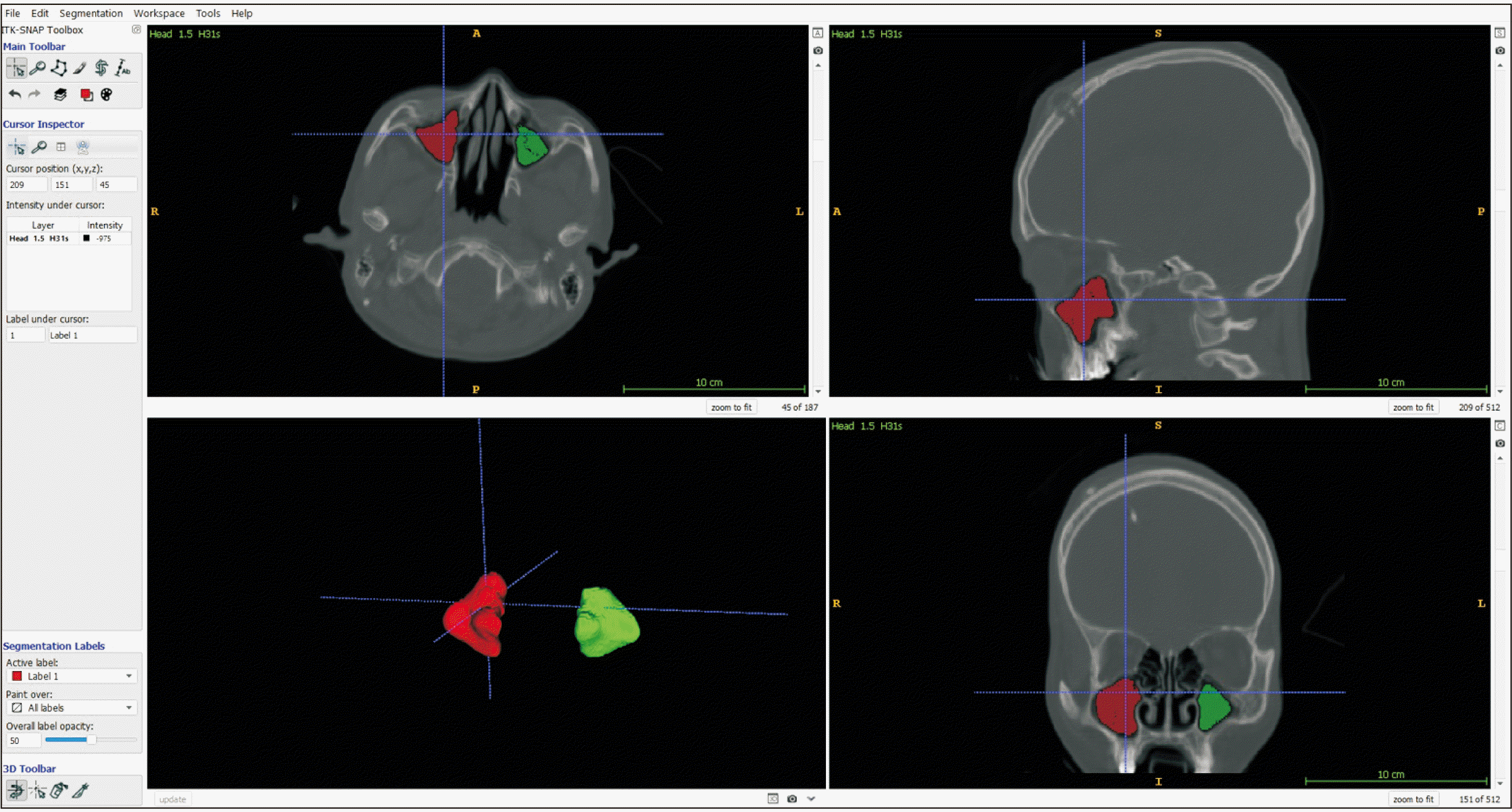
Slice thickness 1.0–1.5 mm of the CT images were used. Digital Imaging and Communications in Medicine files were reconstructed and MSV were extracted using ITK-SNAP 3.0 software (Cognitica, Philadelphia, PA, USA) (https://www.itksnap.org) [15]. Region of Interest (ROI) volume was created using active contour segmentation to selected sinus area ROI from slice axial, coronal and sagittal for semi-automatic segmentation (Fig. 1).
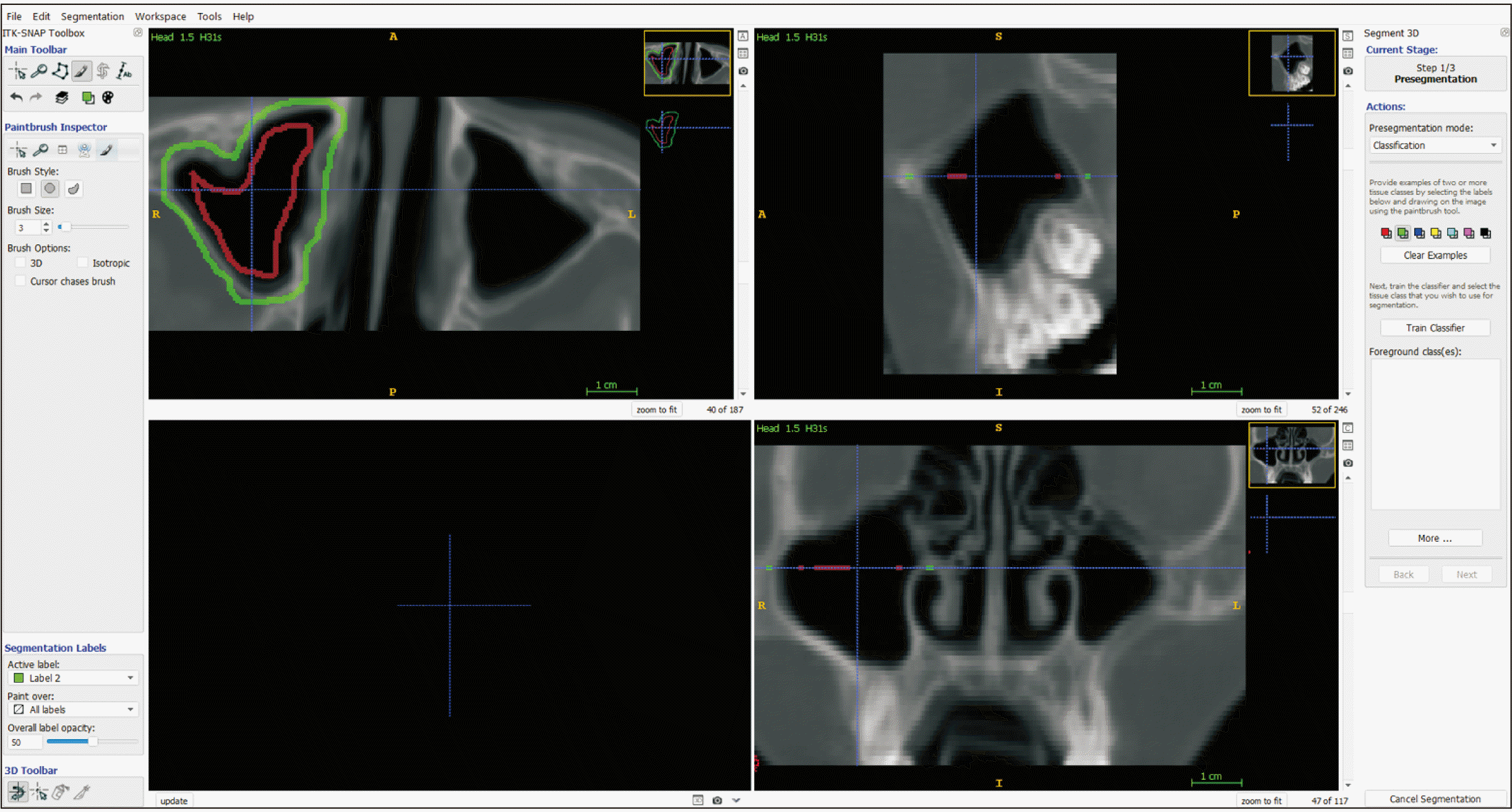
The aired mask was drawn using train classifier: red label for sinus cavity and green label for bone. Then, paint brush mode was used to add manual label for any actives label or erase the excess of anatomy including the airway and the surrounding environment (Fig. 2). Next, the image was placed to initialize the contour of cavity.
The contours evolution was executed. The object rendered was export into 3D STL file. Open segmentation window was opened and volumetric measurement was chosen to export volume data into a spreadsheet. ITK-SNAP software were used to measure MSV as the method of choice in this CT imaging data study, because semi-automatically approach was suggested to be more accurate than automatic segmentation [15].
The data were analyzed using IBM SPSS ver. 25.0 (IBM Corp., Armonk, NY, USA). The age of the subjects were grouped into 5 cohorts: 0–5, 6–10, 11–15, 16–20, and 20–25 years. The descriptive statistics were displayed using frequencies of sex and age, including average and standard deviation of the volumetric measurements. The symmetry of the right and left side MSV were noted, including the difference between genders.
Go to : 
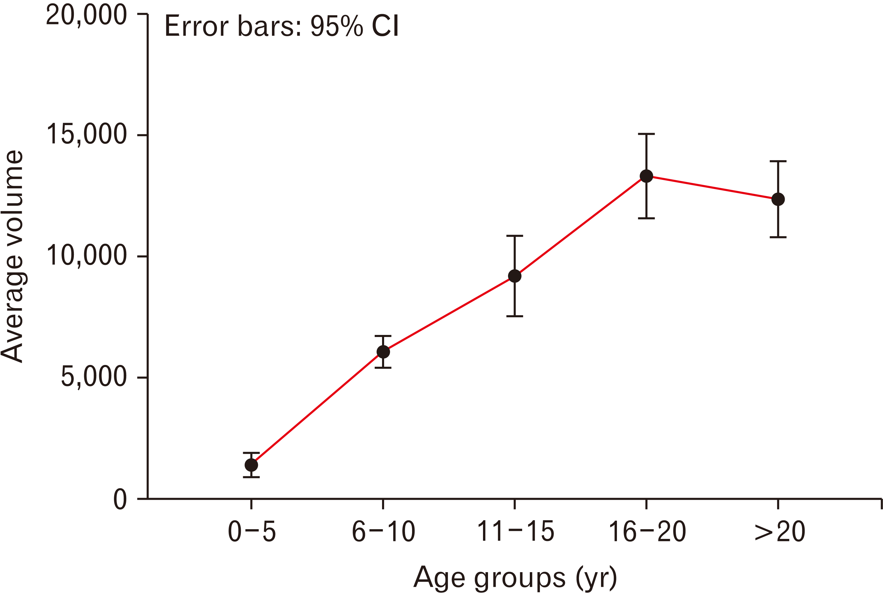
A total of 194 maxillary sinus were examined. Descriptive statistics were displayed in Table 1. The lowest average of MSV were shown between 0–5 years old group (1,361.12 mm3) and the largest MSV were noticed at the 16–20 years old group (13,278.73 mm3). After that, the MSV fell in 20–25 years old group to 12,325.21 mm3. The trend of MSV average value was increasing beginning the first year of life until reaching the 16–20 years old cohort, in which it began to decrease (Fig. 3). There was no significant difference between right and left MSV (Table 2). Moreover, no sexual dimorphism were detected in 16–20 years cohort (male=16, female=14; P=0.608) and 20–25 years old (male=14, female=24; P=0.654) cohort. The earliest pneumatization was observed on a baby boy at 28 days of life at 23.97 mm3. The largest MSV was recorded on a female patient aged 21 years old at 26.450 mm3.
Distribution of patients and volume of maxillary sinuses pooled by age groups
Go to : 
The paranasal sinuses volumetric study is well explored in the East Asian population [14, 16-18]. However, specifically for Indonesian population there is a lack of published paper in the indexed literature. Due to the vast distance between the eastern and western border of Indonesia geographical region, the population reside within this country varied, in which East Asian ancestry group dominated the majority of the population [19].
This study pooled the subjects into 5 chronological age cohorts. The maxillary sinus pneumatization began since the first year of life which support previous findings [5, 20] and peak at the 16–20 years old cohort. Then the average MSV declined at the 20–25 years old group. This growth pattern of maxillary sinus appeared to agree with the previous East Asian studies [16, 17, 21]. This study also demonstrated that there was no difference detected between the MSV right and left side which support the previous studies [4, 16, 18, 20].
The current project served as the pilot study of paranasal sinuses volumetric quantification in Indonesian population. Ideally, MSV growth study should be performed on healthy subjects longitudinally, by following the maxillary sinus as the age progresses. Nevertheless, this approach will expose healthy population to unnecessary radiation exposure. Therefore, although deviate from the ideal, this type of project using the existing archive of CT images is considered optimal as the tool of exploring the paranasal sinuses volume characteristics as age progressing.
Previous published paper described the linear measurements of maxillary sinus using CBCT in Indonesian population [22]. Up to this date, this study was the first in analyzing the volumetric aspect of maxillary sinus in Indonesian population and may have served as an initial database for clinicians. Further research should aim at the accuracy of paranasal sinuses volumetric assessment for age and sex estimation; and also for clinical evaluation; using automated method to accommodate larger sample size with equal contribution within each age and gender. In addition, this paper provided morphological knowledge for otolaryngology surgeons and dental implant practitioners as the foundation for sinus pathology assessment and surgical treatment planning.
In conclusion, The result of the study corroborated that the maxillary sinus pneumatization initialized during infancy. In addition, this study also presented the age-related MSV degeneration after the age of 20 years old and the similarity between right and left MSV. The knowledge of maxillary sinus morphometrics is essential in surgical approach of maxillary sinus pathology and dental implant procedures.
Go to : 
Acknowledgements
This study was funded by the Universitas Padjadjaran Internal Research Grant (RPLK) No.1427/UN6.3.1/LT/2020. This study was conducted under the supervision of Oral Biomaterial Research Centre Universitas Padjadjaran. The authors would like to extend gratitude to the donors of the CT images.
Go to : 
Notes
Author Contributions
Conceptualization: ES, YAL, HGN. Data acquisition: ES, YAL, HGN. Data analysis or interpretation: ES, YAL. Drafting of the manuscript: ES, YAL. Critical revision of the manuscript: ES, YAL, HGN, NM, HYY. Approval of the final version of the manuscript: all authors.
Go to : 
References
1. Maroldi R, Ravanelli M, Borghesi A, Farina D. 2008; Paranasal sinus imaging. Eur J Radiol. 66:372–86. DOI: 10.1016/j.ejrad.2008.01.059. PMID: 18375083.

2. Cellina M, Gibelli D, Cappella A, Toluian T, Pittino CV, Carlo M, Oliva G. 2021; Segmentation procedures for the assessment of paranasal sinuses volumes. Neuroradiol J. 34:13–20. DOI: 10.1177/1971400920946635. PMID: 32757847. PMCID: PMC7868593.

3. Natsis K, Karabatakis V, Tsikaras P, Chatzibalis T, Stangos A, Stangos N. 2004; Frontal sinus anatomical variations with potential consequences for the orbit. Study on cadavers. Morphologie. 88:35–8. DOI: 10.1016/S1286-0115(04)97997-0. PMID: 15208811.

4. Emirzeoglu M, Sahin B, Bilgic S, Celebi M, Uzun A. 2007; Volumetric evaluation of the paranasal sinuses in normal subjects using computer tomography images: a stereological study. Auris Nasus Larynx. 34:191–5. DOI: 10.1016/j.anl.2006.09.003. PMID: 17084569.

5. Aktuna Belgin C, Colak M, Adiguzel O, Akkus Z, Orhan K. 2019; Three-dimensional evaluation of maxillary sinus volume in different age and sex groups using CBCT. Eur Arch Otorhinolaryngol. 276:1493–9. DOI: 10.1007/s00405-019-05383-y. PMID: 30879193.

6. Pirimoglu B, Sade R, Sakat MS, Ogul H, Eren S, Kantarci M. 2017; Comparing subjective and objective image quality at two different radiation exposure ranges of the paranasal sinus CT examinations using a volumetric 320-row detector CT system. Dentomaxillofac Radiol. 46:20170212. DOI: 10.1259/dmfr.20170212. PMID: 28937286. PMCID: PMC5965943.

7. Sahlstrand-Johnson P, Jannert M, Strömbeck A, Abul-Kasim K. 2011; Computed tomography measurements of different dimensions of maxillary and frontal sinuses. BMC Med Imaging. 11:8. DOI: 10.1186/1471-2342-11-8. PMID: 21466703. PMCID: PMC3080316.

8. Stutzki M, Jahns E, Mandapathil MM, Diogo I, Werner JA, Güldner C. 2015; Indications of cone beam CT in head and neck imaging. Acta Otolaryngol. 135:1337–43. DOI: 10.3109/00016489.2015.1076172. PMID: 26313160.

9. Kirmeier R, Arnetzl C, Robl T, Payer M, Lorenzoni M, Jakse N. 2011; Reproducibility of volumetric measurements on maxillary sinuses. Int J Oral Maxillofac Surg. 40:195–9. DOI: 10.1016/j.ijom.2010.10.008. PMID: 21074367.

10. Bui NL, Ong SH, Foong KW. 2015; Automatic segmentation of the nasal cavity and paranasal sinuses from cone-beam CT images. Int J Comput Assist Radiol Surg. 10:1269–77. DOI: 10.1007/s11548-014-1134-5. PMID: 25503593.

11. Humphries SM, Centeno JP, Notary AM, Gerow J, Cicchetti G, Katial RK, Beswick DM, Ramakrishnan VR, Alam R, Lynch DA. 2020; Volumetric assessment of paranasal sinus opacification on computed tomography can be automated using a convolutional neural network. Int Forum Allergy Rhinol. 10:1218–25. DOI: 10.1002/alr.22588. PMID: 32306522.

12. Giacomini G, Pavan ALM, Altemani JMC, Duarte SB, Fortaleza CMCB, Miranda JRA, de Pina DR. 2018; Computed tomography-based volumetric tool for standardized measurement of the maxillary sinus. PLoS One. 13:e0190770. DOI: 10.1371/journal.pone.0190770. PMID: 29304130. PMCID: PMC5755892.

13. Spaeth J, Krügelstein U, Schlöndorff G. 1997; The paranasal sinuses in CT-imaging: development from birth to age 25. Int J Pediatr Otorhinolaryngol. 39:25–40. DOI: 10.1016/S0165-5876(96)01458-9. PMID: 9051437.

14. Park IH, Song JS, Choi H, Kim TH, Hoon S, Lee SH, Lee HM. 2010; Volumetric study in the development of paranasal sinuses by CT imaging in Asian: a pilot study. Int J Pediatr Otorhinolaryngol. 74:1347–50. DOI: 10.1016/j.ijporl.2010.08.018. PMID: 20863577.

15. Yushkevich PA, Piven J, Hazlett HC, Smith RG, Ho S, Gee JC, Gerig G. 2006; User-guided 3D active contour segmentation of anatomical structures: significantly improved efficiency and reliability. Neuroimage. 31:1116–28. DOI: 10.1016/j.neuroimage.2006.01.015. PMID: 16545965.

16. Ariji Y, Kuroki T, Moriguchi S, Ariji E, Kanda S. 1994; Age changes in the volume of the human maxillary sinus: a study using computed tomography. Dentomaxillofac Radiol. 23:163–8. DOI: 10.1259/dmfr.23.3.7835518. PMID: 7835518.

17. Ikeda A, Ikeda M, Komatsuzaki A. 1998; A CT study of the course of growth of the maxillary sinus: normal subjects and subjects with chronic sinusitis. ORL J Otorhinolaryngol Relat Spec. 60:147–52. DOI: 10.1159/000027584. PMID: 9579359.

18. Kawarai Y, Fukushima K, Ogawa T, Nishizaki K, Gunduz M, Fujimoto M, Masuda Y. 1999; Volume quantification of healthy paranasal cavity by three-dimensional CT imaging. Acta Otolaryngol Suppl. 540:45–9. DOI: 10.1080/00016489950181198. PMID: 10445079.
19. Sarilita E, Rynn C, Mossey PA, Black S, Oscandar F. 2018; Nose profile morphology and accuracy study of nose profile estimation method in Scottish subadult and Indonesian adult populations. Int J Legal Med. 132:923–31. DOI: 10.1007/s00414-017-1758-4. PMID: 29260392. PMCID: PMC5919985.

20. Adibelli ZH, Songu M, Adibelli H. 2011; Paranasal sinus development in children: a magnetic resonance imaging analysis. Am J Rhinol Allergy. 25:30–5. DOI: 10.2500/ajra.2011.25.3552. PMID: 21711972.

21. Jun BC, Song SW, Park CS, Lee DH, Cho KJ, Cho JH. 2005; The analysis of maxillary sinus aeration according to aging process; volume assessment by 3-dimensional reconstruction by high-resolutional CT scanning. Otolaryngol Head Neck Surg. 132:429–34. DOI: 10.1016/j.otohns.2004.11.012. PMID: 15746857.

22. Waluyo RF, Priaminiarti M, Yuniastuti M, Soedarsono N, Susilo BT. 2020; Measurements of sex-related differences in maxillary sinus and mandibular canal characteristic using cone beam computed tomography. Forensic Imaging. 21:200371. DOI: 10.1016/j.fri.2020.200371.

Go to : 


 PDF
PDF Citation
 Print
Print


 XML Download