

 PDF
PDF Citation
 Print
Print


Introduction
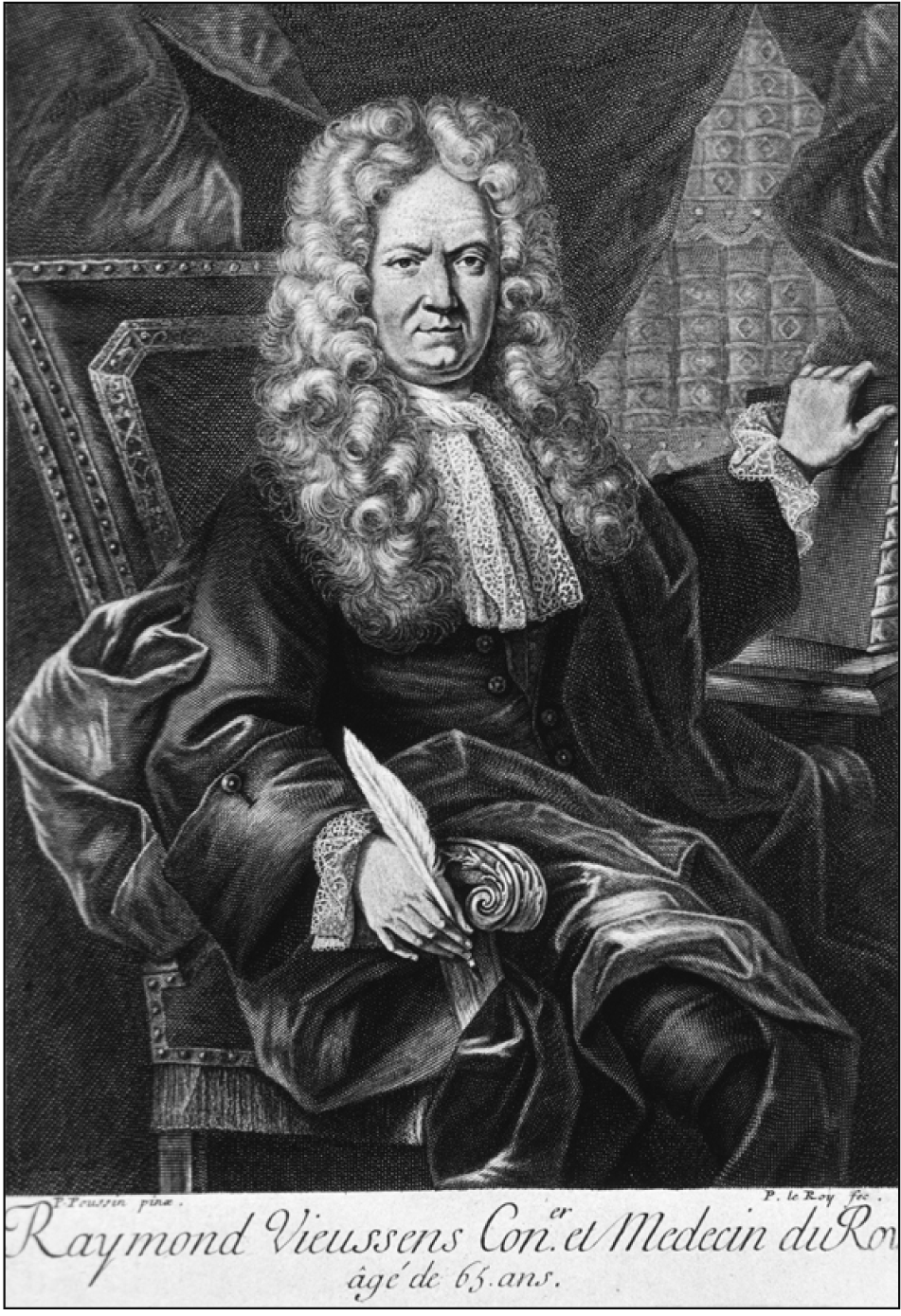
Raymond de Vieussens (1641–1715) was a French anatomist (Fig. 1) who made cardinal contributions in the field of cardiology [1]. He was a pioneer figure of cardiologic anatomy and torch bearer of two classic trends that defined anatomical research in 17th and 18th centuries. He diligently followed the human dissection based experimental methods during the late 17th century and heralded the trend of pathologic anatomy (autopsy based research) which became the signature model of 18th century anatomists [2]. His work is seminal in the field of cardiology and constitutes an important transitional phenomenon in terms of scientific outlook of research methods across centuries [3]. His discoveries and observations contributed significantly towards the advancement of cardiology and paved the way for its emergence as a sovereign discipline within the realm of medical science [4]. It was hypothesized that the seminal contributions of this exemplary anatomist need to be revisited as they constitute significant landmarks in the process involving the metamorphosis of cardiology into its present form as we are familiar with in modern times. Hence the present study was conducted to chronicle the life and achievements of this visionary anatomist.
Methods
An extensive literature search was undertaken for this study and indexed databases such as MEDLINE, PubMed, Scopus, EMBASE, CINAHL, Google Scholar as well as popular search platforms such as Wikipedia and standard Google search engine were referred to relevant published materials. The following terms were used during literature search: “Raymond de Vieussens,” “Vieussens,” “Raymond Vieussens biography,” “Vieussens and cardiology,” “Vieussens and cardiovascular system,” “Vieussens and anatomy,” “Vieussens and heart,” “cardiology in 17th century,” “cardiology in 18th century,” “pathologic anatomy,” and “Vieussens eponyms.” Published texts of Vieussens and their translations in English were consulted from online libraries while conducting the present study and wherever applicable have been appropriately referenced. The images used in the text were procured from the internet and it was ensured that all figures included in this study are in public domain i.e. free from copyright issues. Nevertheless, the source of these images have been duly acknowledged in their respective legends.
Onset of Career in Medicine at Montpellier
Raymond de Vieussens was born in 1641 in the commune of Le Vigan situated in Rouergue province of Southern France [1]. Not much is known about his early days and there is even confusion regarding his actual year of birth as few literature mentions it as 1635 [4]. His father Francois was a high ranking official in the French Army and Vieussens went on to become a first generation physician. During early part of his career Vieussens registered himself as a student of philosophy in Rodez, a small city on Southern France [5]. As destiny would unfold, Vieussens eventually moved to Montpellier and began his eventful journey in medicine and anatomical research in the famed University which is eponymous with the city [6]. Vieussens’s career as an avid researcher in anatomy was shaped during his training as a medical graduate from the prestigious University of Montpellier. It may be mentioned here that the University is among the earliest ones established in Europe (founded in 1220) [7]. It witnessed the first human dissection being conducted by French anatomist and surgeon Henri de Mondeville in 1315. Although the exercise was not legitimized by religious authorities, nevertheless it was a bold step towards breaking the shackles that hindered academic advances in medicine for 1700 years. Luminaries in anatomical sciences like Jacobus Sylvius (15th century), Andreas Vesalius (16th century) and Jean Pecquet (17th century) were associated with this illustrious institution [8]. Therefore, it was no surprize that Vieussens embraced anatomical research as his career path. In accordance with the trend prevailing among anatomists during 17th century, Vieussens also adopted experimental models based on human dissection as his medium of research activities [9, 10]. This aspect of Vieussens’s career was influenced by the academic environment in his alma mater as human dissection was routinely practised for teaching anatomy in Montpellier. More significantly, Vieussens’s journey to explore the unfound anatomical attributes received a major boost from the availability of large number of human cadavers in Montpellier, which was subsequently acknowledged in his communication with peers [11].
Early Venture in Neuroanatomy
As a medical graduate, Vieussens joined in the post of a physician in the Saint-Eloi Hospital in Montpellier in 1670. In addition to his normal duties, Vieussens devoted himself to anatomical research [3]. Like many of his contemporaries, Vieussens was remarkably influenced by the works of British anatomist Thomas Willis (1621–1675), whose seminal works in the domain of brain and nervous system were published around this time. Willis had documented his observations in Cerebri Anatome (published in 1664) and Pathologiae Cerebri et nervosa (published in 1667) [12]. Vieussens tirelessly worked on unravelling the anatomical details related to central and peripheral nervous system for the next ten years. The fact that he dissected 500 human cadavers during this period is a testimony of his hard work and dedication to the advancement of science [11]. The culmination of his humongous efforts was the publication of a compilation of his findings as Neurographia Universalis in 1685. Vieussens gave valuable insights on the structure and function of spinal cord, visual pathway and finer structural details of brainstem components [13]. Subsequent to the publication of his work, Vieussens received recognition in France (he was inducted as a member of the Academy of Sciences in Paris) as well as in Europe as such (he was honoured with the fellowship of Royal Society of London) [11]. However, the turning point in his academic career came in 1688, when his work received appreciation from the royal family of France. King Louis XIV himself was a great admirer of Vieussens and granted him an annual pension of 1,000 livres [1]. It is needless to mention that this economic support further strengthened his resolve to explore the human anatomical details through experiment based observations. His already glittering academic career continued to flourish even further under the royal patronage.
Discoveries in Cardiologic Anatomy
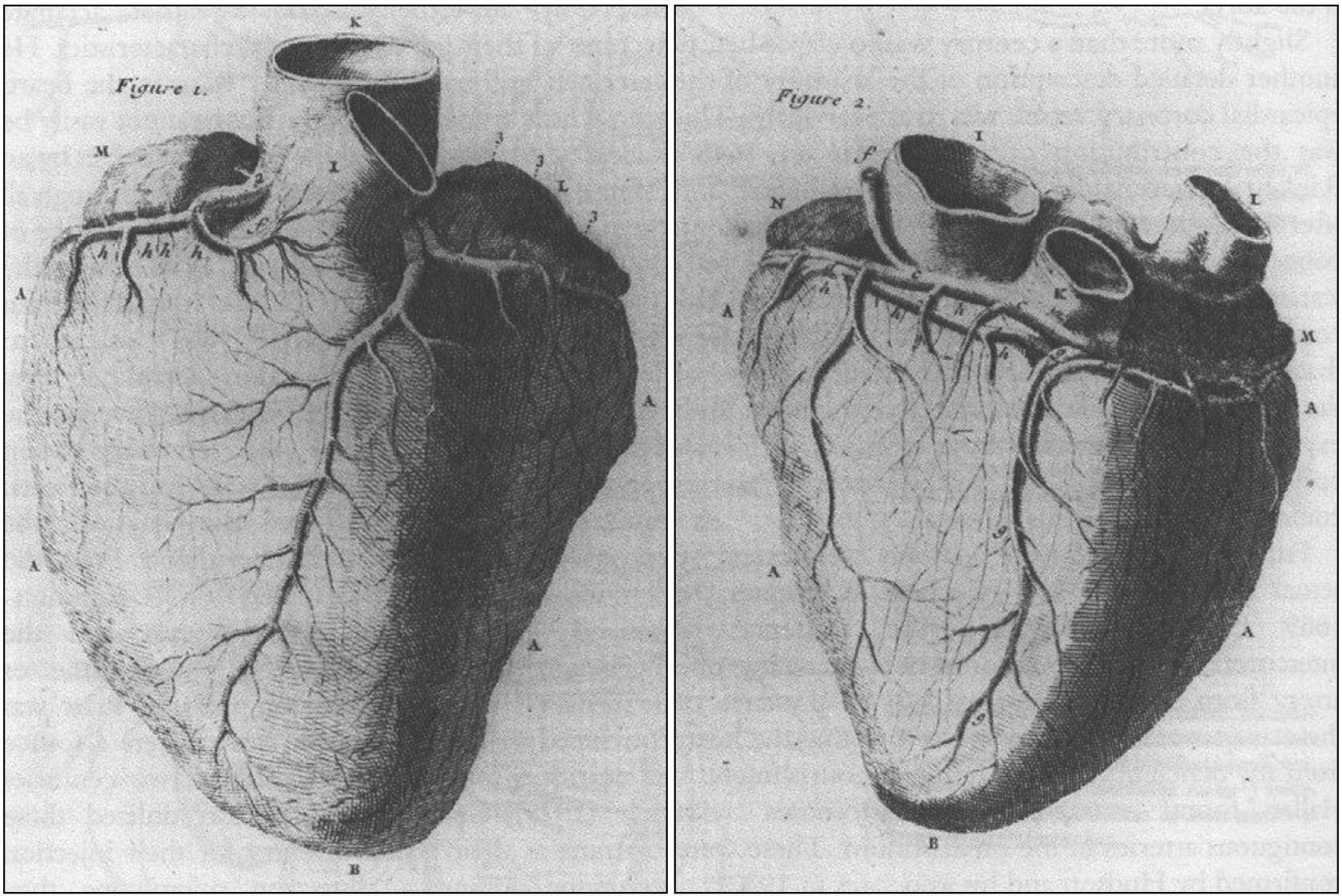
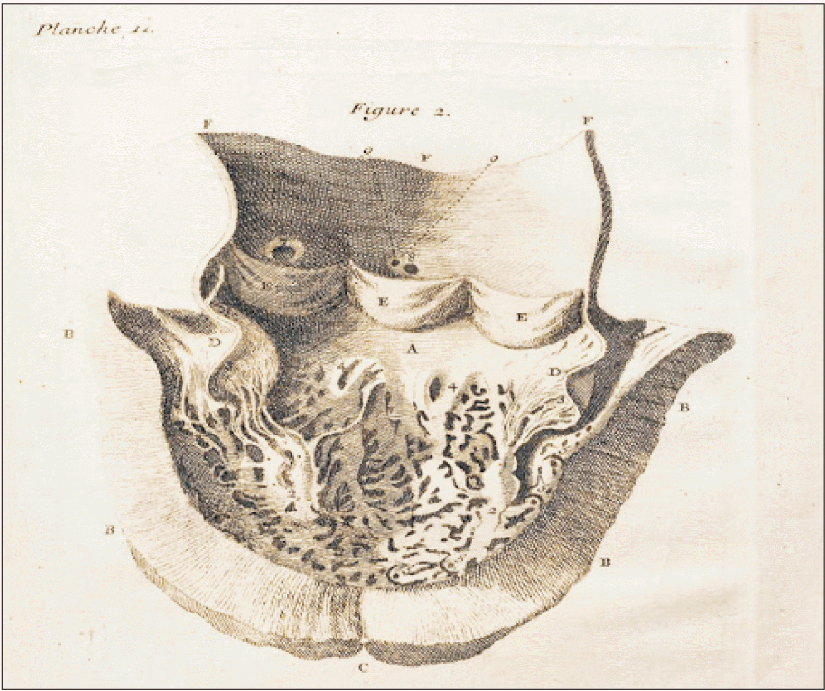
In the following years, Vieussens continued to work in Saint-Eloi Hospital and began his journey in the domain of cardiologic anatomy [3]. His inclination towards the anatomy of heart was possibly fuelled by pivotal observations documented by English physician William Harvey (1578–1657) regarding the anatomy of circulation and heart during the first half of 17th century [14]. Vieussens made a number of fascinating observations (Fig. 2) which he eventually published in 1705 as Novum vasorum corporis humani systema [15]. He was the first to describe the presence of tiny venous tributaries communicating between cardiac veins and chambers of heart [16]. As part of his experimental design, he ligated the superior and inferior vena cava as well as pulmonary veins. This was followed by injecting saffron dye into the coronary arteries. The dye followed its normal anatomical course and entered the coronary capillary network (located in myocardium) and then entered the cardiac veins and eventually into coronary sinus. Surprisingly, a small amount of dye was also observed in the ventricles of heart. Vieussens thus unveiled the minute cardiac venous tributaries and introduced the nomenclature ‘ducti carnosi’ [15]. However, a couple of years later, German anatomist Adam Thebesius (1686–1732) provided a more detailed account on the anatomy of these veins and hence they are eponymous as ‘Thebesian veins’ in present times [17]. The more accepted nomenclature is however ‘venae cordis minimae,’ which is used in contemporary texts [18].
Vieussens reported the existence of a unique arterial pathway linking the conus branch of right coronary artery with the conus branch of left anterior descending coronary artery (branch of left coronary artery). He described the structural anatomy of this arterial pathway in the form of a ring. It has become eponymous as Vieussens Arterial Ring (VAR) in the field of cardiology. Vieussens described this anatomical entity in details in his text Nouvelles Decouvertes sur le coeur which was published in 1706 [19]. Later on it was observed that VAR is present in 48% individuals and assumes significance from clinical perspective as it acts as a collateral circulatory pathway between the right and left coronary arterial system thus serving as a protective measure in case of coronary artery occlusive disease [20].
Vieussens also discovered the presence of a valve at the junction of great cardiac vein and coronary sinus [19]. This anatomical entity is known as the valve of great cardiac vein and is often referred to as ‘valve of Vieussens’ in contemporary literature. This anatomical structure holds significance in the field of clinical cardiology as depending on its morphology it may potentially cause obstruction during cardiac catheterization procedures [21]. Vieussens was the first to note the prominent oval margin of the fossa ovalis (limbus fossa ovalis) in the adult heart [19]. He identified that this anatomical structure develops from a different embryological component (septum secundum) as compared to the fossa ovalis (septum primum). Limbus fossa ovalis or annulus fossa ovalis is also referred to as ‘Vieussens Annulus’ [22]. The exemplary success of Vieussens in terms of unveiling the hitherto unknown details of cardiologic anatomy made him immensely popular as an anatomist across Europe [1]. Unsurprisingly, his achievements got the attention from his royal patrons and his annual pension was enhanced to 3,000 livres in 1707. As a mark of honour, Vieussens was appointed as a Conseiller (advisor) of state by King Louis XIV in the same year [11].
Invigorated by the recognition as well as adulation of his work, Vieussens continued with his experiments in cardiologic anatomy and the mystique associated with this vital organ of the human body was gradually unmasked before him. A considerable number of his findings were published less than one year before his death in 1715 as Traite Nouveau de la structure et de la Cause de Mouvement Naturel du coeur [23]. He described in detail the organization of myocardial fibres in the right and left ventricles of heart. He demonstrated that the myocardium is arranged in three layers: superficial, middle and deep. The muscle fibres are arranged vertically in the superficial and deep layers. The middle layer (more prominent in left ventricle) has horizontal fibres in the right ventricle and oblique fibres in the left ventricle (Fig. 3) [23]. His findings were later validated through advanced techniques in contemporary period albeit with modification of few finer details [24].
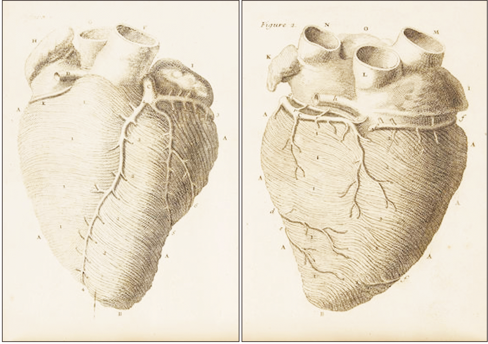
Vieussens was the first anatomist to give a precise description of the location and the course of coronary arteries, their branches and the coronary sinus [23, 25]. They were demonstrated by means of fine illustrations in his published works (Fig. 4). Vieussens described the pericardium of the heart as a bilayered sac like structure covering the organ [23]. It may be mentioned here that the gross anatomical details of pericardium were actually described by Vesalius in the 16th century itself. However, Vieussens accurately charted the clinical scenario associated with significant accumulation of fluid between two layers of pericardium. He hypothesized that the pericardial fluid compresses the heart whereby the chambers cannot dilate to their full capacity thus generating a feeble pulse. His clinical correlation of the anatomo-pathological condition had pin point precision based on which he could accurately diagnose a case of cardiac tamponade where the face of a patient became darker as his pulse became faster but weaker [26].
Trendsetter in Pathologic Anatomy within the Realm of Cardiology
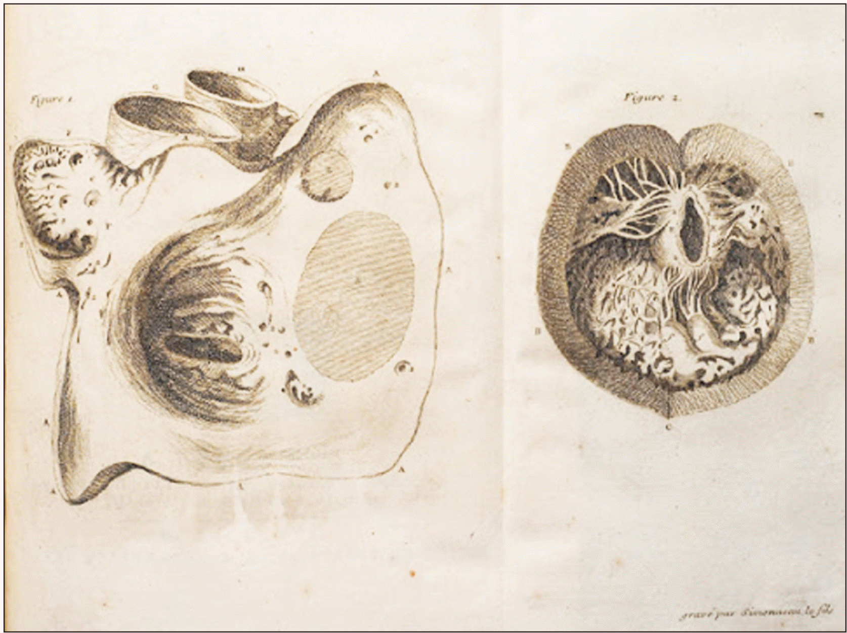
In addition to descriptions of normal anatomy of the heart, Vieussens initiated a new trend of detailing its pathologic anatomy. Vieussens used to correlate the findings in the cardiac tissue during autopsy of diseased patients with clinical manifestations when the same patient was alive [27]. Using this method which was unique in itself and different from the dissection based experiments prevalent during 17th century, Vieussens charted the anatomo clinical correlation of a classical case of mitral stenosis in his text Traite Nouveau (published in 1715) [23]. The observation was made by Vieussens in 1705, when a young man aged 30 was admitted at Saint-Eloi Hospital. The patient lied in a bed with his head propped up, having great difficulty in breathing with his heart beating with violent palpitation, his pulse feeble with eyes sunken and lips dark in appearance. Vieussens concluded a poor prognosis which was validated a week later vas the patient died. Vieussens himself conducted the autopsy and correlated his observations (pathology) with logical sequence of events involving the structures of the heart (anatomy). The principal observations were listed as chest cavity filled with yellowish serous fluid, both the lungs abnormally large in size and soft in consistency, heart became enlarged with dilated pulmonary veins. On opening the heart he noted that the right ventricle and right atrium were inordinately enlarged (Fig. 5). He also observed that the substance of mitral valve became bone like in appearance with consequent narrowing of the lumen into the left ventricle. He unerringly identified the mitral valve as the seat of the pathological condition (mitral stenosis). In a sequential manner he traced that with impairment of circulation the pulmonary veins were dilated eventually leading to stasis of blood in pulmonary vessels resulting in the enlargement of lungs, difficulty in breathing (impaired exchange of gases at alveolar level) and oozing out of serous component of blood in the chest cavity. Vieussens also concluded that the accumulated blood in the pulmonary circulation led to violent contraction of the right ventricle (visible palpation and enlargement in size). Also, as left ventricle was pumping very little blood into circulation, the pulse was weak and peripheries exhibited dark colour [23, 28].
Vieussens also presented the anatomo clinical correlation in a case of severe aortic insufficiency (aortic regurgitation) with remarkable precision [23]. The patient was a young male who presented with a pulse that was violent and whose nature was unknown to Vieussens till then. Vieussens however categorized the condition as catastrophic and associated with a serious pathologic condition of the heart. During autopsy Vieussens observed a markedly dilated left ventricle and the semilunar aortic valves were unusually stretched. He concluded failure of the aortic valve as root cause of the pathologic condition, whereby during each ventricular contraction a portion of the blood that was just pumped into the aorta was sent back to the left ventricle. This event led to the generation of the powerful and anomalous pulse evident at the peripheral arteries [4, 23]. This type of peripheral pulsation is referred to as the ‘Watson’s water hammer pulse’ or ‘Corrigan’s pulse’ or ‘collapsing pulse’ in contemporary literature. The pulse is described as bounding and forceful, rapidly increasing followed by subsequent collapsing and is characteristic of aortic regurgitation [29]. The visionary efforts of Vieussens revolutionized anatomical studies during the 18th century as the concept of pathologic anatomy gained immense popularity in the academic domain.
Pivotal Anatomical Figure at the Transition of 17th and 18th Century
Vieussens role as researcher in anatomy was pivotal and his contributions were critical in shaping the evolutionary pathway of cardiology as a prominent discipline in medical sciences [30-32]. During the initial part of his glowing academic career, particularly with reference to his first ten years in Saint-Eloi Hospital, his research methodology was based on human dissection based experiments [11]. Here he is seen as an anatomist following the footsteps of his luminary predecessors from 17th century like Harvey, Pecquet, Bartholin, and Malpighi [33]. However, in the second half of his career, particularly after receiving royal patronage, he was more inclined to autopsy based findings for his academic pursuits [4]. Maybe this change in perception was a result of his booming confidence, social prestige and also the economic security that comes collaterally with royal patronage. Nevertheless, this proved to be boon not only for cardiologic anatomy but anatomical sciences as such. Pathologic anatomy (autopsy based study of anatomy) emerged as a popular trend and later on it was pioneered by noted anatomists of 18th century like Giovanni Battista Morgagni, William Hunter and his brother John Hunter for significant discoveries in relation to pathologic anatomy of the heart and its valves as well as those of blood vessels in the human body [34, 35]. Hence it may be opined that in the history of anatomy, Vieussens holds his position as a vital link between the scientific revolution of 17th century and remarkable advances during 18th century.
Conclusion
Raymond de Vieussens was an exemplary anatomist of his time and his impeccable experimental methods with subsequent observations thereof brought him to the forefront of cardiologic anatomy during early 18th century. His extensive volume of scientific works essentially comprises of an amalgamation of normal anatomical details and pathological deviations in anatomical structures. His discoveries were pivotal in the advancement of cardiology and triggered its eventual rise as a prominent medical discipline in modern times.
 XML Download