Journal List > Ann Lab Med > v.42(3) > 1148568

Jain, Zhang, Bennett, and Komrokji: Myelodysplastic Syndromes with Bone Marrow Fibrosis: An Update

Abstract

Myelodysplastic syndrome (MDS) is a diverse hematological malignancy with a wide spectrum of presentations and implications. Treatment strategies for patients with MDS heavily rely on prognostic scoring systems, such as the revised international prognostic scoring system (IPSS-R). Bone marrow fibrosis (BMF) has been identified as an independent risk factor for poor survival in patients with MDS, irrespective of the IPSS-R risk category. However, BMF is not widely included in scoring systems and is not always considered by clinicians when making treatment decisions for patients. In this review, we discuss the available literature about the presentation and prognosis of patients with MDS and concurrent BMF. The prognostic impact of BMF should be factored in when deciding on transplant candidacy, especially for intermediate-risk patients.

INTRODUCTION

The stromal microenvironment of the bone marrow (BM) comprises structural fibrils that provide connective tissue structure and support for hematopoietic progenitor cells. Reticulin and collagen type I/III are the most common BM fibers [1]. BM biopsy sections can be assessed for the presence of these stromal fibers to evaluate the extent of fibrosis using a silver impregnation technique called Gomori’s stain [2]. A pathological increase in BM fibrosis (BMF) reportedly has both clinical and prognostic significance in various hematological malignancies. In chronic myeloid leukemia and multiple myeloma, BMF portends decreased responsiveness to most commonly used treatments [3, 4]. In primary myelofibrosis (PMF), the higher the fibrosis grade, the poorer the prognosis [5]. In patients with essential thrombocythemia, increased reticulin fibrosis predicts an increased risk of thrombosis, major bleeding, and transformation into myelofibrosis, and in patients with polycythemia vera, it is associated with worse outcomes [6].
Myelodysplastic syndrome (MDS) is a heterogenous hematological malignancy with a wide spectrum of clinical presentations. MDS is characterized by dysplasia in one or more BM cell lineages and/or ineffective erythropoiesis. In 1981, Sultan, et al. [7] first described eight cases of MDS with BMF at presentation. The disease presentation described was later included in the category of acute myeloid leukemia (AML), not otherwise specified as acute panmyelosis with myelofibrosis [8]. In 1989, Pagliuca, et al. [9] described 10 cases of primary MDS with striking BMF at presentation. Since then, multiple case reports, case series, and retrospective studies have reported MDS with fibrosis (MDS-F). MDS-F accounts for 10%–20% of MDS cases [10-13].
The risk stratification scale of the revised international prognostic scoring system (IPSS-R) for MDS ranges from very low risk to very high risk [14]. The IPSS-R utilizes cytogenetics, BM blasts, and laboratory parameters, including Hb, platelets, and absolute neutrophil count (ANC), at diagnosis [14]. BMF is not included in the IPSS-R for determining the risk category and prognosis. In addition, the clinical relevance of BMF was not recognized in the 2008 WHO classification [15]. The 2016 WHO classification identified MDS-F as an unclassified MDS subtype [8]. Moderate to severe BMF in de novo MDS is closely associated with multilineage dysplasia, transfusion dependence, and severe cytopenia [11, 16, 17]. Overall survival (OS) is inferior in de novo MDS patients with moderate to severe BMF. Poorer OS has been attributed to greater BM failure or a higher rate of leukemic transformation [10]. In patients with MDS who proceed to a hematopoietic allogeneic stem cell transplant (alloSCT), moderate to severe BMF is an independent risk factor for an inferior event-free survival. Delayed engraftment post alloSCT has been reported in MDS patients with any degree of BMF [18]. BMF can be seen in the initial diagnostic marrow or sometimes can develop during the course of MDS or after treatment. In a study by Fu, et al. [12], out of 551 patients with no significant BMF at diagnosis, 17 (4%) patients developed BMF during follow-up, and the median time to development of BMF was 22 months.
While the pathophysiology of BMF is not well understood, some theories suggest increased cytokine production from megakaryocytes and platelets, which reportedly are higher in patients with BMF [19]. Cytokine transforming growth factor-β has been suggested to be a potent stimulator of fibroblast collagen synthesis [19]. Other cytokines implicated in the pathophysiology of BMF include platelet-derived growth factor, calmodulin, and basic fibroblast growth factor [20]. Spliceosome and RAS pathway mutations have been implicated in the pathogenesis of BMF in patients with MDS [21]. However, it is clear that the BMF seen in MDS is a reactive process and that the fibroblasts present in the BM responsible for the fibrosis are not monoclonal [19]. We describe patients with MDS with BMF and their diagnosis, management, and prognosis.

Grading of BMF

Before 2005, the BMF grading was subjective, and pathologists used various grading systems [1]. In 2001, Steensma, et al. [20] attempted to define MDS-F. They used the Manoharan scale and recommended that only patients with grade 3+ or 4+ be diagnosed as having MDS-F [22]. In 2005, the European Myelofibrosis Network framed a consensus-based proposal for semi-quantitative evaluation of BMF to achieve a higher level of reproducibility in clinical practice [23]. Four categories were introduced to simplify the grading of marrow fibrosis (MF). MF-0 or no fibrosis in normal BM is used for patients with scattered linear reticulin without any intersections (cross-overs). Mild fibrosis (MF-1) is defined as a loose network of reticulin fibers with numerous intersections, particularly in the perivascular areas. Moderate fibrosis (MF-2) is graded when diffuse and dense reticulin with extensive intersections is noted, with focal and occasional bundles of collagen and/or osteosclerosis. In severe or grade 3 fibrosis (MF-3), there can be coarse bundles of collagen (on trichrome stain), frequently accompanying significant osteosclerosis in addition to diffuse and dense reticulin with extensive intersections [17]. MF-0/1 is present in up to 50% of MDS cases [7, 9, 13]. In this review, we discuss the significance of moderate to severe fibrosis (MF-2/3).

Phenotype of MDS-F

There are two types of fibers that contribute to BMF. While an increase in reticulin fibers has a limited association with the severity of the underlying malignancy, collagen fibers are strongly linked with abnormal blood counts and poor outcomes [1]. Reticulin fibrosis is frequently reversed after therapeutic intervention, while collagen fibrosis is less likely to alleviate with therapy [1]. Trichrome stain for collagen fibrosis is not commonly used for MDS-F as it is often, though not always, correlated with reticulin fibrosis (≥MF-2). Fig. 1 shows peripheral blood and BM findings in a patient with MDS-F. BMF is often associated with dry tap which could hinder morphologic assessment of dysplasia. Careful examination of peripheral blood and the slides from bone marrow touch imprint would be helpful.
In a study on the clinical significance of moderate to severe BMF in patients with therapy-related (t-)MDS, Fu, et al. [24] found that 47 out of 266 t-MDS patients (17%) had moderate to severe BMF. Clinically, patients with moderate to severe BMF had more severe thrombocytopenia (42×109/L vs. 62×109/L, P=0.039) and tended to have more circulating blasts (P=0.051) than patients with mild BMF, whereas the rates of other clinical symptoms, including anemia, neutropenia, hepatosplenomegaly, transfusion requirements, and constitutional symptoms, were alike. Risk factors for BMF were prior solid tumor (46.8% vs. 28.3% in prior hematological malignancies) and radiation therapy. Fu, et al. [12] found substantial BMF (MF-2/3) in 79 (13%) out of 630 MDS patients. On comparing patients with substantial BMF to patients that did not have MF-2/3 fibrosis, they found that hepatosplenomegaly, constitutional symptoms, and transfusion dependence were more frequently observed in patients with substantial BMF. They also noted that although white blood cell counts, ANCs, and Hb levels were similar between the two groups, thrombocytopenia was more severe in patients with substantial BMF (51 vs. 94×109/L) [12]. BM dysplasia was similar between patients without or with minimal BMF and patients with substantial BMF. As expected, patients with substantial BMF had higher megakaryocyte numbers and lower cellularity. However, they did not note a difference in the number of BM blasts [12].
In a study conducted at our institution by Melody, et al. [25], using our MDS database of 2,624 patients, MF-3 BMF was observed in only 4% of patients. Since preliminary analyses had indicated that only MF-3 affected survival, patients with MF-3 were compared with those with MF-0–2 fibrosis. In line with previous findings [12, 24], we noted that thrombocytopenia and a higher BM blast percentage were common in patients with MF-3 BMF. Interestingly, lactate dehydrogenase levels were higher in MF-3 patients (46% vs. 34%, P=0.02).
Ramos, et al. [26] reported that BMF is positively correlated with ferritin and erythropoietin levels, marrow cellularity, abnormal localization of immature precursors (ALIP), packed red blood concentrate transfusions in the first 16 weeks from diagnosis, p53 immunohistochemistry scores, and peripheral Wilm’s tumor 1 gene expression. Marisavljević, et al. [27] studied 236 patients with de novo MDS and found that immature cells and blasts in the peripheral blood and BM blasts were more frequently present in patients with MDS-F. In the BM, ALIP, dysmegakaryopoesis, and dyserythropoesis were also significantly higher in patients with MDS-F.

Genotype of primary MDS with fibrosis

It is imperative to recognize cytogenetic and mutational profiles that can help in BMF prognosis assessment. Cytogenetic features of MDS-F include monosomy 7 [9, 16, 28]. Complex or other unique cytogenetic features have been reported [7, 9, 16, 28-32]. Fu, et al. [24] studied 266 t-MDS patients and reported that monosomy 5 and monosomy 17 were more commonly detected in t-MDS patients with MF-2/3 (P=0.031 and P=0.043, respectively). In another study including 79 patients with substantial BMF, they noted that the distribution of cytogenetic risk using the new comprehensive cytogenetic scoring system for primary MDS/oligoblastic AML was similar [12]. In contrast to the findings by Fu, et al. [24], we noted poor cytogenetic and IPSS-R risk in patients with MF-3 BMF, but complex karyotypes were more commonly seen in these patients (31% vs. 16%, P=0.002) than in patients with MF-1/2 [25].
JAK2, CALR, or MPL variants leading to the constitutional activation of the JAK-STAT pathway are conspicuous features of myeloproliferative neoplasms. Molecular changes observed in patients with fibrotic MDS may be similar to those observed in patients with myelofibrosis. However, mutations in JAK2, CALR, and MPL are rare in MDS. Hussein, et al. [33] studied fibrosis-related gene transcripts in 119 patients with MDS, 70 of whom had accompanying BMF. They found that BMPR2, SMAD3, and SMAD4 were expressed in both PMF and MDS-F. Although mutational profiles were not available for all patients in the study by Fu, et al. [24], they noted that the JAK2, V617F variant was more frequently detected in patients with BMF (21% vs. 2%; P=0.014) than in patients without BMF. The median allele burden for the JAK2 V617F variant was 22.5% (5.5%–46%). Melody, et al. [25] reported that TP53 and SETBP1 variants are more frequently detected in patients with MF-3. Wang, et al. [21] recently studied 239 patients with MDS or MDS-AML with BMF and reported that U2AF1 variants were more common in patients with MF-2/3 (P=0.029). The inability of ruxolitinib to reduce BMF in patients with myelofibrosis contends against a pathogenic role of the JAK-STAT pathway in fibrosis. Hence, the pathogenic mechanisms of BMF in MDS remain to be fully unraveled.

Management of patients with fibrotic MDS

Patients with MDS with substantial BMF are treated using similar strategies as those without fibrosis. Supportive care, immunomodulatory agents, hypomethylating agents, induction chemotherapy, and/or alloSCT can be used. Fu, et al. [12] reported that treatment with hypomethylating agents did not decrease BMF even in patients who attained complete hematological responses, whereas patients who underwent alloSCT showed a complete resolution of BMF.
AlloSCT is the only potentially curative treatment for MDS. However, alloSCT is associated with a high mortality rate and hence is generally offered only to patients with high- and very-high-risk MDS. As noted before, BMF is not considered when IPSS-R is used to score the prognostic risk, but if BMF accompanies rapidly progressing MDS, alloSCT should be considered. Table 1 lists studies that comparatively investigated the outcomes of patients with MDS-F that underwent alloSCT and MDS patients without BMF. In a study in 721 MDS patients who underwent alloSCT, Kroger, et al. [18] compared patients with MF-3 BMF (N=39) with those with MF-1/2 (N=199) and MF-0 (N=483) BMF. They found that neutrophil engraftment occurred later in patients with MF-1,2/3 than in patients with MF-0 (16 vs. 20 vs. 17 days, P=0.002). Scott, et al. [34] studied 471 patients undergoing alloSCT, including 113 patients with either MDS or AML arising from MDS. They also noted delayed engraftment in patients with BMF (hazard ratio [HR] 0.4; P<0.001). However, they did not find differences in OS, relapse-free survival (RFS), and non-relapse mortality (NRM) between patients with BMF and those without, in their cohort. On subgroup analysis of patients with intermediate or high disease risk according to the IPSS-R, they found that OS, RFS, and NRM were inferior in patients with BMF.
Wang, et al. [21], in their recent study in 239 MDS and secondary AML patients who underwent alloSCT, reported that OS and disease-free survival were significantly lower in patients with MF-2/3 than in patients with MF-0/1 (P=0.018). BMF was an independent predictor for survival in patients with ≥10% BM blasts at diagnosis, and these patients had improved outcomes if they achieved complete remission before alloSCT.

Prognostic value of BMF

The most important implication of BMF in MDS patients is its impact on their prognosis in terms of survival as well as progression. Table 2 compares outcomes of patients with MDS-F with those of patients with MDS. Fu, et al. [24] in their study in 266 t-MDS patients, 17% of whom had MF-2/3 BMF, reported a similar risk of AML transformation (9/39 vs. 48/191, P=0.482) and a comparable OS (8 vs. 9 months, P=0.926) between patients with MF-2/3 BMF and those with MF-1 at the median follow-up of 11.5 months in both univariate and multivariate analyses. In their study in 79 patients with MF-2/3 BMF, Fu, et al. [12] noted that the rate of AML transformation was higher in patients with substantial BMF (37% vs. 26%; P=0.057), and OS (21 vs. 42 months; P=0.000) and leukemia-free survival (LFS; 52 vs. 120 months; P=0.0003) were inferior in patients with BMF than in patients who did not undergo alloSCT. Cox regression analysis revealed that BMF was an independent risk factor for inferior OS and LFS. In their study in 236 de novo MDS patients, Marisavljević, et al. [27] reported that MDS patients with BMF had a significantly inferior OS compared to those without BMF (13 vs. 35 months; P=0.0005), and the average time to AML transformation was significantly shorter in patients with BMF (32 vs. &gt;56 months, P=0.015).
Melody, et al. [25] found that OS was significantly shorter in patients with MF-3 BMF than in those with MF-0–2 BMF (17 vs. 39.4, 36.6, and 24.5 months, respectively, P<0.005). OS was shorter in all IPSS-R risk groups (low, intermediate, and high) when patients with MF-3 BMF were compared with those with MF-0–2 BMF. Multivariate analysis revealed that the HR for death was 1.6 (95% CI, 1.2–1.9) (P<0.005) for MF-3 BMF compared with MF-0 BMF. Although there was no increase in the rate of AML transformation in patients with MF-3 BMF in our patient cohort (29% vs. 28%, P=0.84), the median time to AML transformation was shorter in patients with MF-3 than in those with MF-1/2 (23 vs. 35 months, P=0.001) [25].
Fu, et al. [12] noted that patients who developed BMF during the course of MDS showed evidence of disease progression on BMF development. The median OS for these patients from the time of BMF development was only nine months. For MDS patients with BMF that go on to receive alloSCT, the 3-year relapse rate was higher in patients with BMF than in those without (47% vs. 28%, P=0.04) [34]. Accordingly, Wang, et al. [35] recently reported that mild to moderate BMF is predictive of a poor clinical outcome.

CONCLUSION

When the IPSS-R prognostic criteria were first established, BMF was considered a potential factor for determining the prognostic risk. However, it was discounted as an additive factor for predicting survival in MDS because of its low prevalence and discrepancies in the evaluation of the degree of BMF across institutions. We presented evidence that BMF is a poor prognostic variable, even if it is observed during the course of disease. It is recommended that BMF be integrated into currently used risk classification/stratification systems. The presence of moderate to severe BMF in patients with MDS should be accounted for when considering transplantation as a potential treatment strategy in patients with intermediate-risk disease. Future directions include further improving our understanding of the underlying pathobiology of BMF and assessing the impacts of treatments in patients with MDS-F.

ACKNOWLEDGEMENTS

None.

Notes

AUTHOR CONTRIBUTIONS

Jain AG wrote the manuscript. Zhang L reviewed the manuscript and contributed to the images for the manuscript. Bennet JM and Komrokii R critically reviewed and corrected the manuscript.

CONFLICTS OF INTEREST

Jain AG, Zhang L, and Bennett JM do not have any conflicts of interest to report. Komroji R does not have any conflicts significant for this review. Komroji R: BMSCelgene: Consultancy, Membership on an entity’s Board of Directors or Advisory Committees and Speakers Bureau, Jazz: Consultancy and Speakers Bureau, Novartis: Consultancy, Honoraria and Membership on an entity’s Board of Directors or Advisory Committees, AbbVie: Consultancy, Acceleron: Consultancy, PharmaEssentia: Membership on an entity’s Board of Directors or Advisory Committees, Taiho Oncology: Membership on an entity’s Board of Directors or Advisory Committees, Geron: Consultancy.

RESEARCH FUNDING

None declared.

REFERENCES

1. Kuter DJ, Bain B, Mufti G, Bagg A, Hasserjian RP. 2007; Bone marrow fibrosis: pathophysiology and clinical significance of increased bone marrow stromal fibres. Br J Haematol. 139:351–62. DOI: 10.1111/j.1365-2141.2007.06807.x. PMID: 17910625.
crossref
2. Gömöri G. 1937; Silver impregnation of reticulum in paraffin sections. Am J Pathol. 13:993–1002.5. PMID: 19970363. PMCID: PMC1911151.
3. Kvasnicka HM, Thiele J, Schmitt-Graeff A, Diehl V, Zankovich R, Niederle N, et al. 2001; Bone marrow features improve prognostic efficiency in multivariate risk classification of chronic-phase Ph1+ chronic myelogenous leukemia: a multicenter trial. J Clin Oncol. 19:2994–3009. DOI: 10.1200/JCO.2001.19.12.2994. PMID: 11408494.
crossref
4. Subramanian R, Basu D, Dutta TK. 2007; Significance of bone marrow fibrosis in multiple myeloma. Pathology. 39:512–5. DOI: 10.1080/00313020701570038. PMID: 17886102.
crossref
5. Vener C, Fracchiolla NS, Gianelli U, Calori R, Radaelli F, Iurlo A, et al. 2008; Prognostic implications of the European consensus for grading of bone marrow fibrosis in chronic idiopathic myelofibrosis. Blood. 111:1862–5. DOI: 10.1182/blood-2007-09-112953. PMID: 18029552.
crossref
6. Campbell PJ, Bareford D, Erber WN, Wilkins BS, Wright P, Buck G, et al. 2009; Reticulin accumulation in essential thrombocythemia: prognostic significance and relationship to therapy. J Clin Oncol. 27:2991–9. DOI: 10.1200/JCO.2008.20.3174. PMID: 19364963. PMCID: PMC3398138.
crossref
7. Sultan C, Sigaux F, Imbert M, Reyes F. 1981; Acute myelodysplasia with myelofibrosis: a report of eight cases. Br J Haematol. 49:11–6. DOI: 10.1111/j.1365-2141.1981.tb07191.x. PMID: 7272222.
crossref
8. Arber DA, Orazi A, Hasserjian R, Thiele J, Borowitz MJ, Le Beau MM, et al. 2016; The 2016 revision to the World Health Organization classification of myeloid neoplasms and acute leukemia. Blood. 127:2391–405. DOI: 10.1182/blood-2016-03-643544. PMID: 27069254.
crossref
9. Pagliuca A, Layton DM, Manoharan A, Gordon S, Green PJ, Mufti GJ. 1989; Myelofibrosis in primary myelodysplastic syndromes: a clinico-morphological study of 10 cases. Br J Haematol. 71:499–504. DOI: 10.1111/j.1365-2141.1989.tb06309.x. PMID: 2640542.
crossref
10. Della Porta MG, Malcovati L, Boveri E, Travaglino E, Pietra D, Pascutto C, et al. 2009; Clinical relevance of bone marrow fibrosis and CD34-positive cell clusters in primary myelodysplastic syndromes. J Clin Oncol. 27:754–62. DOI: 10.1200/JCO.2008.18.2246. PMID: 19103730.
crossref
11. Buesche G, Teoman H, Wilczak W, Ganser A, Hecker H, Wilkens L, et al. 2008; Marrow fibrosis predicts early fatal marrow failure in patients with myelodysplastic syndromes. Leukemia. 22:313–22. DOI: 10.1038/sj.leu.2405030. PMID: 18033321.
crossref
12. Fu B, Jaso JM, Sargent RL, Goswami M, Verstovsek S, Medeiros LJ, et al. 2014; Bone marrow fibrosis in patients with primary myelodysplastic syndromes has prognostic value using current therapies and new risk stratification systems. Mod Pathol. 27:681–9. DOI: 10.1038/modpathol.2013.187. PMID: 24186132.
crossref
13. Lambertenghi-Deliliers G, Orazi A, Luksch R, Annaloro C, Soligo D. 1991; Myelodysplastic syndrome with increased marrow fibrosis: a distinct clinico-pathological entity. Br J Haematol. 78:161–6. DOI: 10.1111/j.1365-2141.1991.tb04411.x. PMID: 1712222.
crossref
14. Greenberg PL, Tuechler H, Schanz J, Sanz G, Garcia-Manero G, Solé F, et al. 2012; Revised international prognostic scoring system for myelodysplastic syndromes. Blood. 120:2454–65. DOI: 10.1182/blood-2012-03-420489. PMID: 22740453. PMCID: PMC4425443.
crossref
15. Vardiman JW, Thiele J, Arber DA, Brunning RD, Borowitz MJ, Porwit A, et al. 2009; The 2008 revision of the World Health Organization (WHO) classification of myeloid neoplasms and acute leukemia: rationale and important changes. Blood. 114:937–51. DOI: 10.1182/blood-2009-03-209262. PMID: 19357394.
crossref
16. Verhoef GE, De Wolf-Peeters C, Ferrant A, Deprez S, Meeus P, Stul M, et al. 1991; Myelodysplastic syndromes with bone marrow fibrosis: a myelodysplastic disorder with proliferative features. Ann Hematol. 63:235–41. DOI: 10.1007/BF01698371. PMID: 1958747.
crossref
17. Della Porta MG, Malcovati L. 2011; Myelodysplastic syndromes with bone marrow fibrosis. Haematologica. 96:180–3. DOI: 10.3324/haematol.2010.039875. PMID: 21282718. PMCID: PMC3031682.
crossref
18. Kröger N, Zabelina T, van Biezen A, Brand R, Niederwieser D, Martino R, et al. 2011; Allogeneic stem cell transplantation for myelodysplastic syndromes with bone marrow fibrosis. Haematologica. 96:291–7. DOI: 10.3324/haematol.2010.031229. PMID: 20971823. PMCID: PMC3031698.
crossref
19. Tefferi A. 2005; Pathogenesis of myelofibrosis with myeloid metaplasia. J Clin Oncol. 23:8520–30. DOI: 10.1200/JCO.2004.00.9316. PMID: 16293880.
crossref
20. Steensma DP, Hanson CA, Letendre L, Tefferi A. 2001; Myelodysplasia with fibrosis: a distinct entity? Leuk Res. 25:829–38. DOI: 10.1016/S0145-2126(01)00055-8. PMID: 11532514.
crossref
21. Wang J, Wang Q, Zhang H, He Y, Huang Y, Zhang R, et al. 2021; Moderate to severe marrow fibrosis as a more advanced risk factor for MDS and MDS-AML patients with excess of blasts receiving allogeneic hematopoietic stem cell transplantation. Transplant Cell Ther. 27:666.e1–9. DOI: 10.1016/j.jtct.2021.05.006. PMID: 34020086.
crossref
22. Manoharan A, Horsley R, Pitney WR. 1979; The reticulin content of bone marrow in acute leukaemia in adults. Br J Haematol. 43:185–90. DOI: 10.1111/j.1365-2141.1979.tb03740.x. PMID: 508627.
crossref
23. Thiele J, Kvasnicka HM, Facchetti F, Franco V, van der Walt J, Orazi A. 2005; European consensus on grading bone marrow fibrosis and assessment of cellularity. Haematologica. 90:1128–32. PMID: 16079113.
24. Fu B, Ok CY, Goswami M, Xei W, Jaso JM, Muzzafar T, et al. 2013; The clinical importance of moderate/severe bone marrow fibrosis in patients with therapy-related myelodysplastic syndromes. Ann Hematol. 92:1335–43. DOI: 10.1007/s00277-013-1776-3. PMID: 23660629. PMCID: PMC4190057.
crossref
25. Melody M, Al Ali N, Zhang L, Ramadan H, Padron E, Sallman D, et al. 2020; Decoding bone marrow fibrosis in myelodysplastic syndromes. Clin Lymphoma Myeloma Leuk. 20:324–8. DOI: 10.1016/j.clml.2020.01.003. PMID: 32044274.
crossref
26. Ramos F, Robledo C, Izquierdo-García FM, Suárez-Vilela D, Benito R, Fuertes M, et al. 2016; Bone marrow fibrosis in myelodysplastic syndromes: a prospective evaluation including mutational analysis. Oncotarget. 7:30492–503. DOI: 10.18632/oncotarget.9026. PMID: 27127180. PMCID: PMC5058695.
crossref
27. Marisavljević D, Rolović Z, Cemerikić V, Bosković D, Colović M. 2004; Myelofibrosis in primary myelodysplastic syndromes: clinical and biological significance. Med Oncol. 21:325–31. DOI: 10.1385/MO:21:4:325. PMID: 15579916.
crossref
28. Ohyashiki K, Sasao I, Ohyashiki JH, Murakami T, Iwabuchi A, Tauchi T, et al. 1991; Clinical and cytogenetic characteristics of myelodysplastic syndromes developing myelofibrosis. Cancer. 68:178–83. DOI: 10.1002/1097-0142(19910701)68:1<178::AID-CNCR2820680131>3.0.CO;2-Q. PMID: 2049740.
crossref
29. Allen EF, Lunde JH, McNally R, Branda RE. 1996; A case of acute myelofibrosis with complex karyotypic changes: a type of myelodysplastic syndrome. Cancer Genet Cytogenet. 90:24–8. DOI: 10.1016/0165-4608(96)00037-4. PMID: 8780742.
crossref
30. Reilly JT, Dolan G. 1991; Proposed classification for the myelodysplasia/myelofibrosis syndromes. Br J Haematol. 79:653. DOI: 10.1111/j.1365-2141.1991.tb08099.x. PMID: 1772789.
crossref
31. Narayanan MN, Benaim ME, Geary CG, Harrison CJ. 1991; Hyperfibrotic myelodysplasia. Br J Haematol. 79:653–5. DOI: 10.1111/j.1365-2141.1991.tb08100.x. PMID: 1772790.
crossref
32. Maschek H, Georgii A, Kaloutsi V, Werner M, Bandecar K, Kressel MG, et al. 1992; Myelofibrosis in primary myelodysplastic syndromes: a retrospective study of 352 patients. Eur J Haematol. 48:208–14. DOI: 10.1111/j.1600-0609.1992.tb01587.x. PMID: 1592101.
33. Hussein K, Stucki-Koch A, Kreipe H. 2018; Profile of fibrosis-related gene transcripts and megakaryocytic changes in the bone marrow of myelodysplastic syndromes with fibrosis. Ann Hematol. 97:2099–106. DOI: 10.1007/s00277-018-3411-9. PMID: 29961094.
crossref
34. Scott BL, Storer BE, Greene JE, Hackman RC, Appelbaum FR, Deeg HJ. 2007; Marrow fibrosis as a risk factor for posttransplantation outcome in patients with advanced myelodysplastic syndrome or acute myeloid leukemia with multilineage dysplasia. Biol Blood Marrow Transplant. 13:345–54. DOI: 10.1016/j.bbmt.2006.10.030. PMID: 17317588.
crossref
35. Wang N, Xu H, Li Q, Fang X, Liu J, Sui X, et al. 2020; Patients of myelodysplastic syndrome with mild/moderate myelofibrosis and a monosomal karyotype are independently associated with an adverse prognosis: long-term follow-up data. Cancer Manag Res. 12:5881–91. DOI: 10.2147/CMAR.S258875. PMID: 32765089. PMCID: PMC7371562.
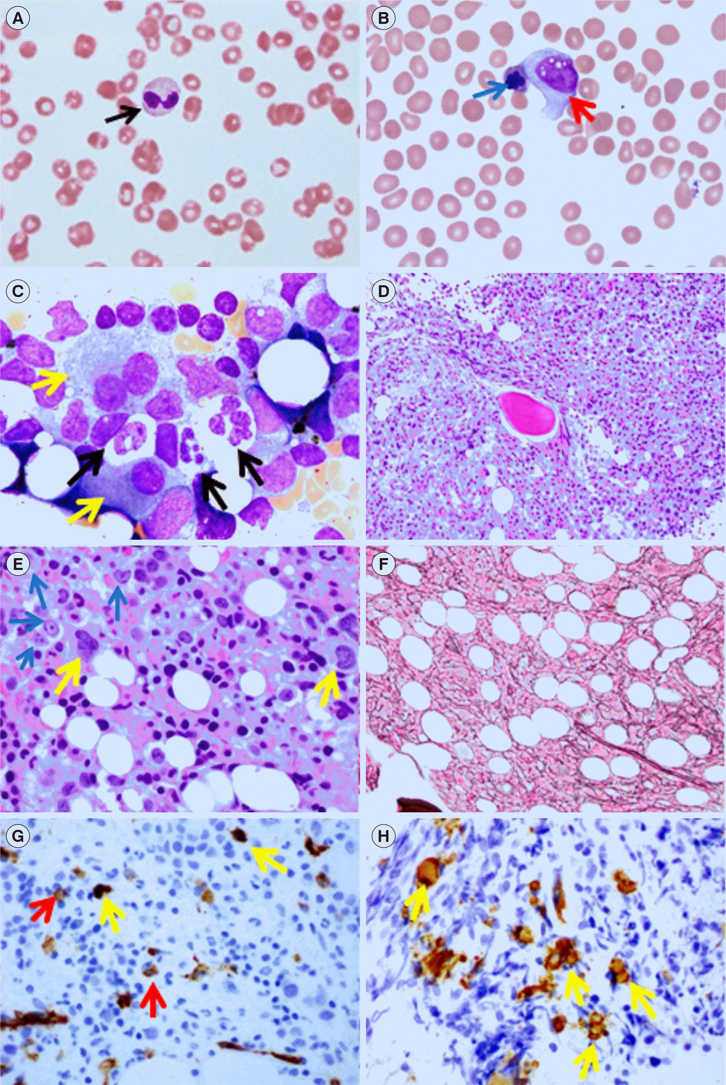
Fig. 1
Fibrotic MDS. (A, B) Peripheral blood smears revealing dysplastic neutrophils with pseudo-Pelger–Huet changes (black arrow–bilobed neutrophil, usually indicates an underlying BM disorder) and circulating dysplastic erythroid precursors with multilobulated nuclei (blue arrow) and a circulating blast (red arrow) - leukoerythroblastosis (Wright stain, ×1,000). (C) BM touch imprint smear showing hypogranulated neutrophils (black arrows) and a bilobed megakaryocyte (yellow arrow) (Wright–Giemsa stain, ×1,000). (D, E) BM core biopsy showing hypercellularity (>90%) associated with increased left-shifted erythroid precursors (blue arrow), dysplastic megakaryocytes (yellow arrow), decreased myeloid precursor numbers, and patchy crush artifact (hematoxylin and eosin stain, ×200 [D], and ×600 [E]). (F) Reticulin stain (×200) showing moderate reticulin fibrosis (MF-2/3). (G) CD34-stained (brown colored) occasional myeloblasts (red arrows) and occasional small megakaryocytes/megakaryoblasts (yellow arrows) and (H) CD61-stained megakaryocytes, including small forms (immunoperoxidase stain, ×200).
Abbreviation: MDS, myelodysplastic syndrome; BM, bone marrow.
alm-42-3-299-f1.tif
Table 1
Studies in patients with MDS with BMF undergoing BM transplantation
Reference Patients (N) Patients with fibrosis, N (%) OS AML transformation 3-yr RFS Engraftment (day)
Wang, et al. 2021 [21] 239 (MDS + MDS-AML) MF-1 81 (33.9) MF-2/3 37 (15.5) 3-yr OS rate 72% vs. 67.5% vs. 41.3% (P = 0.018) MF-2/3 vs. MF-1 vs. MF-0 8.8% vs. 39.9% vs. 50%, P = 0.028 72.8% vs. 68.8% vs. 44.8%; P = 0.018 Neutrophils, 13 vs. 13 vs. 14; P = 0.031 Platelets, 17 vs. 17.5 vs. 20.5; P = 0.05
Scott, et al. 2007 [34] 471 113 HR, 1.21; P = 0.16 Platelets, 17 vs. 28; P < 0.0001
Kroger, et al. 2011 [18] 721 MF-1/2 199 MF-3 39 HR, 1.13 vs. 1.94; P = 0.002 HR 1.13 vs. 1.88 (P = 0.003) Platelets, 16 vs. 17 vs. 20; P = 0.002

Abbreviations: BM, bone marrow; BMF, bone marrow fibrosis; OS, overall survival; AML, acute myeloid leukemia; RFS, relapse free survival; MDS, myelodysplastic syndrome; MF-1/2/3, BMF grade 1/2/3: HR, hazard ratio.

Table 2
Studies comparing MDS patients with BMF with those without BMF
Reference Patients (N) Patients with fibrosis, N (%) OS (months) AML transformation (%) DFS
Wang, et al. 2020 [35] 157 34 (21.7) 24 MF-1 10 MF-2 17.7 vs. 47.6; P = 0.001 20.3 vs. 41; P = 0.013 13.5 vs. 42.0 months, P = 0.002
Fu, et al. 2014 [12] 630 MF-2/3 79 (13) Control 166 21 vs. 42; P = 0.000 LFS 52 vs. 120; P = 0.003
Della Porta, et al. 2009 [10] 298 MF-1 128 (43) MF-2 45 (15) MF-3 7 (2) Inferior in 2/3 vs. 0/1; P < 0.0001 Inferior in 2/3 vs. 0/1; P < 0.0001
Maschek, et al. 1992 [32] 352 61 (17.3) 10 vs. 28.9 36.6 in patients with BMF
Marisavljević, et al. 2004 [27] 236 126 (53.4) 13 vs. 35; P = 0.00055 24.1 vs. 18.9
Melody, et al. 2020 [25] 2,624 MF-0–2 2,517 MF-3 107 19 vs. 56; P = 0.02 (alloSCT) 29 vs. 28; P = 0.84

Abbreviations: MDS, myelodysplastic syndrome; BMF, bone marrow fibrosis; OS, overall survival; AML, acute myeloid leukemia; DFS, disease-free survival; MF-1/2/3, BMF grade 1/2/3; LFS, leukemia-free survival; MF, myelofibrosis; alloSCT, allogeneic stem cell transplantation.

TOOLS
Similar articles