Journal List > J Korean Foot Ankle Soc > v.25(4) > 1148560

Irreducible Spacer Subluxation after Total Ankle Replacement Arthroplasty in a Patient with Rheumatoid Arthritis: A Case Report

Abstract

End-stage ankle arthritis is a debilitating condition that causes functional limitations and consequently a poor quality of life. Total ankle replacement arthroplasty is a good alternative to arthrodesis for preserving the ankle’s range of motion. However, many complications can occur in patients with rheumatoid arthritis and with poor soft tissue and bone conditions. A 61-year-old female experienced spacer subluxation after surgery, which was not reduced by medial soft tissue release and spacer change. Buttress plating was found to be a good treatment option to prevent spacer subluxation and can be considered in patients with rheumatoid arthritis with bone erosion and soft tissue damage.

Rheumatoid arthritis is a systemic disease that commonly involves multiple joints and has numerous complications. Ankle joint arthritis occurs in 15% to 52% of adult-onset rheumatoid arthritis patients.1) Patients usually have osteoporosis, fragile skin, and poor soft tissue around the ankle.2) The difference between rheumatoid and osteoarthritic arthritis is that the coexistence of severe degenerative changes in the ankle and hindfoot can occur in the former.3) Therefore, the ankle and hindfoot joints could be damaged simultaneously, as another study mentioned that 90% of patients had obvious radiological arthritic changes in the hindfoot, and 30% had spontaneous fusion of the subtalar joint.2,3) Standard treatment for an ankle with rheumatoid arthritis is arthrodesis with acceptable outcomes and fewer complications.4) However, arthrodesis makes climbing steps and walking on uneven surfaces difficult.3) For the present case, we planned total ankle arthroplasty to preserve ankle motion as other peritalar joints were all spontaneously fused. However, irreducible spacer subluxation occurred after the operation, even after revision with additional soft tissue release and spacer change. Our report focuses on the reduction of subluxation with buttress plating after total ankle arthroplasty in a patient with rheumatoid arthritis.

CASE REPORT

The relevant institutional review board approved this study. A 61-year-old female presented with chronic pain in both ankle joints. The patient had a 30-year history of rheumatic arthritis and a satisfactory result in the left ankle joint after total ankle arthroplasty. Due to the severe progression of her rheumatoid arthritis, her right midfoot joints and subtalar joint were spontaneously fused (Fig. 1). We believed that total ankle arthroplasty would be more suitable for preserving the range of motion in the ankle joint, which was the remaining mobile joint among the patient’s middle and hindfoot joints. The operation was performed under spinal anesthesia with a HINTEGRA (Integra lifesciences, Plainsboro Township, NJ, USA) total ankle replacement system. The soft tissue around the ankle joint was severely tight after the distal tibia was cut. Medial and lateral soft tissue release was performed with deltoid ligament release, ostectomy of the tip of the medial malleolus, and soft tissue release around the lateral malleolus. An old avulsed bony fragment in the anterior tibiofibular ligament fibular attachment was missed during the procedure. Stability was checked after the insertion of the trial implant and polyethylene spacer.
The spacer was mildly subluxated laterally with hindfoot inversion and foot supination. We thought that excessive hindfoot inversion and foot supination would be postoperatively diminished with soft tissue closure and spontaneous soft tissue adhesion. Careful sutures and a short leg splint were applied to prevent spacer subluxation. However, lateral subluxation of the spacer (Fig. 2) was observed on immediate postoperative plain radiography. An increased anterior slope (16°) of the distal tibia was also observed.
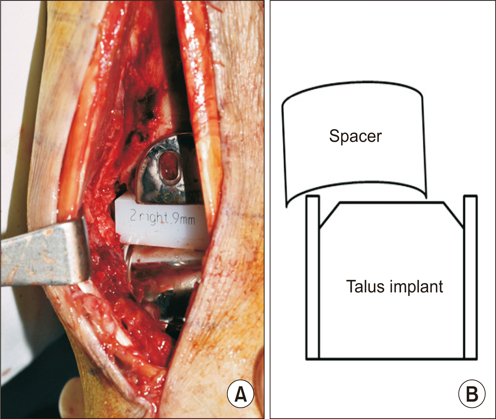
A revision operation was planned for spacer reduction and soft tissue management. We rechecked the gap balance, and a tight medial joint was observed. To reduce excessive force around the medial ankle joint, additional soft tissue release was performed. However, this was not sufficient in creating a balanced joint gap. Additional Z-plasty on the posterior tibialis tendon was performed, which resulted in a balanced and widened joint space. The polyethylene spacer was changed from 6 mm to 9 mm in height to adapt to and realign with the widened joint space. However, the spacer was still subluxated with hindfoot inversion and forefoot supination. The subluxated spacer was not reduced spontaneously when the edge of the spacer was locked outside the ridge of the talar implant (Fig. 3). A buttress plate was fixed on the anterolateral side of the distal tibia to prevent the crossing of the spacer edge over the ridge of the talus implant. Spacer subluxation was not provoked by hindfoot inversion or foot supination (Fig. 4). At the 2-year follow-up, the ankle joint showed symmetrical space, and the polyethylene spacer did not show recurrent subluxation or wear in standing ankle plain radiographs (Fig. 5).

DISCUSSION

Ankle arthritis is a common condition that causes chronic disability and decreased quality of life.5) Total ankle arthroplasty has become a popular treatment option with better outcomes with advanced implant designs.6,7) However, rates of reoperation, revision, and other complications are still concerns associated with this operation.8) A meta-analysis has reported that the early failure and revision rate of total ankle arthroplasty after an average of 5 years, ranges from 1.3% to 32.3% (mean, 9.8%).9)
Polyethylene spacer dislocation is a complication. It can occur in various conditions, such as a preoperative coronal malalignment greater than 5°, thinner insert height, and unaddressed ankle ligament laxity at the time of surgery.10-12) The edge loading of the prosthesis is frequent in ankles treated with mobile-bearing prosthesis and coronal plane deformity.10,13) Total ankle arthroplasty in coronal plane deformity showed a lower survival rate in inflammatory joint disease (48%) compared to that of neutrally aligned ankles (90%) after 8 years.14) Deltoid ligament release or lateral ligament reconstruction was introduced to manage coronal plane deformities. Some authors recommended medial malleolar osteotomy after skin necrosis with deltoid release and recurrent laxity with lateral ligament reconstruction.15)
In our patient, the main reason for spacer subluxation was a tight medial joint gap due to insufficient medial soft tissue release. However, lateral subluxation of the spacer still existed with hindfoot inversion and foot supination after medial soft tissue release and spacer change. We thought that this recurrent subluxation was mostly due to spontaneous hindfoot and midtarsal fusion. In this situation, the range of motion normally achieved by the hindfoot and midtarsal joints might be concentrated only in the ankle joint. Furthermore, a decreased elasticity of soft tissue due to multiple adjacent degenerative changes in rheumatoid arthritis could be a contributing factor to this motion concentration. Additionally, the absence of fibular bone stock to which the anterior tibiofibular ligament is attached could be another reason.16) This bony stock could act as a lateral static stabilizer to prevent lateral movement of the spacer. Finally, the increased anterior slope of the distal tibia cutting could also be another reason. An anterior slope of approximately 7° is recommended, as this is perpendicular to the tibial axis.17) In these patients, an increased anterior slope (16°) was observed on postoperative plain radiography. However, the slope was not corrected because spacer subluxation was managed with buttress plating. Crossing of the spacer edge to the talus implant ridge was effectively prevented.
Soft tissue management for ligament balancing is critical for preventing polyethylene complications. Careful assessment of subtalar and midtarsal joint motion to prevent prosthesis subluxation and dislocation is also necessary in patients with rheumatoid arthritis with poor soft tissue and bone condition.2,3) Buttressing plating could be one of the options to prevent polyethylene spacer subluxation in cases wherein almost the whole range of motion, except that of the ankle, is restricted in patients with rheumatoid arthritis. Complications such as polyethylene wear were a concern in this patient because friction between the plate and spacer was anticipated. However, there are insufficient studies to date on wear in total ankle arthroplasty.18) Therefore, careful long-term follow-up is needed with concerns of polyethylene fracture, wear, and osteolysis.

Notes

Financial support

None.

Conflict of interest

None.

REFERENCES

1. Su EP, Kahn B, Figgie MP. Total ankle replacement in patients with rheumatoid arthritis. Clin Orthop Relat Res. 2004; (424):32–8. doi: 10.1097/01.blo.0000132181.46593.82. DOI: 10.1097/01.blo.0000132181.46593.82. PMID: 15241141.
crossref
2. Ng SY, Crevoisier X, Assal M. 2012; Total ankle replacement for rheumatoid arthritis of the ankle. Foot Ankle Clin. 17:555–64. doi: 10.1016/j.fcl.2012.08.004. DOI: 10.1016/j.fcl.2012.08.004. PMID: 23158370.
crossref
3. Wood PL, Crawford LA, Suneja R, Kenyon A. 2007; Total ankle replacement for rheumatoid ankle arthritis. Foot Ankle Clin. 12:497–508. viidoi: 10.1016/j.fcl.2007.05.002. DOI: 10.1016/j.fcl.2007.05.002. PMID: 17765841.
crossref
4. Cracchiolo A 3rd, Cimino WR, Lian G. 1992; Arthrodesis of the ankle in patients who have rheumatoid arthritis. J Bone Joint Surg Am. 74:903–9. DOI: 10.2106/00004623-199274060-00012. PMID: 1634581.
crossref
5. Glazebrook M, Daniels T, Younger A, Foote CJ, Penner M, Wing K, et al. 2008; Comparison of health-related quality of life between patients with end-stage ankle and hip arthrosis. J Bone Joint Surg Am. 90:499–505. doi: 10.2106/JBJS.F.01299. DOI: 10.2106/JBJS.F.01299. PMID: 18310699.
crossref
6. Lefrancois T, Younger A, Wing K, Penner MJ, Dryden P, Wong H, et al. 2017; A prospective study of four total ankle arthroplasty implants by non-designer investigators. J Bone Joint Surg Am. 99:342–8. doi: 10.2106/JBJS.16.00097. DOI: 10.2106/JBJS.16.00097. PMID: 28196036.
crossref
7. Stavrakis AI, SooHoo NF. 2016; Trends in complication rates following ankle arthrodesis and total ankle replacement. J Bone Joint Surg Am. 98:1453–8. doi: 10.2106/JBJS.15.01341. DOI: 10.2106/JBJS.15.01341. PMID: 27605689.
crossref
8. Daniels TR, Younger AS, Penner M, Wing K, Dryden PJ, Wong H, et al. 2014; Intermediate-term results of total ankle replacement and ankle arthrodesis: a COFAS multicenter study. J Bone Joint Surg Am. 96:135–42. doi: 10.2106/JBJS.L.01597. DOI: 10.2106/JBJS.L.01597. PMID: 24430413.
9. Gougoulias N, Khanna A, Maffulli N. 2010; How successful are current ankle replacements?: a systematic review of the literature. Clin Orthop Relat Res. 468:199–208. doi: 10.1007/s11999-009-0987-3. DOI: 10.1007/s11999-009-0987-3. PMID: 19618248. PMCID: PMC2795846.
crossref
10. Brunner S, Barg A, Knupp M, Zwicky L, Kapron AL, Valderrabano V, et al. 2013; The Scandinavian total ankle replacement: long-term, eleven to fifteen-year, survivorship analysis of the prosthesis in seventy-two consecutive patients. J Bone Joint Surg Am. 95:711–8. doi: 10.2106/JBJS.K.01580. DOI: 10.2106/JBJS.K.01580. PMID: 23595069.
11. Mulcahy H, Chew FS. 2015; Current concepts in total ankle replacement for radiologists: complications. AJR Am J Roentgenol. 205:1244–50. doi: 10.2214/AJR.14.14171. DOI: 10.2214/AJR.14.14171. PMID: 26587931.
crossref
12. Scott AT, Nunley JA. 2009; Polyethylene fracture following STAR ankle arthroplasty: a report of three cases. Foot Ankle Int. 30:375–9. doi: 10.3113/FAI.2009.0375. DOI: 10.3113/FAI.2009.0375. PMID: 19356364.
crossref
13. Wood PL, Deakin S. 2003; Total ankle replacement. The results in 200 ankles. J Bone Joint Surg Br. 85:334–41. doi: 10.1302/0301-620x.85b3.13849. DOI: 10.1302/0301-620X.85B3.13849. PMID: 12729104.
14. Doets HC, Brand R, Nelissen RG. 2006; Total ankle arthroplasty in inflammatory joint disease with use of two mobile-bearing designs. J Bone Joint Surg Am. 88:1272–84. doi: 10.2106/JBJS.E.00414. DOI: 10.2106/JBJS.E.00414. PMID: 16757761.
crossref
15. Doets HC, van der Plaat LW, Klein JP. 2008; Medial malleolar osteotomy for the correction of varus deformity during total ankle arthroplasty: results in 15 ankles. Foot Ankle Int. 29:171–7. doi: 10.3113/FAI.2008.0171. DOI: 10.3113/FAI.2008.0171. PMID: 18315972.
crossref
16. Beumer A, Valstar ER, Garling EH, Niesing R, Ginai AZ, Ranstam J, et al. 2006; Effects of ligament sectioning on the kinematics of the distal tibiofibular syndesmosis: a radiostereometric study of 10 cadaveric specimens based on presumed trauma mechanisms with suggestions for treatment. Acta Orthop. 77:531–40. doi: 10.1080/17453670610012557. DOI: 10.1080/17453670610012557. PMID: 16819696.
crossref
17. Buechel FF Jr, Pappas MJ. 2003; Ten-year evaluation of cementless Buechel-Pappas meniscal bearing total ankle replacement. Foot Ankle Int. 24:462–72. doi: 10.1177/107110070302400603. DOI: 10.1177/107110070302400603. PMID: 12854666.
crossref
18. Mujukian A, Ho NC, Day MJ, Ebramzadeh E, Sangiorgio SN. 2020; A systematic review of unsystematic total ankle replacement wear evaluations. JBJS Rev. 8:e0091. doi: 10.2106/JBJS.RVW.19.00091. DOI: 10.2106/JBJS.RVW.19.00091. PMID: 32149932.
crossref

Figure. 1
Preoperative plain radiographs show narrowed ankle joint space and spontaneous fusion of subtalar and midtarsal joints (A: standing anterior-posterior; B: standing lateral).
jkfas-25-4-181-f1.tif
Figure. 2
Immediate postoperative anterior-posterior ankle plain radiograph shows asymmetrical ankle joint space, absence of bone fragment of anterior tibiofibular ligament attachment, and laterally subluxated polyethylene spacer.
jkfas-25-4-181-f2.tif
Figure. 3
Polyethylene spacer was subluxated laterally. It was not reduced when the edge of space was locked on the ridge of talus implant (A: intraoperative photo; B: a schematic diagram).
jkfas-25-4-181-f3.tif
Figure. 4
Polyethylene spacer was not subluxated with buttress plating when ankle inversion and foot supination force was applied (A: intraoperative photo; B: a schematic diagram).
jkfas-25-4-181-f4.tif
Figure. 5
At the two-year follow-up plain radiographs, spacer was located appositely in the ankle joint (A: standing anterior-posterior; B: standing lateral).
jkfas-25-4-181-f5.tif
TOOLS
Similar articles