

 PDF
PDF Citation
 Print
Print


INTRODUCTION
Paraganglioma is a tumor that arises from the paraganglion. It can occur in various organs associated with sympathetic and parasympathetic nerves. Gallbladder paraganglioma (GP) is rare, with only 12 cases reported in the literature to date [1-11]. According to reported cases, GP usually presents non-specific symptoms such as abdominal pain owing to associated cholecystitis, although it does occasionally present symptoms of gastrointestinal bleeding [4]. Laboratory and imaging studies also present non-specific findings. Thus, most GP cases are diagnosed incidentally based on histopathological findings after cholecystectomy [10].
Because the disease is rare, clinical information about GP is insufficient. Herein, we describe a case of GP in a 48-year-old female along with a literature review of all GP cases described to date.
Go to : 
CASE
A 48-year-old female visited the hospital because of an intermittent abdominal pain that had occurred 3 to 4 months previously. She had no remarkable issues in her medical or family history. There were no abnormalities in her vital signs or physical examinations. All laboratory findings were within normal limits except that hemoglobin level was low at 9.0 g/dL (range, 12–18 g/dL). Tumor markers were within their normal ranges, CA19-9 level < 2.00 U/mL (range, 0–37 U/mL) and carcinoembryonic antigen level 0.82 ng/mL (range: 0–5 ng/mL).
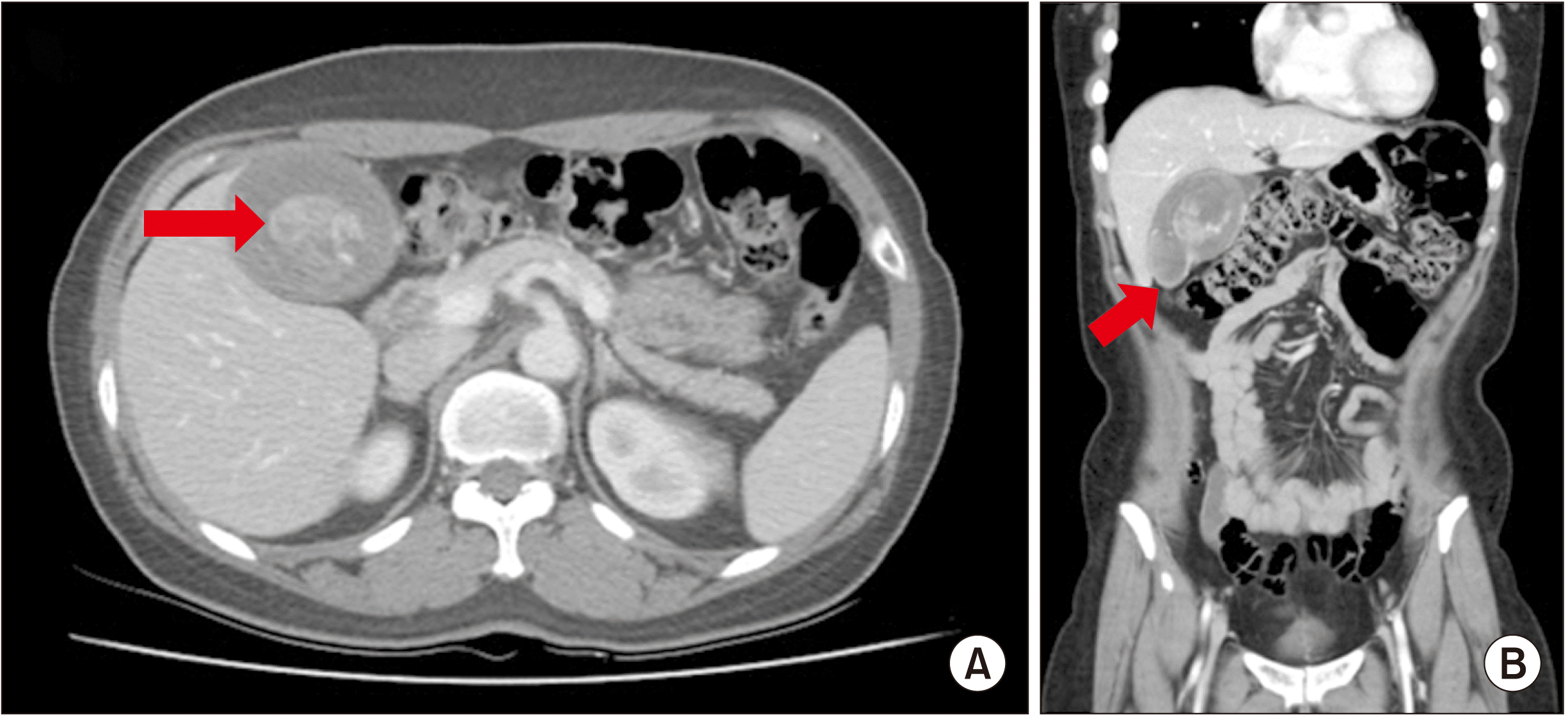
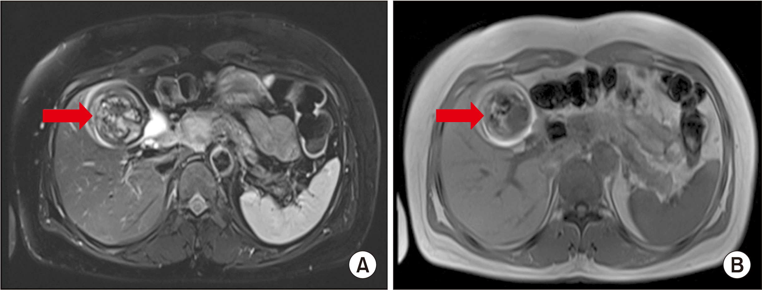
Abdominal ultrasonography (US) revealed a single uniformly shaped mass measuring 8.0 cm × 5.0 cm in the gallbladder lumen (Fig. 1). Abdominal computed tomography (CT) showed a uniformly contrasted mass measuring 8.7 cm × 5.3 cm and a small area of calcification in the gallbladder (Fig. 2). Abdominal magnetic resonance imaging (MRI) showed an 8-cm-sized mass in the gallbladder body and fundus with uneven high signal intensity on T1-weighted imaging. T2-weighted image showed low signal intensity of the mass with a linear dark signal intensity within the mass suggesting hemosiderin deposition in the hematoma (Fig. 3). These MRI findings suggested that the intraluminal mass was s gallbladder hematoma rather than a malignancy.
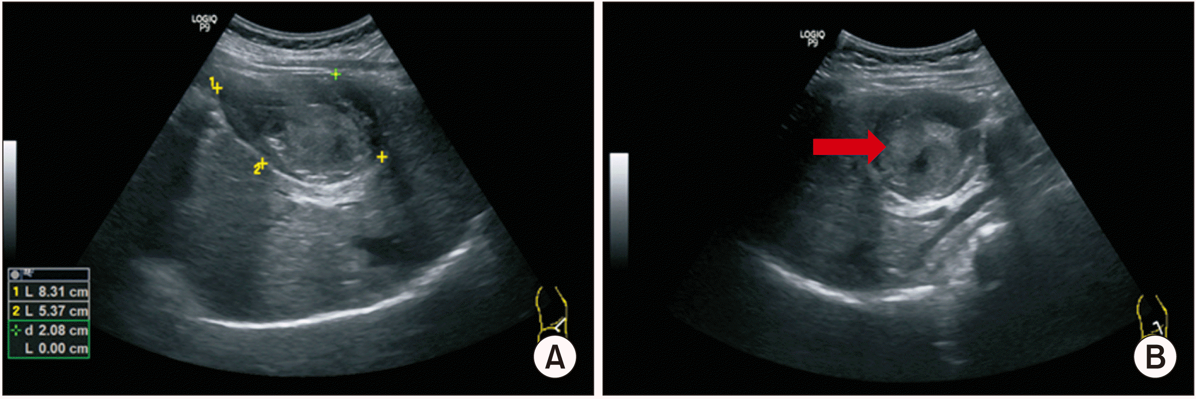 | Fig. 1Abdominal ultrasonography showing gallbladder distention (A) and an 8.0-cm × 5.0-cm sized heterogenous mass in the gallbladder lumen (arrow in B).
|
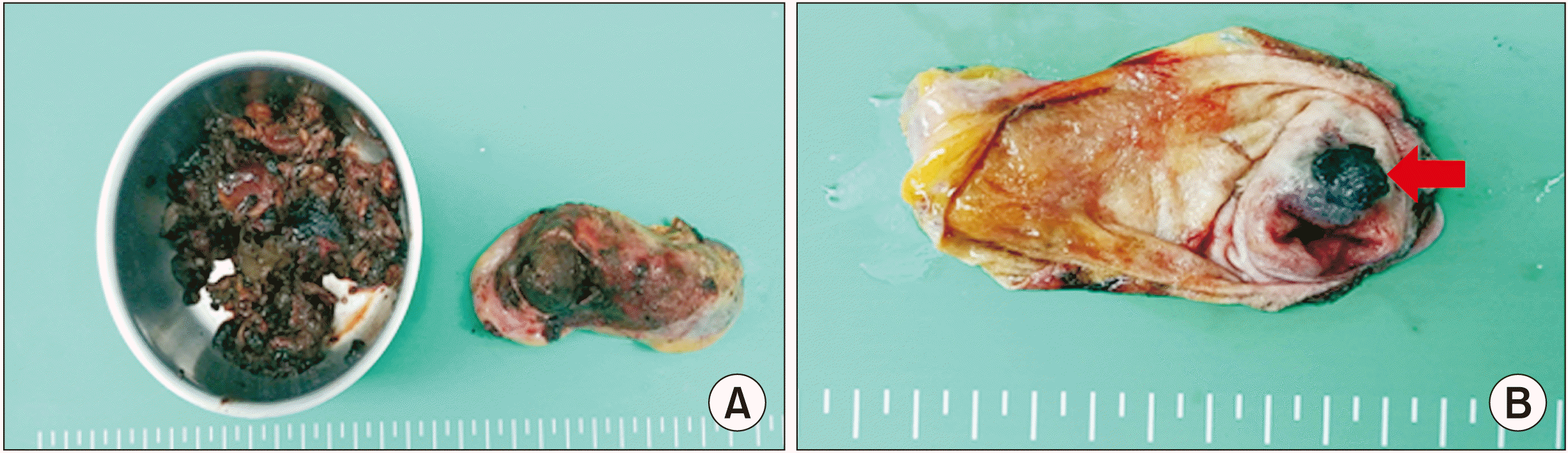
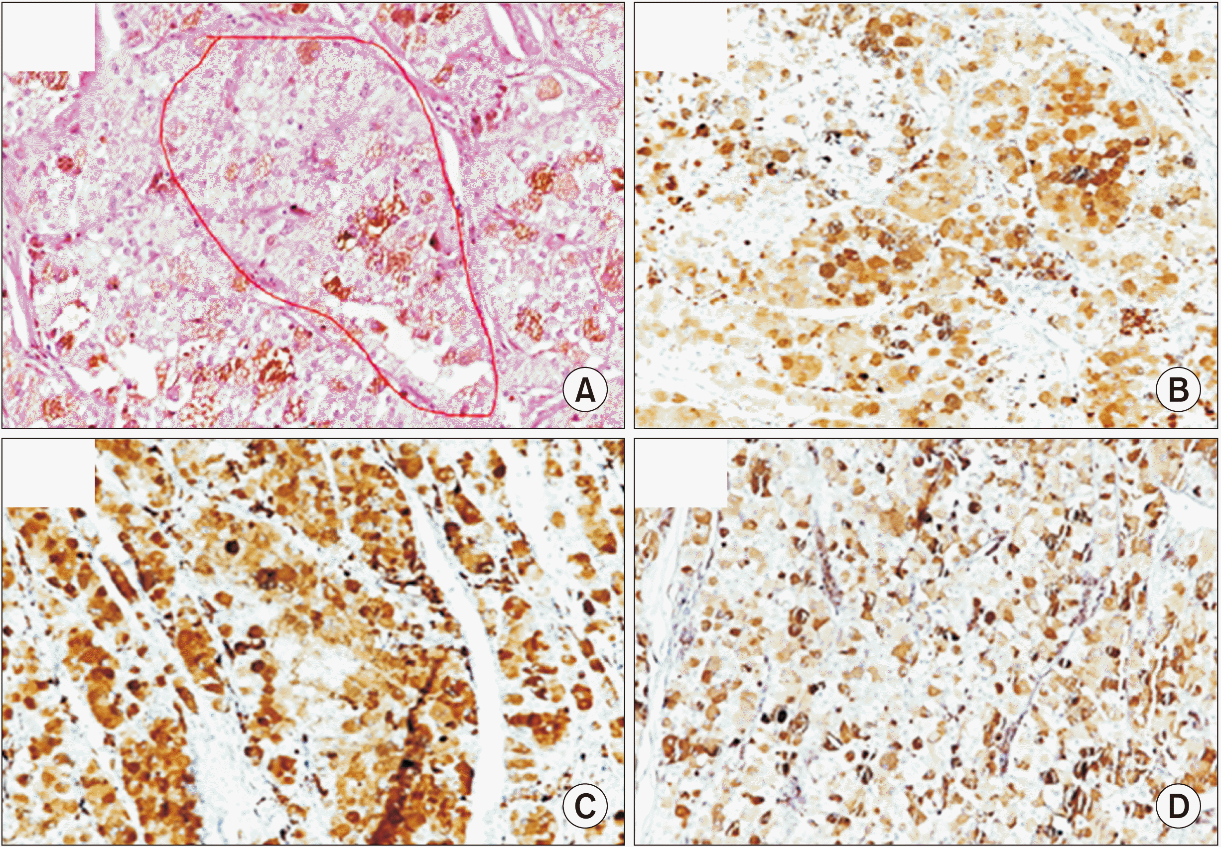
Laparoscopic cholecystectomy was tried under the impression of gallbladder stone and hematoma due to unknown cause. There were no specific findings other than gallbladder distention during the operation. After removal of the gallbladder, gross examination revealed large amounts of hematomas and multiple small stones measuring less than 1 cm in the gallbladder lumen. Gallbladder wall was thickened and a 1.6-cm-sized polypoid lesion was detected at the gallbladder fundus (Fig. 4). The polypoid lesion was well demarcated with smooth margin suggesting a benign lesion. The operation was finished with the plan to identify the nature of the polypoid lesion through a biopsy later. Microscopy of the polypoid mass showed a zellballen appearance, wherein chief cells showed copious eosinophilic granules gathered to form cell groups separated by blood vessels and fibrous tissues. Immunohistochemical analysis showed that the mass was positive for synaptophysin, CD56, and chromogranin (Fig. 5). The polypoid mass of the gallbladder was confirmed as GP based on histopathological findings. The final diagnosis of the patient was GP with multiple gallbladder stones, chronic cholecystitis, and hemorrhage.
The patient had an uneventful recovery after the operation. Abdominal CT performed at six weeks after surgery showed non-specific findings without tumor recurrence.
Go to :
DISCUSSION
Paragangliomas are subdivided into pheochromocytoma developing in the adrenal gland and extra-adrenal paraganglioma developing in the extra-adrenal paraganglion system. Paraganglioma can occur in various organs and tissues, including the carotid area, vagus nerve, mediastinum, retroperitoneum, organ of Zuckerkandl, orbit, ear, nasopharynx, duodenum, urinary bladder, prostate, and thyroid. Pheochromocytoma presents symptoms such as high blood pressure, headache, palpitation, and sweating caused by hypersecretion of sympathetic hormones. In contrast, extra-adrenal paraganglioma usually has no functional activity. It presents as an asymptomatic painless mass [12].
Paraganglioma rarely occurs in the biliary system. Only 12 cases of GP have been reported in the literature so far. In 1972, Miller et al. [1] reported the first case of GP. The patient was a 67-year-old male who was admitted to the hospital mainly for recurrent hematemesis. Surgical findings revealed neoplastic bleeding in the gallbladder and cholecystoduodenal fistula. Cholecystectomy was performed [1]. In 1973, Wolff [2] reported about a patient with cholelithiasis and chronic cholecystitis who was diagnosed with GP based on histopathology following cholecystectomy. In 2000, Hirano [3] reported about a patient with GP who presented with right upper abdominal pain and a single gallbladder mass in an imaging examination, which was confirmed as GP after cholecystectomy. In 2001, Cho et al. [4] reported about a patient with right upper abdominal pain and surgical findings of a mass combined with bleeding in the gallbladder fundus. Mehra and Chung-Park [5] and D’John and Jabbar [10] each described patients presenting with abdominal pain who were diagnosed with GP after cholecystectomy.
According to the literature, GP is more common in females than in males (9 females vs. 3 males). It usually occurs in middle-aged patients (range, 32–67 years) (Table 1). In terms of tumor size, GP is usually small, measuring 1–2 cm in diameter (range, 0.25–3 cm).
Table 1
Summary of reported cases of gallbladder paraganglioma
| Reference | Case no. | Age (yr)/sex | Clinical finding | Imaging study | Size (cm) | Treatment |
|---|---|---|---|---|---|---|
| Miller et al. (1972) [1] | 1 | 67/M | Recurrent hematemesis/tumor bleeding & cholecystoduodenal fistula | Duodenal ulcer next to the scarred duodenal bulb | 3 | Cholecystectomy |
| Wolff (1973) [2] | 2 | 32/F | Cholelithiasis/ chronic cholecystitis | NM | NM | Cholecystectomy |
| Wolff (1973) [2] | 3 | 52/F | Cholelithiasis/ chronic cholecystitis | NM | NM | Cholecystectomy |
| Wolff (1973) [2] | 4 | 59/F | Cholelithiasis/ chronic cholecystitis | NM | NM | Cholecystectomy |
| Hirano (2000) [3] | 5 | 58/F | Right hypochondrial pain | Lesion in the neck of the gallbladder | 1.3 × 0.9 | Cholecystectomy |
| Cho et al. (2001) [4] | 6 | 45/F | Right upper quadrant pain/tumor bleeding with chronic cholecystitis | Fundal mass with diffusely thickened wall | NM | Cholecystectomy |
| Mehra and Chung-Park (2005) [5] | 7 | 36/M | None/chronic cholecystitis | Normal imaging | 1.5 | Cholecystectomy |
| Rodríguez-Merchán et al. (2006) [6] | 8 | 50/F | Right upper quadrant pain/cholelithiasis | Intra- and extrahepatic biliary dilatation | 1.0 | Cholecystectomy |
| Ece et al. (2014) [7] | 9 | 57/F | Right upper quadrant pain | Hypoechoic solid mass at the neck of the gallbladder | 1.8 | Cholecystectomy |
| AlMarzooqi et al. (2018) [8] | 10 | NM | Right upper quadrant pain | Early cholecystitis | 0.25 | Cholecystectomy |
| Abdul Sater et al. (2019) [9] | 11 | 36/M | Mild hypertension, tinnitus, bilateral carotid masses | T2 hyperintense lesion with arterial enhancement | 2.2 | Cholecystectomy |
| D'John and Jabbar (2020) [10] | 12 | 63/F | Recurrent biliary colic exacerbation | Mildly dilated gallbladder | < 1.0 | Cholecystectomy |
| Present case | 13 | 48/F | Intermittent abdominal pain | Hemorrhage onthe gallbladder body and fundus | 1.6 | Cholecystectomy |
![]()
Clinical manifestations of GP are usually non-specific findings of gallbladder diseases, such as abdominal pain associated with cholelithiasis or chronic cholecystitis. In one case, there was a recurrent hematemesis with a cholecystoduodenal fistula caused by tumor bleeding [1]. Gallbladder hemorrhage was found in three cases of GP, including our case. The cause of hemorrhage was tumor bleeding in the two previous cases [1,6]. However, in our case, there was no definitive finding of bleeding from the tumor itself. Hemorrhagic cholecystitis is a rare disease, with an estimated incidence of 7% among all cases of cholecystitis [13]. The etiology of hemorrhagic cholecystitis is considered to be associated with cholecystitis, trauma, tumors, and anticoagulant use. In hemorrhagic cholecystitis caused by inflammation, transmural inflammation is considered to cause infarction and necrosis of the gallbladder mucosa, resulting in destruction of vessel walls and subsequent hemorrhage [14]. In contrast, in hemorrhagic cholecystitis caused by tumor bleeding, microscopic findings reveal vascular invasion around the tumor [4]. In our case, gallbladder hemorrhage might have been caused by cholecystitis rather than tumor bleeding.
Mehra and Chung-Park [5] have reported a case of GP with a family history of pheochromocytoma. They suggested that GP might occur as a part of multiple endocrine neoplasm syndrome and recommended careful search for possible association with multiple endocrine neoplasm syndrome. However, other cases of GP including our case were not associated with multiple endocrine neoplasm syndrome.
Laboratory examinations and imaging studies such as abdominal US, CT, and MRI are usually unhelpful because GP presents no specific findings. According to a 2006 study by Lee et al. [15] on imaging examinations of extra-adrenal paraganglioma, the tumor appears similar to hemangioma on CT owing to its hyper-vascular characteristics. On MRI, particularly on T2-weighted imaging, the high and low signal intensity regions of the tumor repeatedly appear in a “salt and pepper” pattern. Nonetheless, GP might be difficult to diagnose if a hematoma owing to intraluminal bleeding is present, as in the present case. Therefore, almost all GP cases were diagnosed incidentally based on histopathological findings after cholecystectomy.
In terms of histologic findings, paraganglioma has unique features such as the presence of well-defined nests made up of round or polygonal epithelioid cells with a zellballen pattern in the tumor [5]. In immunohistochemical staining, paraganglioma is positive for chromogranin and synaptophysin, suggesting a neuroepithelial origin of the tumor.
Cholecystectomies were performed for all reported cases of GP. No tumor recurrence or metastasis was noted after operations. Moreover, in our case, there was no tumor recurrence or abnormal findings in follow-up abdominal CT at six weeks after the operation.
In summary, GP is a rare tumor that is difficult to diagnose before surgery. It can be diagnosed based on histopathological findings after cholecystectomy. Simple cholecystectomy is suggested as an appropriate treatment for GP. However, further clinical studies with a large number of GPs are needed in the future.
Go to :
 XML Download