Abstract
Since the inception of the associating liver partition and portal vein ligation for staged hepatectomy (ALPPS) procedure, many centres have used this technique for patients who would otherwise be considered unresectable due to insufficient future liver remnant. In this report, we presented the case of a paediatric patient with recurrent hepatocellular carcinoma who underwent monosegment ALPPS (M-ALPPS) hepatectomy preserving segment 1 as the sole liver remnant using indocyanine green (ICG) as a fluorescence guide.
Associating liver partition and portal vein ligation for staged hepatectomy (ALPPS) is used for patients who otherwise would be considered unresectable due to an insufficient future liver remnant (FLR) [1]. We have previously described the case of a 3-year-old female with a PRETEXT IV Hepatoblastoma who underwent a monosegment (segment 6) ALPPS hepatectomy [2].
In this report, we present the case of a paediatric patient with recurrent hepatocellular carcinoma (HCC) who underwent a monosegment ALPPS (M-ALPPS) hepatectomy leaving segment 1 as the only liver remnant.
In September 2019, a fourteen-year-old female (height, 1.64 m; weight, 40 kg; body mass index [BMI], 14.9 kg/m2) was admitted due to weight loss and a painful liver tumour. The computed tomography (CT) scan showed a multifocal liver tumour (Li-rad V) involving segments 2, 3, 4, 5, and 8. The alpha-fetoprotein (AFP) level was 175,870 ng/mL, and no evidence of hepatitis B virus infection was demonstrated. A left trisectionectomy (segment 2, 3, 4, 5, 8 + 1) was performed because no extra-hepatic disease was found. The pathology report showed a multifocal, classic HCC (largest tumour diameter: 85 mm), with negative surgical margins. The patient had an uneventful postoperative recovery.
However, her AFP levels remained above the normal threshold (145.2 ng/mL) but no evidence of tumour recurrence was evidenced in the CT scan two months after surgery. Adjuvant treatment with doxorubicin and carboplatin was administered.
Eight months after surgery, the CT scan showed a multifocal recurrence involving segments 6 and 7 but segment 1 was negative for the disease. The patient was treated with thalidomide for seven months and AFP levels were lowered to a minimum of 27 ng/mL.
At this time the patient was asymptomatic and had an increase in weight and height (52.3 kg and 169 cm respectively, BMI 18.3 kg/m2). Since no extrahepatic disease was detected, resection of segments 6 and 7 was considered leaving only segment 1 during a multidisciplinary team meeting. A rescue orthotopic liver transplant, on the other hand, was excluded out since data in this clinical scenario was limited, and liver donors were scarce in our country.
Given that the volume of segment 1 was only 35 mL following resection of segments 6 and 7, an ALPPS to promote rapid FLR hypertrophy was recommended. The parents were informed about the consensus and the respective signed informed consent was obtained.
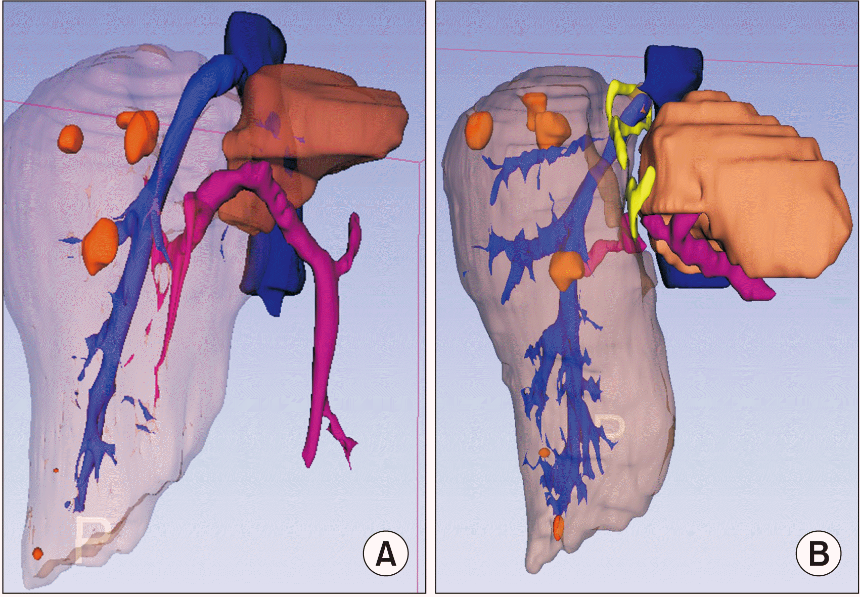
The anatomy was meticulously studied in a three-phase liver CT and the anatomical boundaries of segment 1 were delineated. Using the 3D slicer software (3D Slicer, RRID:SCR_005619) version 4.10.2 (Brigham and Women’s Hospital, Boston, MA, USA), volumetric quantification and anatomical rendering models were obtained.
Pre-operative assessment showed haemoglobin (125 g/L), platelets (200 × 109/L), prothrombin time (14.4 sec), international normalized ratio (INR) (1.1), bilirubin (7.2 µmoL/L), albumin (46.4 g/L). Regarding our institutional practice, the indocyanine green (ICG) retention test was not performed.
In December 2020, an exploration of the abdominal cavity was carried out, after confirming that there was no extrahepatic disease, an L-inverted incision was completed. A two-surgeon technique (ER and RF) was carried out. First, dissection of the porta hepatis was hampered by the firm adhesions of the greater omentum. Thus, we decided to prioritize the dissection of the right posterior portal vein which was ligated and severed. After demarcation of the ischemic area, the liver was mobilized by incising the right triangular and coronary ligaments to expose the bare area of the liver, the vena cava, right adrenal gland, and right hepatic vein. Thereafter, the liver was transected upwards from the boundary of segment 6 and the caudate process to the left border of the hepatic vein using the Cavitron Ultrasonic Aspirator (Söring GmbH, Quickborn, Germany). The right hepatic vein and right posterior glissonean pedicle were encircled with a vessel loop for the second stage. No drains were placed in the abdominal cavity. Operative time was 240 minutes, estimated blood loss (EBL) was 1,400 mL and two Pringle manoeuvres were used (total time: 30 minutes). The patient received two units of packed red blood cells (RBC) and two units of fresh frozen plasma (FFP). During the postoperative phase, the patient had tachycardia and no other abnormal signs were evidenced.
On postoperative day (POD) 14, the volume of segment 1 was 288 mL (Fig. 1B).
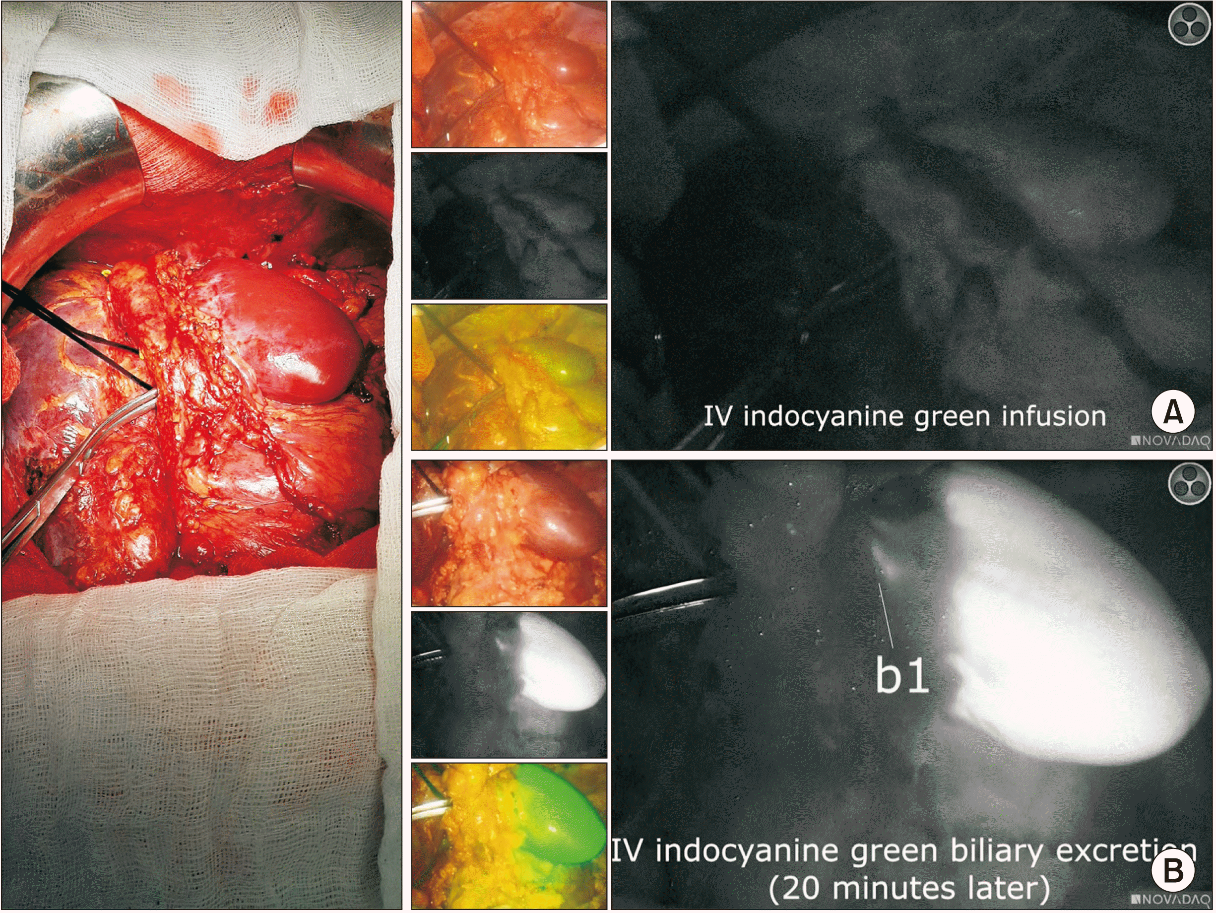
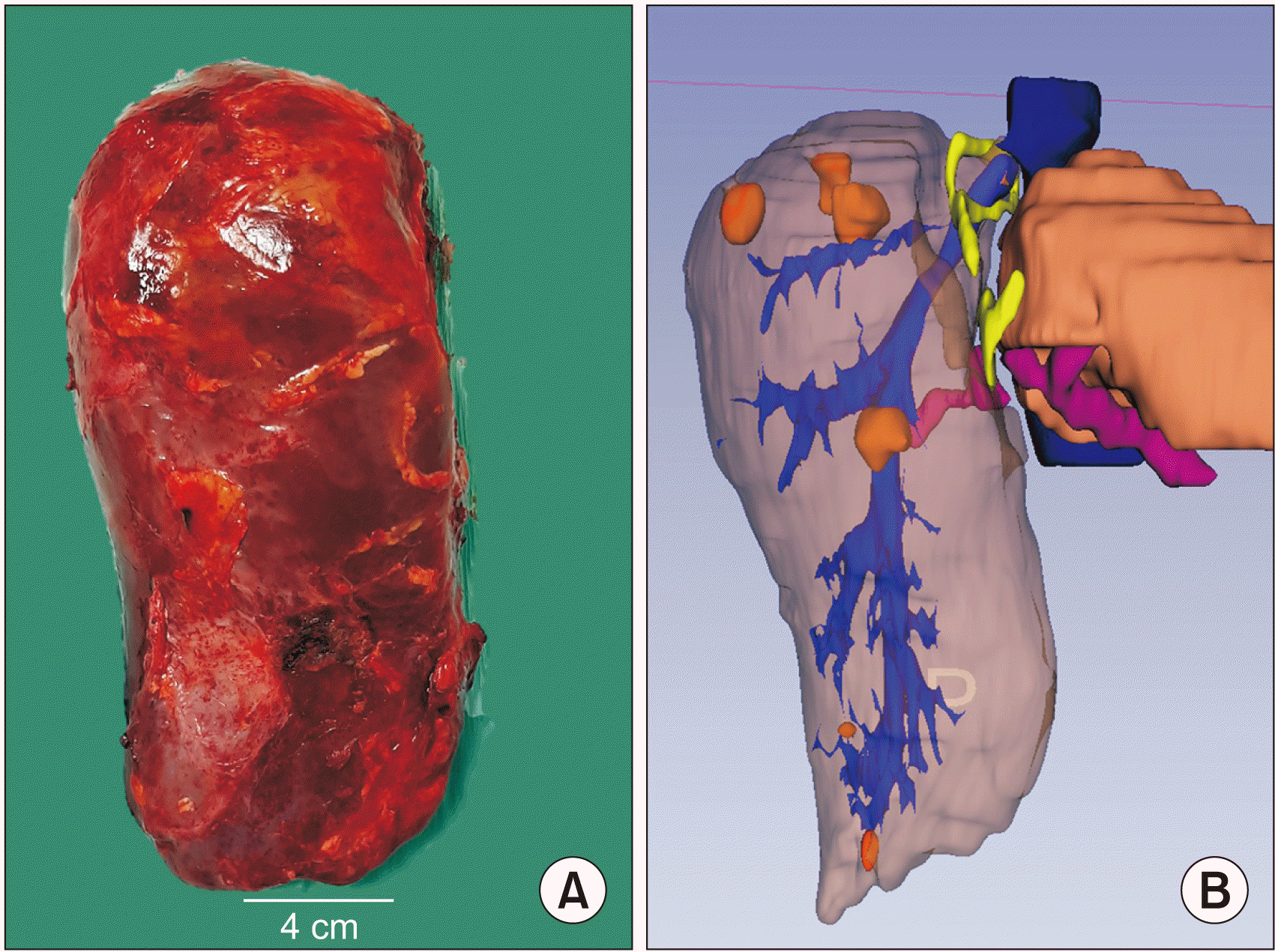
On POD 16, the previous incision was re-opened, firm adhesions were found between the transverse colon and the abdominal wall. Since the biliary tract could be completely identified neither in the magnetic resonance cholangiography nor during the first ALPPS stage (as a result of the firm omental adhesion to the porta hepatis), we used intraoperative fluorescence guidance to: a) identify the main bile duct and avoid any injury and b) ensure adequate perfusion to segment 1. Hence, a Lees clamp was applied at the level of the right posterior glissonean pedicle and ICG (0.05 mg/kg) was administered intravenously. Using the PINPOINT system (Novadaq, Mississauga, ON, Canada) adequate perfusion of segment 1 was confirmed. Twenty minutes later, biliary excretion along with identification of the main bile duct was confirmed (Fig. 2). Thereafter, the right posterior glissonean pedicle along with the right hepatic vein was severed, and the specimen was removed (Fig. 3). No drains were placed, operative time and EBL were 250 minutes and 700 mL, respectively. The patient received one unit of RBC and two of FFP.
Five days after ALPPS stage 2, a post-hepatectomy liver failure (PHLF) [3] grade A along with mild ascites, was diagnosed (both classified as Dindo-Clavien II). The patient was discharged on POD nine. On POD 19, the patient was readmitted with a symptomatic abdominal fluid collection (370 mL) and no signs of sepsis or PHLF was reported, percutaneous drainage was placed, and the effluent was caused by a bile leak. The pathology report demonstrated multifocal classic HCC; surgical margins were negative.
One month later, AFP level was 5.7 ng/mL. Six months later, the AFP level was 1.78 ng/mL and no radiological signs of recurrence were showed in CT scans. The liver function was normal, prothrombin time: 16.1 sec and the INR 1.24.
Recently, thrombocytopenia (74 × 109/L), epistaxis, and hypermenorrhoea were detected, the CT scan showed a slight increase in the spleen maximum diameter (preoperative: 8 cm and postoperative control: 10 cm). Thus far, no evidence of portal hypertension was observed neither in the CT scan nor as clinical stigmata.
In our paper, we showed that segment 1 can be used as the only liver remnant after an ALPPS procedure. To the best of our knowledge, this is the first patient-reported case in the literature.
Caudate lobe-sparing subtotal hepatectomies have been previously described as feasible treatments in hepatolithiasis [4,5], hamartoma [6], and HCC [7]. However, in these cases, hypertrophy of segment 1 was achieved by the natural history of the disease with no previous interventions performed to remodel the FLR.
As previously stated, the use of the ALPPS technique to obtain an adequate monosegment 1 liver remnant was feasible, provided a very stringent selection of the patient. We agree with Lai et al. [8] in that the paediatric population has a better regenerative capacity. Nevertheless, this procedure was not exempted from complications (biliary fistula). The aetiology of the thrombocytopenia must be elucidated.
In conclusions, monosegment 1 ALPPS hepatectomy can be considered as an acceptable option to achieve an R0 resection for recurrent HCC. The use of ICG fluorescence is mandatory to confirm adequate inflow and biliary excretion. ALPPS is not exempted from short and long-term complications and should be considered in tertiary centres familiar with this procedure.
REFERENCES
1. Schadde E, Malagó M, Hernandez-Alejandro R, Li J, Abdalla E, Ardiles V, et al. 2015; Monosegment ALPPS hepatectomy: extending resectability by rapid hypertrophy. Surgery. 157:676–689. DOI: 10.1016/j.surg.2014.11.015. PMID: 25712199.

2. Ruiz Figueroa EF, Fernández-Placencia RM, Berrospi Espinoza FE, Gomez HF, Chávez Passiuri IK. 2019; Monosegmental ALPPS: a long-term survival alternative to liver transplant in PRETEXT IV hepatoblastoma. J Surg Case Rep. 2019:rjz144. DOI: 10.1093/jscr/rjz144. PMID: 31086654. PMCID: PMC6507791.

3. Rahbari NN, Garden OJ, Padbury R, Brooke-Smith M, Crawford M, Adam R, et al. 2011; Posthepatectomy liver failure: a definition and grading by the International Study Group of Liver Surgery (ISGLS). Surgery. 149:713–724. DOI: 10.1016/j.surg.2010.10.001. PMID: 21236455.

4. Wang W, Zhang Z, Wang J. 2020; Subtotal (segment II-VIII) hepatectomy for bilateral diffuse hepatolithiasis with compensatory caudate lobe hypertrophy: a report of two cases. BMC Gastroenterol. 20:350. DOI: 10.1186/s12876-020-01503-9. PMID: 33081716. PMCID: PMC7576830.

5. Dong J, Lau WY, Lu W, Zhang W, Wang J, Ji W. 2012; Caudate lobe-sparing subtotal hepatectomy for primary hepatolithiasis. Br J Surg. 99:1423–1428. DOI: 10.1002/bjs.8888. PMID: 22961524.

6. Shuto T, Kinoshita H, Yamada C, Hirohashi K, Shiokawa C, Kubo S, et al. 1993; Bilateral lobectomy excluding the caudate lobe for giant mesenchymal hamartoma of the liver. Surgery. 113:215–222. PMID: 8430370.
7. Aijun L, Jiamei Y, Qinhe T, Mengchao W. 2014; Caudate lobe as the sole remnant liver following extended liver resection for hepatocellular carcinoma. Int J Surg Case Rep. 5:462–464. DOI: 10.1016/j.ijscr.2014.05.003. PMID: 24973528. PMCID: PMC4147654.

8. Lai Q, Mennini G, Larghi Laureiro Z, Rossi M. 2021; Uncommon indications for associating liver partition and portal vein ligation for staged hepatectomy: a systematic review. Hepatobiliary Surg Nutr. 10:210–225. DOI: 10.21037/hbsn-20-355. PMID: 33898561. PMCID: PMC8050569.

Fig. 1
(A) Preoperative 3-dimensional volume rendering. The tumours are coloured in orange. Segment 1 is demonstrated in darker brown. (B) After 1st stage, segment 1 shows an important increase in volume. Yellow vessel loops are shown to further guide the right portal pedicle and hepatic vein during the 2nd stage of associating liver partition and portal vein ligation for staged hepatectomy (ALPPS).


 PDF
PDF Citation
 Print
Print


 XML Download