

 PDF
PDF Citation
 Print
Print


INTRODUCTION
Intraductal tubulopapillary neoplasms (ITPNs) of the pancreas and bile duct represent rare premalignant neoplasms that were first recognized by the World Health Organization (WHO) in 2010 [1]. The first case of ITPN in the pancreas (p-ITPN) was described in 1992 by Shahinian et al. [2], while the first biliary ITPN (b-ITPN) case was reported in 2010 by Park et al. [3]. Typically, ITPN presents with jaundice, abdominal pain, and weight loss, resembling pancreatic adenocarcinoma or cholangiocarcinoma, and is most commonly found in patients over 50 years old, with a female overrepresentation. ITPN represents less than 1% of all pancreatic exocrine neoplasms, 3% of pancreatic intraductal neoplasms [4], and 15% of intraductal neoplasms of the bile duct [5]. What distinguishes ITPN from other intraductal neoplasms, such as intraductal papillary mucinous neoplasms (IPMN), is their uniquely favorable prognosis.
Given the obscurity of these lesions and the heterogeneity of presentation, we report two cases of ITPN from our center with unusual clinical findings and review the literature on this rare entity.
Go to : 
CASES
Pancreatic intraductal tubulopapillary neoplasm
A 76-year-old male with a previous history of coarctation repair as a child, followed by triple bypass with aortic valve replacement, and eventually a heart transplant, presented to the emergency room with a complaint of severe diffuse abdominal pain located in the upper quadrants. The pain had been present for about a week at the time of his presentation. He reported associated nausea without vomiting. He did not have any jaundice or change in the color of his urine or stools. He did not report any weight loss or recent change in appetite. In the emergency room, a computed tomography (CT) scan was performed, which demonstrated a dilated pancreatic duct measuring 9 mm, a common bile duct measuring 1.3 cm, and a 1.3 cm × 1.3 cm hypodense area in the pancreatic head described as a cystic neoplasm (Fig. 1A). He was then transferred to our institution for further care and evaluation of this newly found mass. Magnetic resonance imaging (MRI) revealed a pancreatic duct diffusely dilated to 1.3 cm, and a 3-cm mass in the head of the pancreas without vessel involvement (Fig. 1B, 1C). Subsequently, an endoscopic retrograde cholangiopancreatography (ERCP) with endoscopic ultrasound (EUS) was performed and the fine needle aspirate results were consistent with IPMN. He had a pancreaticoduodenectomy for a presumed diagnosis of main duct IPMN. An intraoperative frozen section of the pancreatic neck was consistent with IPMN and mild atypia.
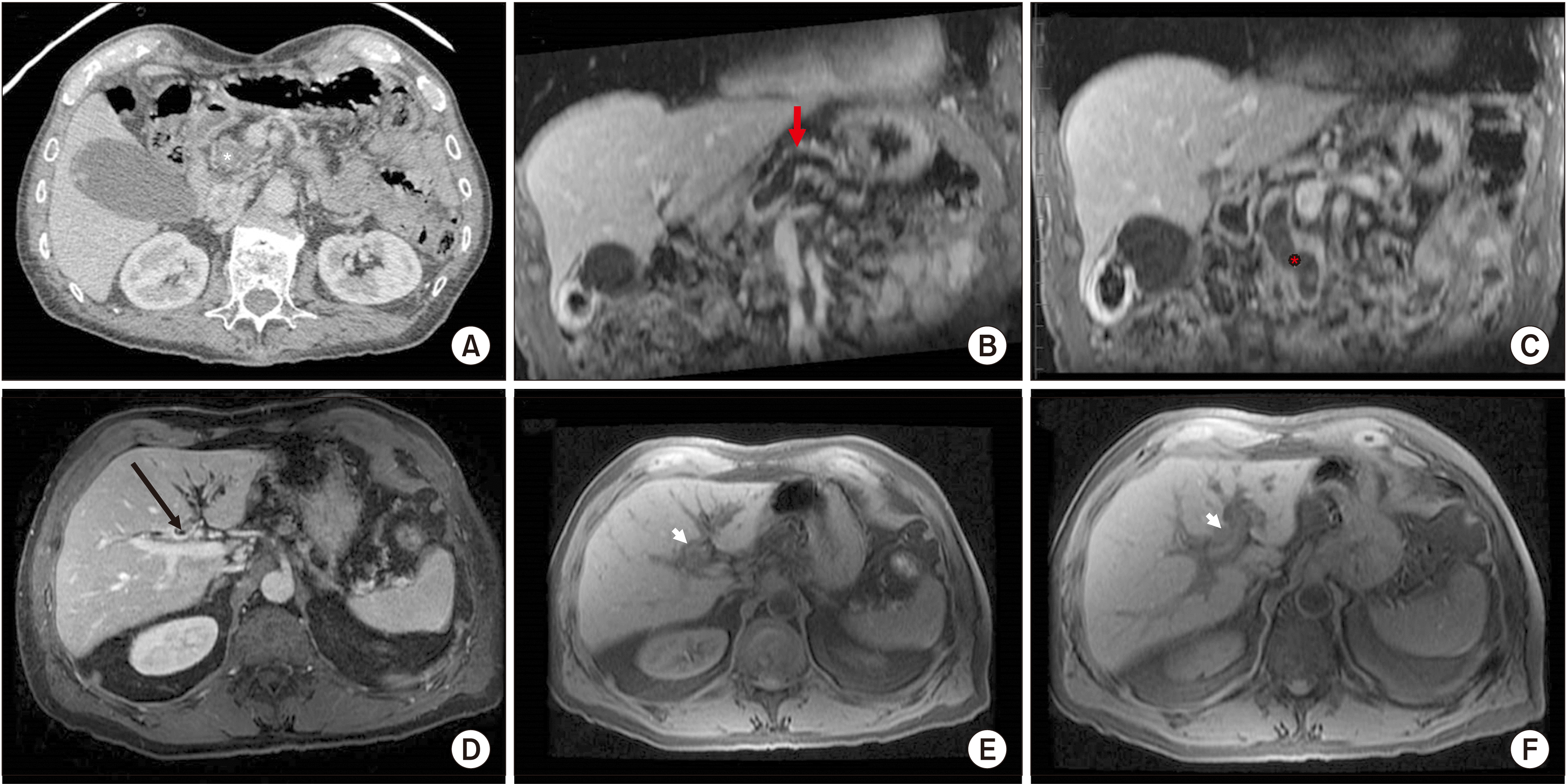 | Fig. 1(A) Cross-sectional computed tomography (CT) imaging demonstrating an atrophic pancreas with a cystic neoplasm in the pancreatic head, measuring 1.3 cm × 1.3 cm (white asterisk). (B) Coronal magnetic resonance imaging (MRI) imaging demonstrated a prominent dilated pancreatic duct, measuring 9 mm, which tapered in caliber at the pancreatic head (arrow). (C) Additionally, the proximal dilated intrapancreatic duct demonstrated increased T1 and decreased T2 signals within the duct in the region of the dilated pancreatic head (red asterisk), suggesting debris or mucin, with more simple-appearing T2 bright fluid in the more proximal duct. (D) Cross-sectional CT imaging demonstrating significant biliary dilatation, particularly of the left lobe intrahepatic biliary radicles. At the level of the common hepatic duct upstream from the cystic duct insertion is a circumferential mass (black arrow). (E) Cross-sectional MRI imagining confirmed a circumferential mass, which was hyper-enhanced (white arrowhead) and retained contrast on delayed imaging, consistent with cholangiocarcinoma. (F) The mass (white arrowhead) measured up to 11 mm in diameter and extended to the hepatic hilum where it extended into the left hepatic duct just beyond the secondary to a biliary radical duct.
|
The pancreatoduodenectomy specimen was bivalved to reveal a tan solid mass within the pancreatic duct (Fig. 2A), with no cystic structure or associated mucin identified. The lesion completely occluded the lumen and measured 3.5 cm longitudinally within the pancreatic duct. Histological low-power microscopic examination showed an intraductal lesion with tubulopapillary architecture and conspicuously, no mucin production (Fig. 2B, 3C). The high-power evaluation showed high-grade nuclei atypia and immunohistochemical findings typical of ITPN of the pancreas. The lesion was found to be positive for cytokeratins 7 (Fig. 3B) and 19, and CA19-9. Neuroendocrine markers synaptophysin, chromogranin, beta-catenin, carcinoembryonic antigen (CEA), MUC5AC, and p53 were all negative. MUC1 showed luminal expression (Fig. 3C). The typical immunohistochemical findings for ITPN of the pancreas include positive MUC1, cytokeratin 7, and/or cytokeratin 19, in addition to negative trypsin, MUC2, MUC5AC, and fascin [4].
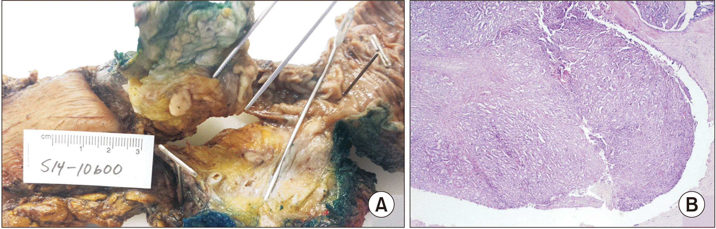 | Fig. 2(A) Whipple specimen showing intraductal tubulopapillary neoplasm (ITPN) of the pancreas within the common pancreatic duct. (B) Low-power microscopic view of H&E-stained slide at 20× magnification showing ITPN filling the pancreatic duct (duct lining).
|
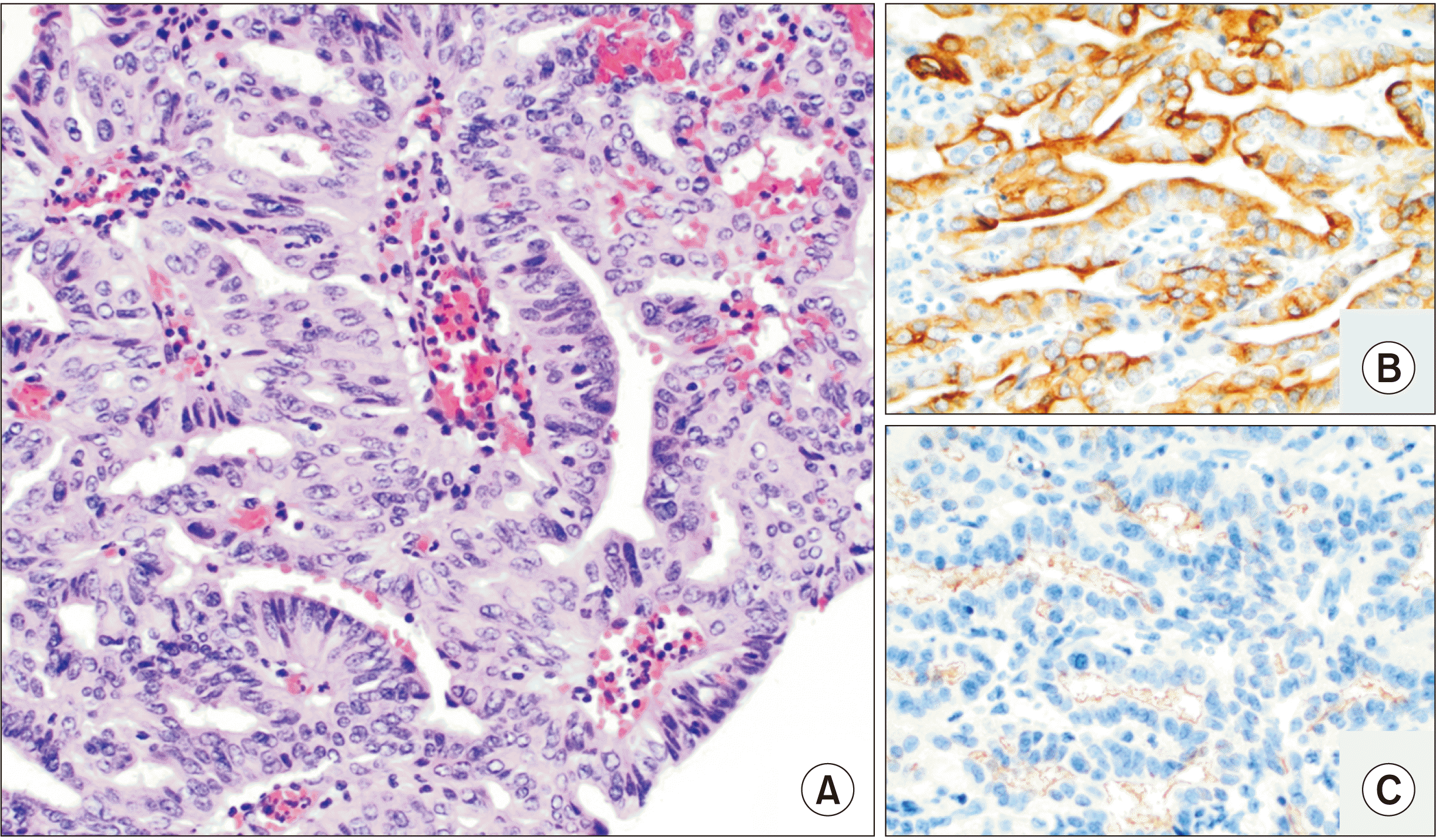 | Fig. 3(A) High-power microscopic image of H&E-stained slide (400× magnification) showing tubular architecture and high-grade nuclear features with an absence of mucin production. (B) Immunohistochemical staining for CK7 (200× magnification) showing positive membranous and cytoplasmic expression. (C) Immunohistochemical stain for MUC1 (200× magnification) showing membranous luminal expression.
|
His postoperative course was complicated by the development of an ileus, which eventually resolved, and he was discharged to home without further complications. Since his surgery, he has been followed with yearly MRIs and has remained disease-free. It is important to note that the literature does not favor MRI over CT for surveillance imaging of the pancreas. In fact, several studies in the literature suggest the non-inferiority of CT over MRI [6]. However, our center prefers pancreatic imaging in the remnant pancreas with MRI since some studies suggest a higher sensitivity and reader confidence in MRI for the detection of individual morphologic features such as duct communication compared to CT [7,8].
Biliary intraductal tubulopapillary neoplasm
A 68-year-old male with a history of hypertension was in his usual state of health when he noticed his urine had turned orange, his stool clay-colored, and he was experiencing intermittent epigastric pain. He also had a recent unintentional 7 kg (15 1b) weight loss in the prior six weeks. He was a former smoker, having quit 10 years ago. He had no significant alcohol or illicit substance use history. He had a paternal family history of alcoholism and unspecified cancer in his brother. He was first seen by his primary care physician, at which time, blood work showed elevated liver function test values. A CT scan of the abdomen showed dilation of the intrahepatic ducts and rounded, elongated density in the common hepatic duct measuring 13 mm in diameter (Fig. 1D). There was no downstream ductal dilation. He was referred to a gastroenterologist, who performed an ERCP, which showed malignant-appearing stenoses in the common bile duct. A sphincterotomy was performed and a stent was placed in the left hepatic duct. Intraductal brushings showed occasional groups of bile duct epithelium with prominent nucleoli and nuclear crowding and overlap consistent with adenocarcinoma, presumed to be cholangiocarcinoma.
Further preoperative imaging included magnetic resonance cholangiopancreatography (MRCP), which showed a Bismuth IIIB lesion. Tumor markers CEA and CA19-9 were normal. He was taken to the operating room and had a left hepatectomy with bile duct resection and Roux-en-Y hepaticojejunostomy. His postoperative course was uneventful, and he left the hospital shortly after regaining bowel function on postoperative day six.
His final pathology demonstrated a b-ITPN with high-grade dysplasia measuring 3.5 cm within the lumen of the left hepatic duct. All margins were negative. The remaining liver parenchyma showed cholestasis indicative of mechanical obstruction. The hepatectomy specimen revealed a tan, solid, and irregular polypoid lesion within a bifurcation of the hepatic duct, which measured 3.5 cm (Fig. 4A). The lesion was entirely contained within the duct with a focal area of attachment identified and no associated mucin. Low-power histologic examination revealed tubulopapillary architecture and the absence of mucin production (Fig. 4B, 4C). High-power microscopic examination showed high-grade nuclear atypia without an invasive component or necrosis and the immunohistochemical findings were consistent with b-ITPN (Fig. 5A, 5B). The lesion demonstrated strong positivity for cytokeratin 7, luminal expression for MUC1 (Fig. 5C, 5D), and patchy positivity for IMP3 and an albumin probe in in situ hybridization. Focal and weak positivity was observed for MUC5AC. The expression of MUC6, MUC2, HepPar-1, CK20, and CDX2 in the lesion was negative. The immunohistochemical expression in b-ITPN has been observed to have characteristics similar to those of p-ITPN, and include the positive expression of MUC1 and MUC6, and the absence of MUC2 and MUC5AC. b-ITPN has also been reported to have positive expressions of CK19 and CA19-9, while synaptophysin, chromogranin, and CA-125 were negative in all cases [4]. In this case, a next-generation sequencing (NGS) solid tumor combined variant plex and RNA fusion panel was performed, and no DNA variants or RNA fusions were detected.
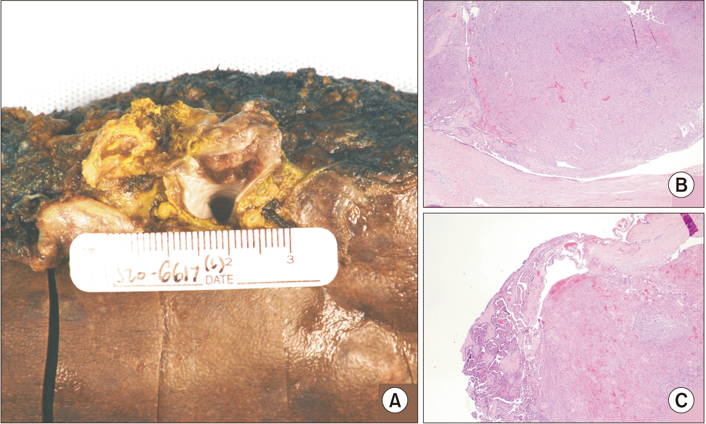 | Fig. 4(A) Resected partial hepatectomy specimen showing intraductal tubulopapillary neoplasm (ITPN) in the left hepatic duct. (B, C) Low-power microscopic examination of H&E-stained slide at 20× magnification showing ITPN filling the left hepatic duct (duct lining), with solid and papillary architecture, and a focally attached stalk.
|
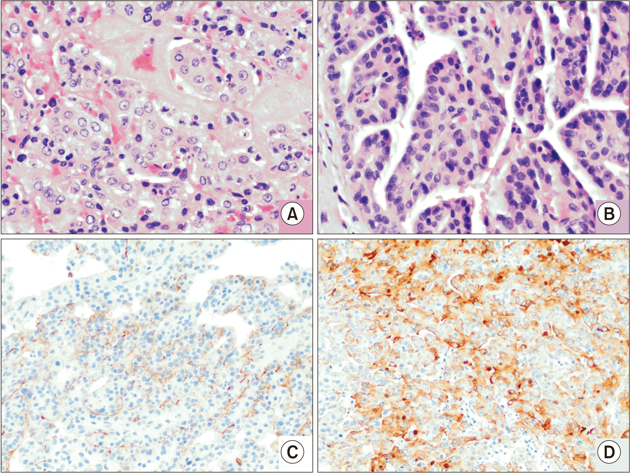 | Fig. 5(A) High-power microscopic image of H&E-stained slide at 400× magnification, which shows tubular architecture and high-grade nuclear features, and papillary architecture with fibrovascular cores, high nuclear grade, and an absence of mucin production. (B, C) Immunohistochemical stain for MUC1 (B: 400× magnification, C: 200× magnification) showing membranous luminal expression. (D) Immunohistochemical stain for CK7 (200× magnification) showing positive membranous and cytoplasmic expression.
|
At a multidisciplinary tumor board, curative surgery was decided and the patient did not require any further treatment. He will be followed every three months for two years to evaluate any potential recurrence.
Go to :
DISCUSSION
ITPN is a rare premalignant entity that is often not diagnosed until after surgical resection. As presented herein, preoperative radiologic, endoscopic, and biopsy studies have proven unreliable for a definitive diagnosis. Standardized guidelines are difficult to establish as the incidence of ITPN is too low for proper evidence-based evaluation.
When ITPN is associated with an invasive component, it is difficult to differentiate it from adenocarcinoma. As of 2018, six cases of the local recurrence of p-ITPN were reported [9]. There is a significant difference in pathophysiology and prognosis, which makes differentiation at an earlier stage even more imperative. Pancreatic-ITPN is defined as an intraductal, grossly visible, tubule-forming epithelial neoplasm, usually with a minor papillary component, demonstrating high-grade dysplasia and ductal differentiation without mucin production [10]. Grossly, these lesions appear solid, occupying a dilated duct and without associated mucin [1]. On imaging, p-ITPN typically resembles IPMN or pancreatic adenocarcinoma with low attenuation and a dilated pancreatic duct or double duct sign. An intraductal lesion with the so-called cork in bottle sign may sometimes be detected as well.
The tumor sites for b-ITPN are intrahepatic (70%), perihilar (20%), and extrahepatic (10%), and appear similar to cystic lesions or cholangiocarcinoma on imaging [5]. In the largest analysis to date of 20 patients with b-ITPN, invasive carcinoma was identified in 80% of the cases. Despite this, the survival rate was 90% for b-ITPN versus 54% for cholangiocarcinoma [5]. Similarly, an analysis of 33 patients with p-ITPN showed a 5-year survival rate of 71% for patients without an invasive component; however, 2 of the 33 patients had an invasive cancer [11]. The pathologic diagnostic criteria for b-ITPN include growth within the intra- or extrahepatic bile duct, preinvasive histology, exophytic growth, and predominantly non-mucinous tubular units with minimal to no papillary growth [5]. In one review, the 5-year survival rate was 80.7%, compared to 37% in patients with early-stage adenocarcinoma after resection [12]. Therefore, it is important to consider this when counseling patients before surgery on an ill-defined pancreaticobiliary lesion.
Thus, how can the diagnostic accuracy of preoperative studies be improved? Perhaps a more constructive approach would be to evaluate the radiologic differences between ITPN and IPMN since there is such radiologic overlap between these two entities. Pancreatic ITPN presents a low-density lesion on pancreas-dedicated CT with a dilated pancreatic duct and potentially a “cork in bottle” sign, indicating the presence of a lesion inside the duct. MRCP is the most effective imaging modality to demonstrate this finding. One case series of p-ITPN suggested that EUS was more accurate than cross-sectional imaging [13]. Two out of the seven patients in this series had intraductal tumors identified on EUS that were not seen on CT or MRI. Despite these identifiers, atypical imaging findings such as diffuse ductal dilation can mimic chronic autoimmune pancreatitis or ductal adenocarcinoma, which may be due to long-standing disease and/or the presence of pancreatic duct stones.
Biliary-ITPN shows ductal dilation and visible intraductal soft tissue with peribiliary liver parenchyma enhancement. In b-ITPN, there is less often a dilated duct downstream given its lack of mucin production, which differentiates it from IPMN, wherein the upstream and downstream ducts are dilated, as was illustrated in our patient [14]. In a comparison of biliary IPMN and ITPN, the radiologic characteristics of ITPN were an intraductal soft tissue proportion of greater than 60%, peribiliary liver parenchyma enhancement, and mild dilation of the upstream biliary system with no downstream dilation. Typically, there is less liver capsule retraction or parenchymal atrophy compared to IPMN, although in the analysis in this study these differences were not statistically significant [14]. In addition, the EUS appearance may not be characteristic and the cytologic features of the lesion without gross pathology and histologic examination may be difficult and lead to misdiagnosis.
Furthermore, as illustrated in our b-ITPN case, the diagnostic accuracy of preoperative fine-needle aspirate biopsies could be improved. In ITPN, the histologic evaluation reveals a tubulopapillary growth pattern, high-grade nuclear atypia, and the absence of mucin production [15]. Focal areas of necrosis are frequently identified, and the lesion is often attached to the duct lining by a fibrous stalk. Immunohistochemically, these neoplasms are characterized by the expression of MUC1 and MUC6 versus MUC2 and MUC5AC, which are more often found in mucinous neoplasms, consistent with our findings. While ITPNs of the pancreas and bile ducts represent premalignant lesions, they do not share molecular features with either pancreatobiliary adenocarcinomas or other premalignant lesions, such as IPMN. The most commonly reported mutations of ITPN are PIK3CA, CDKN2A/p16, and TP53, while other commonly reported mutations identified in IPMN and pancreatobiliary adenocarcinomas (KRAS, GNAS, RNF43, BRAF, EGFR, HER2, and beta-catenin) are not typically identified [1]. In this setting, the cytogenetic analysis of fine-needle aspirates may be of value in ascertaining a diagnosis preoperatively.
Finally, it is important to point out that hepatic resection for hilar cholangiocarcinoma should be limited as much as possible to achieve a curative resection. It has been suggested to perform limited hepatic resection according to the individual tumor extent of each patient [16]. Because S1 resection is known to be a prerequisite for the curative resection of hilar cholangiocarcinoma [17-19], resection of the entire S1 can be the most limited parenchymal-preserving hepatectomy in resection for hilar cholangiocarcinoma.
ITPN of the pancreas and biliary tree are rarely described entities with an elusive diagnostic workup. Resection remains the standard of care, with survival rates that are far superior to other potential hepatobiliary diagnoses. Herein, we discussed two illustrative cases and radiologic identifiers of these pre-malignant tumors to improve future preoperative diagnostic accuracy, which can help better prepare surgeons and patients for the eventual diagnosis.
Go to :
 XML Download