

 PDF
PDF Citation
 Print
Print


INTRODUCTION
Large hepatic tumors can invade the retrohepatic inferior vena cava (IVC) directly. Thus, resecting the involved IVC wall is necessary to achieve complete tumor resection. Partial excision of the IVC wall requires patch venoplasty to repair the wall defect unless the defect is small enough to enable primary repair. The IVC is a large-caliber vessel with low pressure, low velocity, but high flow. Thus, any imprudent handling can result in massive bleeding and air embolism, which can lead to a catastrophic event.
The development of living donor liver transplantation (LDLT) in Korea has contributed to the reciprocal advancement in aggressive hepatobiliary surgeries, especially regarding vascular reconstruction of the IVC and portal vein [1]. Clamping of the IVC such as total hepatic vascular exclusion (THVE) is not frequently performed during hepatobiliary surgery [2-7]. However, it is an essential procedure for liver transplantation. With high-volume experience on LDLT, we herein present detailed surgical procedures of IVC resection and patch venoplasty in two patients who underwent aggressive surgery for hepatic tumors.
Go to : 
CASES
Case no. 1
The patient was a 55-year-old male with advanced intrahepatic cholangiocarcinoma. A 10-cm-sized infiltrative tumor was located in the right lobe of the liver. It invaded the right portal vein, the duodenum, the right adrenal gland, and the retrohepatic IVC (Fig. 1). Considering his relatively young age of 55 years, we thought that macroscopic curative resection combined with adjuvant chemotherapy would provide a survival benefit. Thus, we decided to perform aggressive margin-free resection.
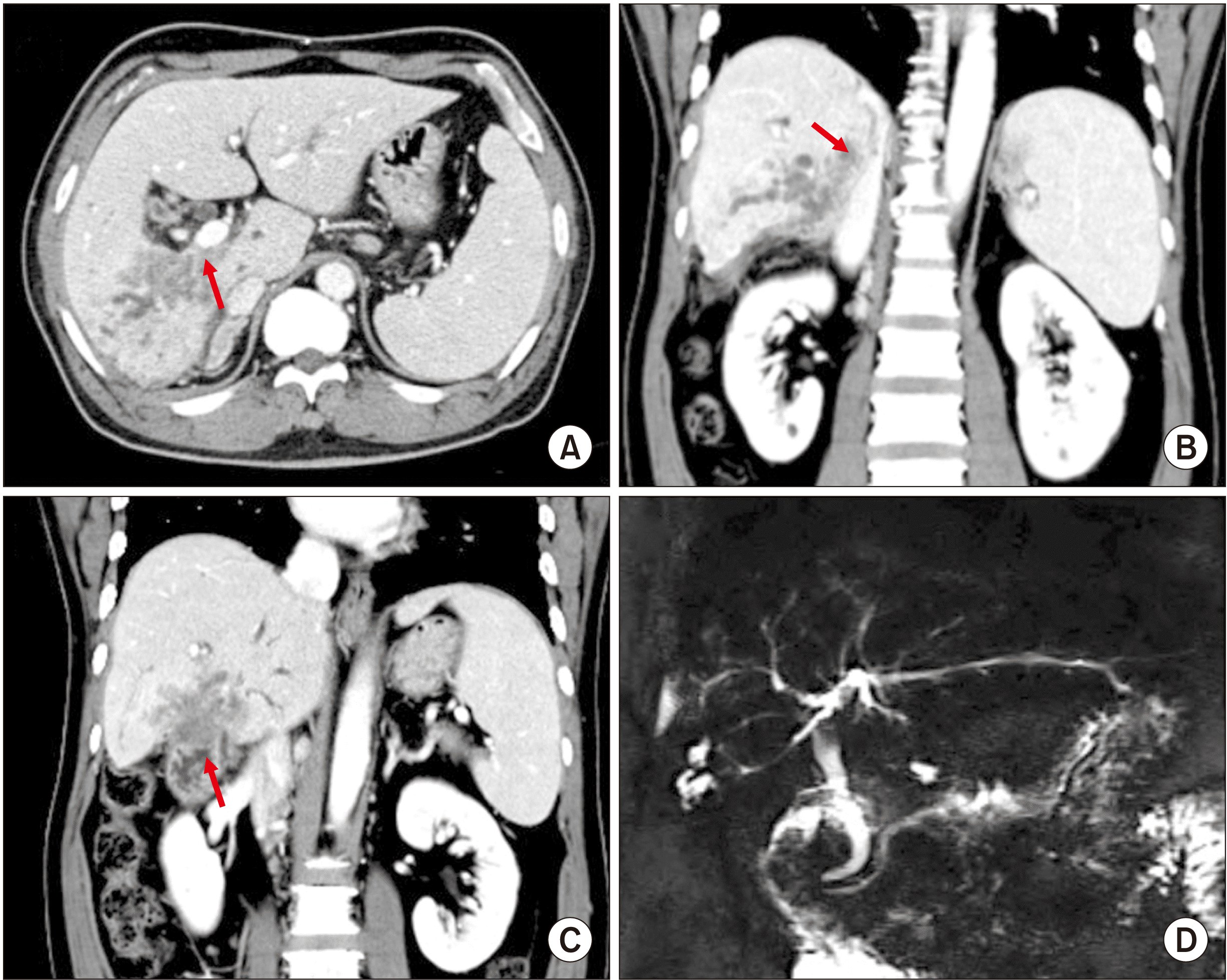 | Fig. 1Preoperative radiologic findings of Case no. 1. (A–C) Computed tomography scan shows a 10-cm-sized infiltrative tumor located at the right liver and invasion of the right portal vein (arrow), the duodenum (arrow), the right adrenal gland, and the retrohepatic inferior vena cava (arrow). (D) Magnetic resonance cholangiopancreatography shows invasion to the right hepatic duct.
|
During the operation, neither peritoneal seeding nor distant metastasis was found. The primary tumor locally invaded the first portion of the duodenum. Thus, wedge resection of the duodenum and primary repair were performed to mobilize the tumor. The right liver was partially mobilized with en bloc resection of the right adrenal gland as there was direct tumor invasion to the retrohepatic IVC. The extrahepatic bile duct was resected. Frozen-section biopsy confirmed that the left hepatic duct margin was tumor-negative. Since the right portal vein was invaded and occluded by the tumor, the right hepatic artery was isolated and clamped to determine the discoloration border at the liver surface. Hepatic parenchymal transection was initiated to perform extended right hepatectomy and caudate lobectomy. After completion of hemihepatic transection, the tumor-involved portion of the main portal vein close to the right portal vein was segmentally resected and reconstructed primarily in an end-to-end fashion. The caudate lobe was completely dissected through a right-sided approach. After the right hepatic vein was transected, it was found that the right side of the liver with caudate lobe was still attached to the IVC through tumor invasion. The retrohepatic IVC was totally clamped at two levels, one just caudal to the insertion site of the middle-left hepatic vein trunk and another at the level of the renal vein insertion using a modified THVE technique. Blood inflow and outflow at the remnant left liver were maintained during this IVC clamping. The invaded IVC wall was excised elliptically to make the resection margin tumor-negative. The IVC wall defect was repaired by applying an expanded polytetrafluoroethylene (ePTFE) patch, which was 50% larger than the IVC wall defect (Fig. 2).
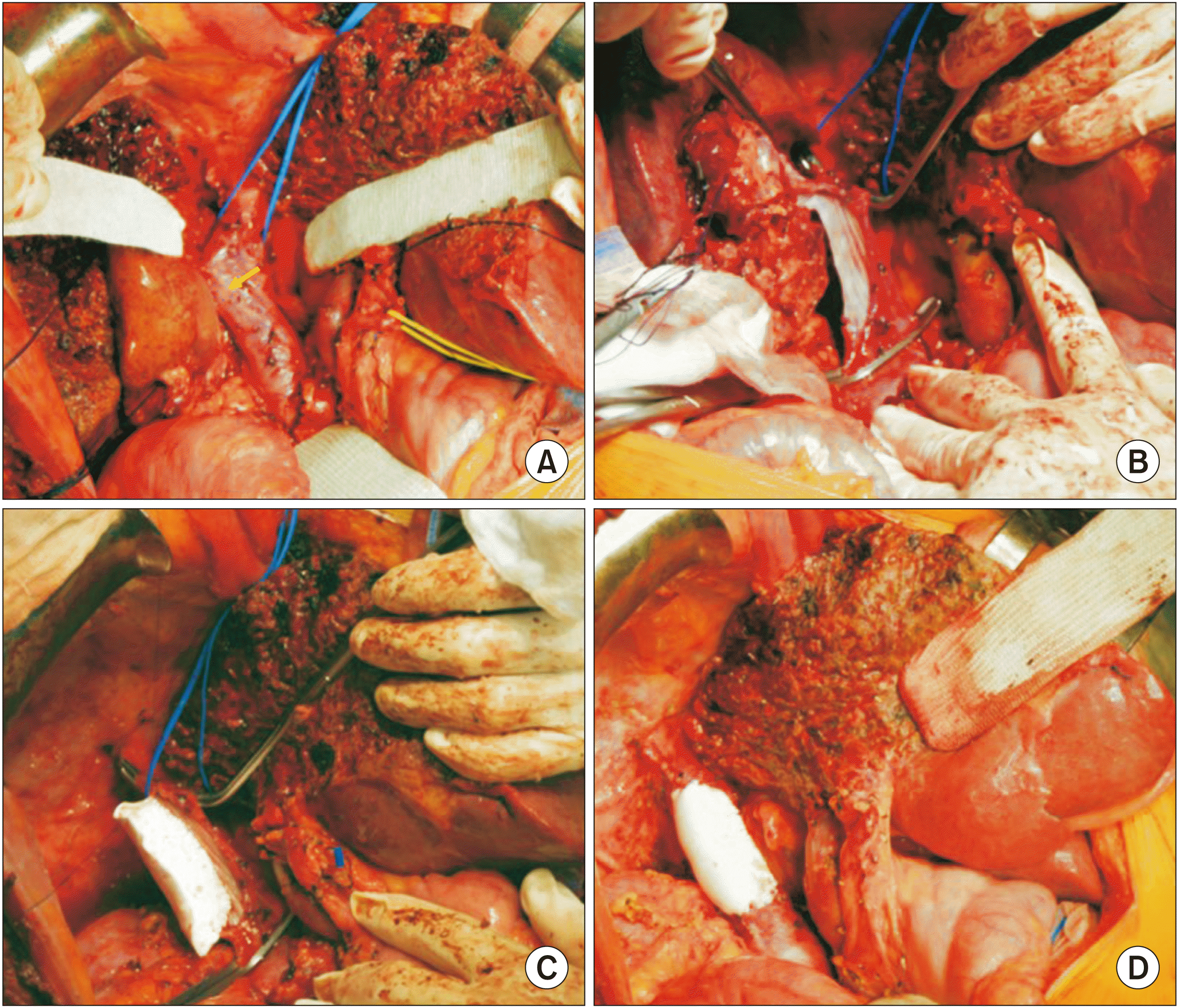 | Fig. 2Intraoperative photographs of the wedge resection of the retrohepatic inferior vena cava (IVC) in Case no. 1. (A) The tumor-invaded IVC is isolated (arrow). (B) The invaded IVC wall was excised. (C, D) The wall defect in the IVC wall was repaired with an expanded polytetrafluoroethylene patch under modified total hepatic vascular exclusion.
|
The extent of resection was extended right hepatectomy with caudate lobe resection, right adrenalectomy, portal vein segmental resection-anastomosis, IVC partial excision-ePTFE patch repair, duodenal wedge resection-primary repair, and bile duct resection. Roux-en-Y hepaticojejunostomy was performed for biliary reconstruction.
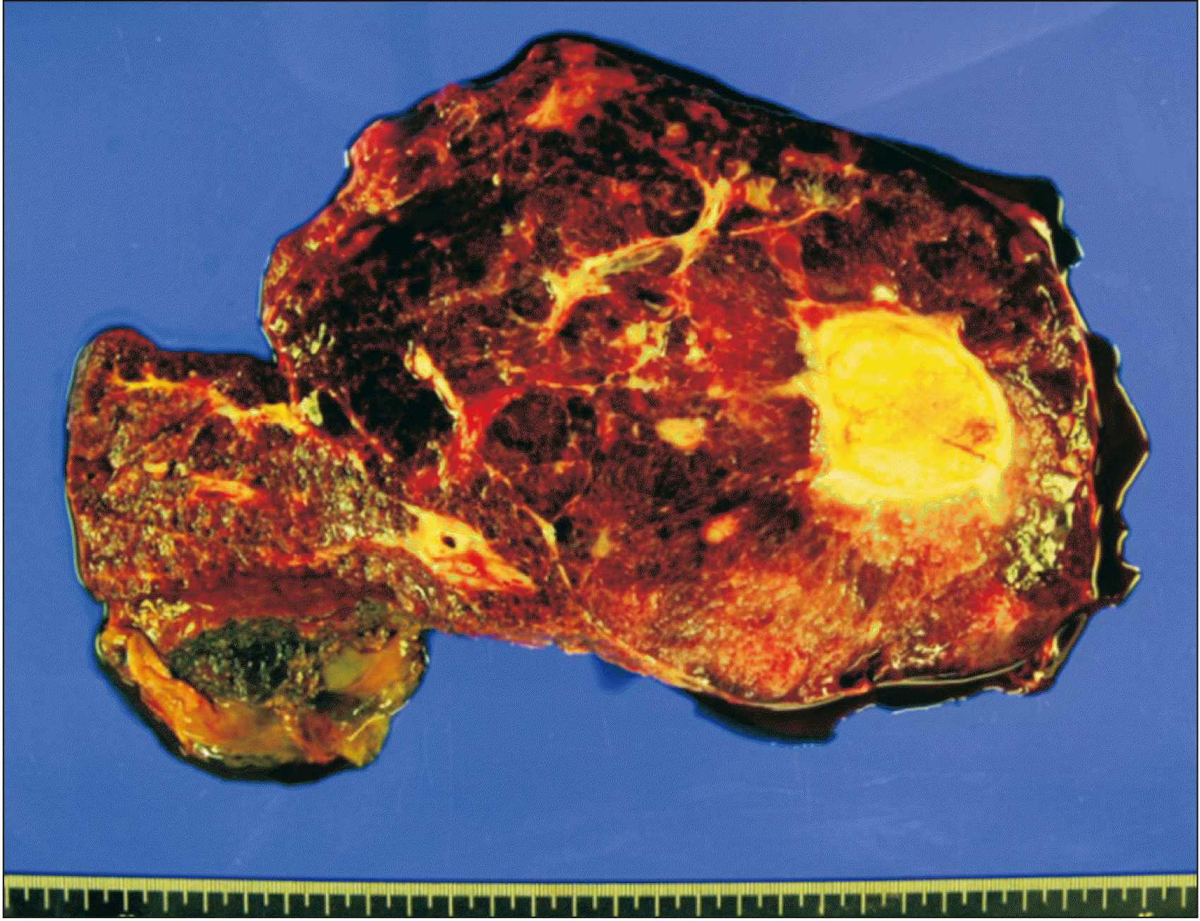
The pathology report revealed that the tumor was a 9 cm-sized mass-forming moderately differentiated intrahepatic cholangiocarcinoma in the background of intraductal papillary neoplasm (Fig. 3). The extent of the perihepatic tumor invasion was the perihepatic soft tissue and IVC. There was no direct involvement of the adrenal gland. Lymphovascular invasion was absent. There was no metastasis in 11 lymph nodes including the retropancreatic and aortocaval nodes. According to the 7th edition of the American Joint Committee on Cancer (AJCC) staging system, the extent of the tumor was pT3N0M0, which was regarded as stage III.
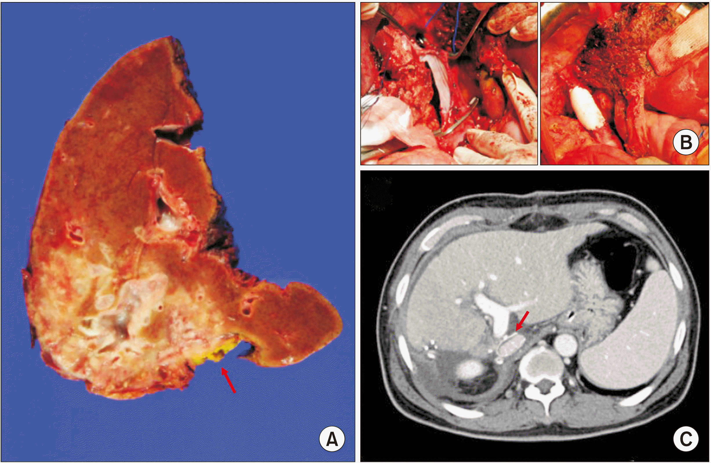 | Fig. 3Gross photograph of Case no. 1 specimen and excision-reconstruction of the retrohepatic inferior vena cava (IVC). (A) A gross photograph of the right liver is visible. The arrow indicates the excised IVC wall. (B) Intraoperative photographs show resection and reconstruction of the IVC wall. (C) Computed tomography taken 1 week after right hepatectomy shows a good patency of the IVC (arrow).
|
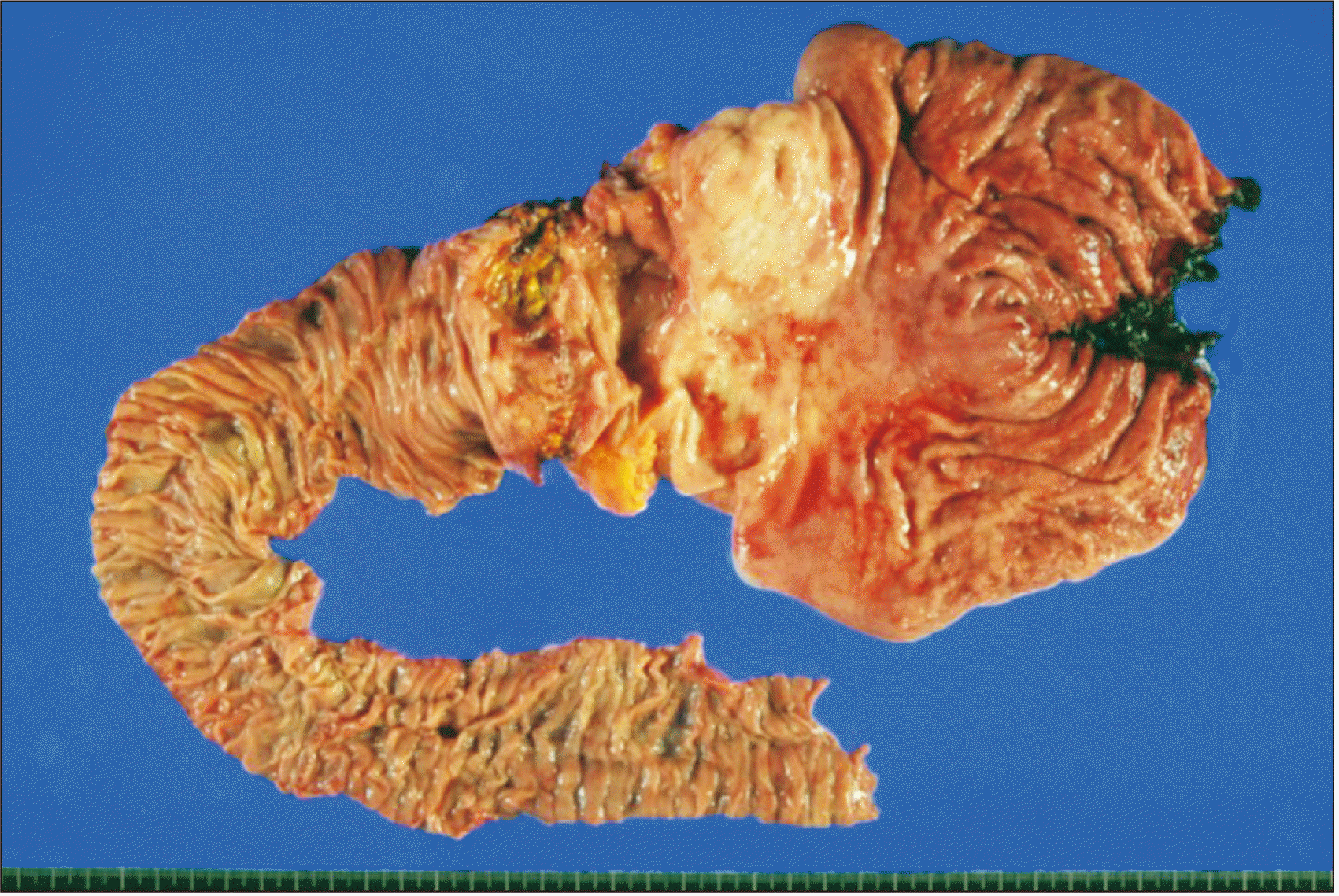
This patient recovered uneventfully from the surgery. He underwent adjuvant chemotherapy at another hospital. Ten months after the surgery, the patient was again referred to our center due to duodenal adenocarcinoma biopsied from a duodenal ulcer. The endoscopic biopsy finding revealed well-differentiated adenocarcinoma in tubular adenoma. This tumor was a second primary tumor because it was different from the first tumor. There was no evidence of local recurrence at the operation site. Preoperative imaging studies revealed advanced duodenal cancer with gastric extension. The general condition of the patient was not so good following systemic chemotherapy. However, the tumor was a second primary tumor with resectability. Thus, we decided to perform pancreaticoduodenectomy.
Pancreaticoduodenectomy was performed with preservation of the pre-existing hepaticojejunostomy. A new jejunal limb was used for pancreatojejunostomy and gastrojejunostomy. Pathology report revealed the presence of two tumors at the duodenum and the gastric antrum. The duodenal mass was a 7 cm-sized moderately differentiated adenocarcinoma with extension to the periduodenal adipose tissue (Fig. 4). Lymphovascular invasion was present. However, perineural invasion and lymph node metastasis were absent. The stomach mass was a 4 cm-sized moderately differentiated adenocarcinoma with extension to the subserosa. The extent of the tumor was pT4N0M0, which was regarded as stage IIB.
This patient recovered uneventfully during the first two weeks. However, pleural effusion and atelectasis developed. Finally, pulmonary edema and pneumonia occurred. His pneumonia progressed despite ventilator support. His general condition gradually deteriorated. This patient passed away due to pneumonia-associated sepsis at 6 weeks after the pancreaticoduodenectomy.
Case no. 2
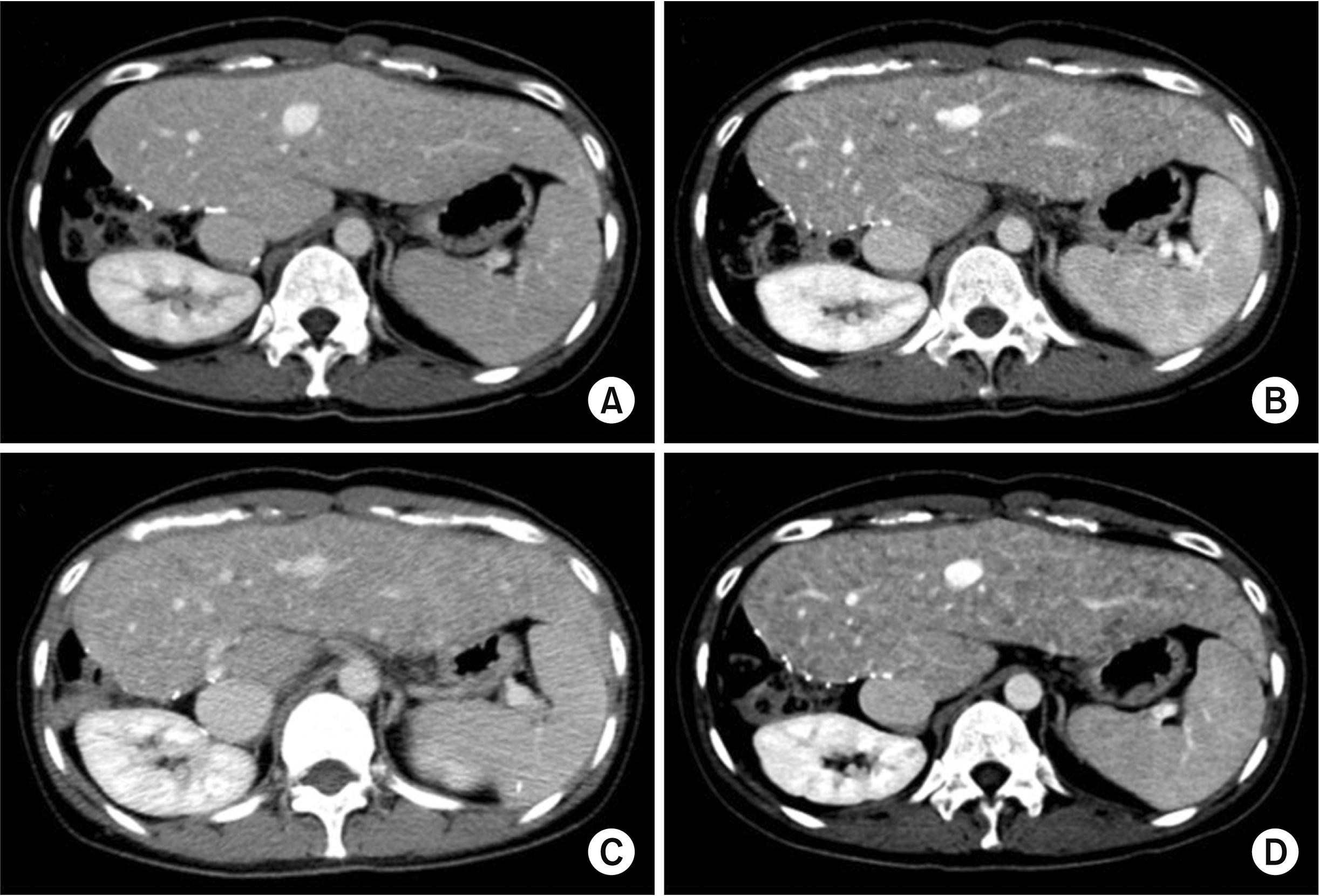
The patient was a 35-year-old female with giant cavernous hemangioma. The hemangioma had progressively grown and compressed the IVC and the left liver with displacement of the right kidney and pancreas (Fig. 5). The patient had undergone transarterial embolization at another hospital. However, the treatment was ineffective in controlling the mass growth.
 | Fig. 5Preoperative computed tomography of Case no. 2. (A–C) Computed tomography scan shows a huge cavernous hemangioma compressing the inferior vena cava and the left liver with displacement of the right kidney and pancreas. (D) Three-dimensional reconstruction of the hepatic arterial system shows the enlarged right hepatic artery.
|
During the operation, the hepatic hilum was dissected to induce hemihepatic inflow block. Several enlarged short hepatic veins and the right hepatic vein were consecutively transected with primary closure. Thereafter, the enlarged right liver was further mobilized. However, a large draining short hepatic vein from the hemangioma-bearing right liver was encountered. Because separating the right liver from the retrohepatic IVC appeared to be infeasible using conventional dissection techniques, total vascular exclusion was performed. The supra- and infra-hepatic portions of the IVC were dissected and occluded with vascular clamps under inflow occlusion with Pringle maneuver according to the usual procedure of THVE. A short hepatic vein appeared to be too large to close it directly without risk of IVC stenosis. Thus, a cryopreserved iliac vein allograft patch was applied to the enlarged defect at the IVC wall. After confirming the absence of bleeding from the retrohepatic IVC, the total vascular exclusion was released (Fig. 6). The longest session of the total hepatic ischemic time was around 15 minutes. After completion of right liver mobilization, right hepatectomy was performed according to standard procedures.
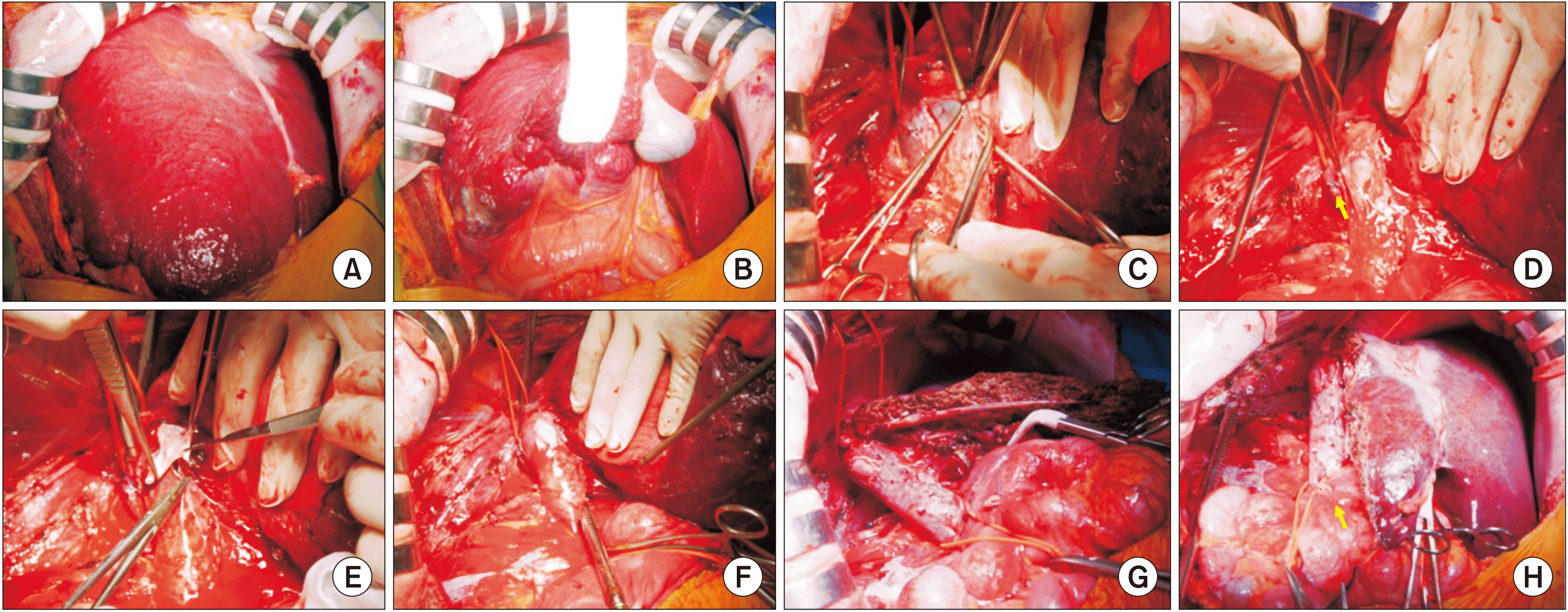 | Fig. 6Intraoperative photographs of right hepatectomy in Case no. 2. (A, B) The enlarged right liver is visible. (C, D) An enlarged short hepatic vein was transected under the standard total hepatic vascular exclusion. (E, F) The wall defect in the inferior vena cava was repaired with a cryopreserve iliac vein allograft patch. (G, H) The remnant left liver is visible with full exposure of the inferior vena cava.
|
The pathology report revealed that the mass was a 29 cm-sized cavernous hemangioma with extensive necrosis (Fig. 7). The weight of the resected liver specimen was 1,569 g. The patient recovered uneventfully from the operation. However, small hemangiomas developed in the remnant left liver at one year after the surgery. There were progressive increases in the number and size of hemangiomas. Thus, her condition was diagnosed as progressive hemangiomatosis (Fig. 8). The patient is currently doing well for the past 6 years after surgery without any abnormal symptoms.
Go to :
DISCUSSION
Liver tumors involving IVC or its junction of major hepatic veins represent situations in which THVE is needed for curative resection. For resection and repair of the retrohepatic IVC, dissecting the IVC to enable clamping is the most important step. In Case no. 1, modified THVE with an oblique cranial cross-clamping technique was used. This technique can be used when the space between the right and the middle hepatic veins is free from involvement so that venous drainage of the residual liver can be preserved during vascular exclusion. For the application of this technique, the retrocaval space must be dissected sufficiently in advance. Hepatic parenchymal transection was completed to facilitate visualization of the anterior aspect of the retrohepatic IVC and the vascular clamp was inserted behind the IVC obliquely, thereby preserving the outflow orifice of the hepatic vein of the liver remnant. By applying the modified THVE, hepatic perfusion was maintained, thereby prolonging the IVC clamping time over hours. A Japanese study has reported that the duration of IVC clamping using modified THVE ranged from 7 minutes to 26 minutes in five cases [2].
In Case no. 2, the standard technique of THVE was used. When the liver cannot be detached from the IVC in the case when the tumor involves the IVC, the retrocaval space behind the retrohepatic IVC should be dissected broadly to facilitate the performance of the standard THVE. The THVE procedure was applied at the last step of combined resection and reconstruction of IVC, with the involved part of the IVC excised en bloc with the liver specimen. Theoretically, no bleeding should occur from the IVC under THVE. However, a significant amount of backflow bleeding often emerges from the cut orifice of the IVC during THVE due to the presence of some small veins around the diaphragmatic crura. To prevent such back bleeding, complete isolation of the IVC from the diaphragm or additional clamping around the diaphragmatic crura is necessary. To reduce the duration of IVC clamping, THVE can be switched to outflow hepatic vein clamping as selective THVE. A Japanese study has reported that the duration of IVC clamping using the standard THVE and switching to selective THVE ranged from 5 to 23 minutes in three cases [2].
To date, only a limited number of studies on THVE for resection of liver tumors involving the IVC have been reported in the literature. The mean duration of THVE was 29 to 78 minutes in reports from very experienced institutes [8-10]. In the literature, the mortality rate of hepatectomy with IVC resection was 4.5% to 25%. Major causes of mortality were liver failure and sepsis [8-12], both of which were likely to be relevant to ischemic injury to the liver and intestinal congestion since they might facilitate bacterial translocation. Damages associated with THVE include both systemic circulatory instability (due to the absence of venous return via the IVC) and total cessation of hepatic blood flow. These conditions can cause congestion of the kidney and intestine, which may explain why the damage and morbidity after THVE are much higher than those experienced after hepatic inflow occlusion alone [9]. Reduction in the duration of THVE might contribute to reduced postoperative complications [2].
Regarding the excision of the IVC wall, it is important to make the wall defect as small as possible to enable primary repair. If primary repair is infeasible, a patch can be attached to repair the wall defect. The patch size should be at least 50% larger than the defect to prevent anastomotic stenosis. Since the IVC is a low-pressure, low-velocity, but high-flow large-caliber vessel with a thick wall, a large wall defect can be repaired with prosthetic vascular grafts, as shown in Case no. 1. In LDLT, we have preferentially used polyethylene terephthalate (Dacron) for IVC replacement [13-15]. On the contrary, a cryopreserved iliac vein allograft was used in Case no. 2 primarily because the IVC wall defect was small. Because the iliac vein is a relatively thin-walled vein, it is not suitable for repairing a large-sized IVC wall defect. However, if the patient is a child, a sizable vein allograft should be used for later physical growth [16,17].
Case no. 1 patient recovered uneventfully from the first operation, but died after the second operation due to the development of pneumonia-associated sepsis. We presume that his relatively poor general condition might be associated with such fatal infectious complication, although there were no detectable differences in perioperative mortality among pancreaticoduodenectomy patients treated with various types of neoadjuvant therapy in the literature [18]. In Case no. 2, small hemangiomas developed in the remnant left liver at 1 year after surgery with progressive increases in the number and size of hemangiomas apparently. This condition was diagnosed as progressive hemangiomatosis. Such post-hepatectomy recurrence of hemangiomatosis has been rarely reported in the literature [19].
In conclusion, standard and modified THVE techniques are proposed as useful techniques to achieve complete tumor resection in patients with large liver tumors invading the retrohepatic IVC.
Go to :
 XML Download