

 PDF
PDF Citation
 Print
Print


INTRODUCTION
Gallbladder carcinoma (GBC) is the commonest biliary tract cancer, ranking the fifth amongst all gastrointestinal malignancies [1]. There is a wide variation in its prevalence globally. Annual incidence of GBC in Western countries is less than 2 per 100,000 people. However, its prevalence is very high in the East (Japan, Poland, and North India) and Chile, with an annual incidence of above 10 per 100,000. It carries a dismal prognosis. Curative surgery after an early recognition is the only effective treatment [2].
Scores of factors have been linked to the development of GBC. A strong association of GBC with the presence of stones has been well established. Four out of five GBC patients have gallstones. On the other hand, 1% to 3% of all cholecystectomy specimens for gallstone disease harbor malignancies [2,3]. Diagnosis of GBC following a cholecystectomy for a benign disease (gallstones, polyps or cholecystitis) is termed as incidental or unsuspected carcinoma gallbladder. There exists a considerable overlap in the definition used to characterize an “incidental GBC (IGBC)” which in true sense implies that diagnosing malignancy is a ‘histo-pathological surprise’ (i.e., no signs of the same on preoperative imaging, surgical findings, or gross examination of the specimen, but the final biopsy suggests malignancy). This has also been described by various authors as unsuspected/unapparent/occult malignancy of the gallbladder [4-6]. With the advent of laparoscopy in the field of surgery and increase in the number of elective cholecystectomies, a rise in the number of IGBC has been reported in recent studies. Of all cases of GBC presenting to a health care center, 25% to 40% are IGBCs [7,8]. Completion re-resection remains the standard of care for a localized disease. Of all patients with IGBC, less than 50% undergo a curative surgery [9].
Detection of distant metastasis (discontinuous liver lesion, peritoneal or mesenteric nodule, pulmonary nodule, distant nodes) or signs of unresectability (vascular involvement, insufficient remnant liver) on imaging or surgical exploration can rule out resection [10]. The use of staging laparoscopy (SL) in IGBC is debatable. Many authors have challenged its utility in detecting metastasis in IGBC [11].
Various factors dictate the probability of a curative resection in IGBC. The timing of completion radical cholecystectomy for optimal results remains a matter of debate. Some suggest an early intervention to improve the resectability rate while others advocate delaying the definitive surgery for biological selection [12,13]. Presentations of IGBC are variable, ranging from intraoperative recognition/suspicion to unexpected pathological finding. This diversity in presentation along with other social and economic factors affect the interval between the index surgery and confirmation of the malignancy for referral of a possible curative resection, especially for patients undergoing cholecystectomy at a non-hepatobiliary center. Besides the referral gap, attributes that might affect outcomes of these patients include patient factors (age, comorbidities) and tumor characteristics (primary tumor stage and differentiation of tumor). The objective of the present study was to evaluate preoperative determinants of a curative resection with special reference to the timing of presentation and the final management of patients presenting with IGBC following an elective cholecystectomy. The utility of SL in IGBC was also evaluated.
Go to : 
PATIENTS AND METHODS
This was a retrospective analysis of prospectively collected data of 91 patients with IGBC who were evaluated in the Department of Surgical Gastroenterology at a North Indian tertiary care centre over 10 years (2009 to 2018). Being a retrospective data analysis not affecting the treatment course or outcome, ethical approval was not needed. This retrospective study was exempted from review by the Ethics Committee (IEC) of the Institute. Study variables included patient’s demographic details, clinical features at presentation (pain, jaundice, lump and weight loss), particulars of index cholecystectomy, primary histopathology (tumor stage and differentiation), and preoperative investigation. Jaundice was defined as the presence of icterus or a total bilirubin level > 2 mg/dL. Weight loss of more than 5% within one month was considered significant. All specimen blocks and/or slides following index surgery were re-evaluated at our institute to validate the diagnosis as per our institutional protocol. In case of discrepancy in the histopathology, in-house review findings overruled. All patients underwent preoperative contrast-enhanced computed tomography (CECT) scans of abdomen and pelvis to rule out metastasis. A few patients with high-risk features (persistent weight loss, jaundice, CECT showing large residual disease, late presentation) underwent fluorodeoxyglucose-positron emission tomography (FDG-PET) scans. Preoperative workup was completed for the majority of patients before admission. Cases judged to be resectable on preoperative clinical and radiological assessment were taken for an intent of curative resection (R0/R1 resection) as early as possible. The majority of patients underwent a SL before laparotomy. It was done using one or two ports. A third port was used when a biopsy was desirable.
The interval between the index cholecystectomy and the definitive surgery at our institute or the day of final staging (for those who were judged to be metastatic on preoperative investigation) was divided into three groups: Early (E), < 4 weeks; Intermediate (I), > 4 weeks and < 12 weeks; and Late (L), > 12 weeks. For practical purposes, this was termed as “time to treatment”. Patient characteristics, preoperative investigations, and intraoperative findings of the three groups were analyzed. Tumors judged to be resectable were considered operable (O) while those judged to be non-resectable due to local advancement or presence of metastasis at any point were considered as inoperable (IO). Various parameters including time to treatment were analyzed by univariate and multivariate analyses to evaluate factors affecting the probability of a curative resection.
Statistical analysis
Normality was assessed for all variables. A variable was considered to be normally distributed when Z score was within ± 3.29 (n ≥ 50). Continuous data are presented as median (range) while categorical data are expressed in frequency (%). Kruskal–Wallis H test was used to compare medians among three groups. Chi-square test was used when expected count of each cell was 5 or more, while Fisher’s exact test was used for the rest to compare categorical data. Predictors of operability were assessed by Binary logistic regression analysis. Variables found to be significant in univariate analysis were included for multivariate analysis. Statistical significance was considered when p-value was less than 0.05. Statistical package for social sciences ver. 23 (IBM SPSS ver. 23; IBM Corp., Armonk, NY, USA) and MedCals software were used for all data analysis.
Go to :
RESULT
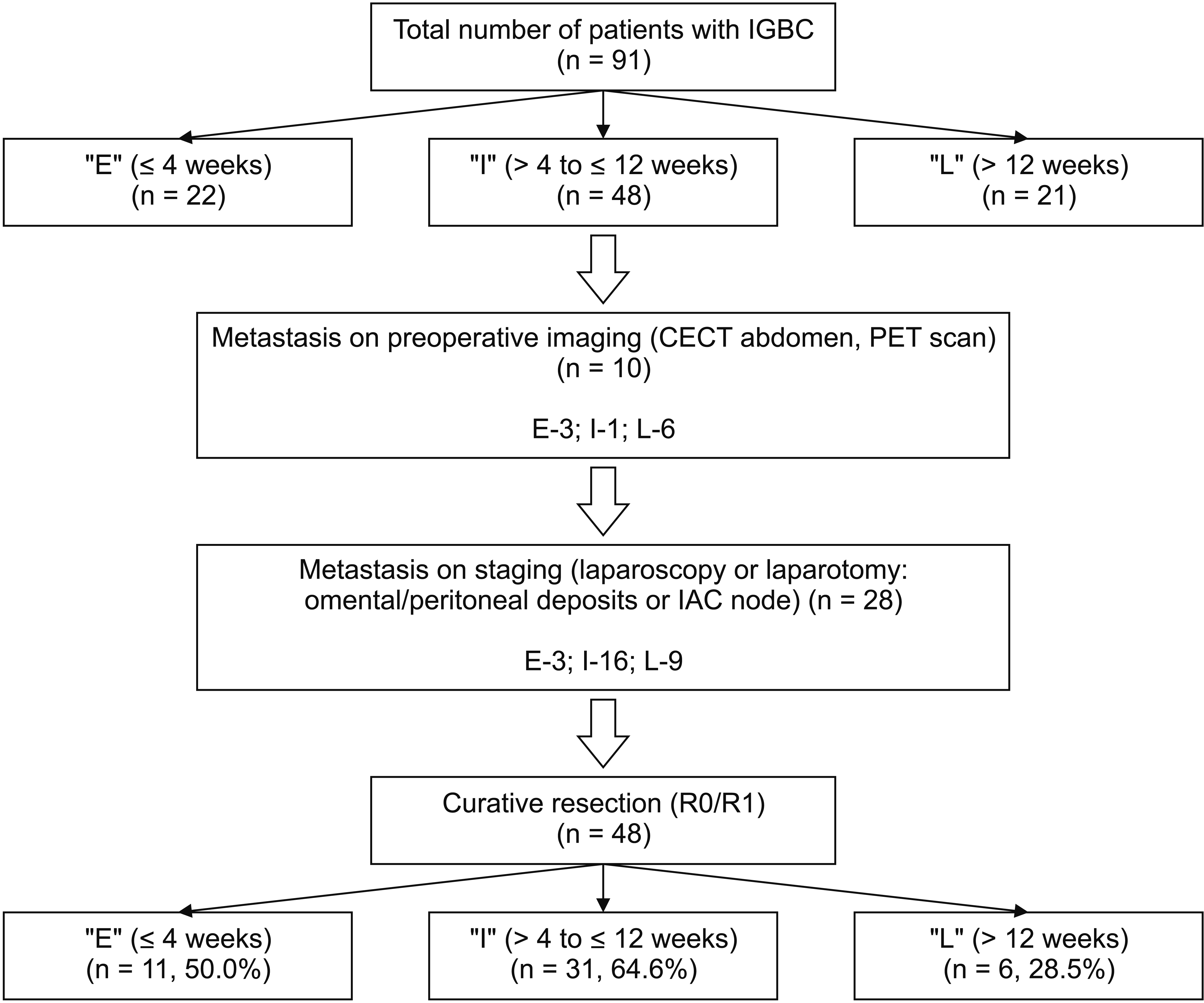
During the study period, 91 patients with the diagnosis of IGBC (referred from other hospitals) following cholecystectomy for a presumed benign disease were admitted for further management. In 57 (62.7%) patients, the index surgery was attempted with a laparoscopic technique, necessitating conversion to open for four patients due to obscure anatomy. Thirty-four (37.3%) patients underwent an open cholecystectomy. The majority (n = 76; 83.5%) of the index surgery were performed electively except for 15 (16.5%) patients who underwent emergency cholecystectomy for acute cholecystitis.
Patients (n = 91) were divided into three groups (‘E’, ≤ 4 weeks; ‘I’, > 4 to ≤ 12 weeks; and ‘L’, > 12 weeks). The ‘I’ group had the most patients (n = 48; 52.7%), followed by ‘E’ (n = 22; 24.2%) and ‘L’ (n = 21; 23.1%) groups. The median age at presentation was 53 years (range, 30–77 years), with a female preponderance (n = 67; 73.6%). There was no significant difference in the age or gender among the three groups (all p > 0.05) (Table 1).
Table 1
Distribution of demographic and clinical characteristics according to time to treatment (n = 91)
| Variable | Early (E) (n = 22; 24.2%) | Intermediate (I) (n = 48; 52.7%) | Late (L) (n = 21; 23.1%) | Total (n = 91) | p-value |
|---|---|---|---|---|---|
| Age (yr)a) | 56.5 (32–75) | 53 (30–77) | 50 (31–76) | 53 (30–77) | 0.154 |
| Sex (female) | 13 (59.1) | 37 (77.1) | 17 (80.9) | 67 (73.6) | 0.22 |
| Preoperative clinical diagnosis | |||||
| Symptomatic gallstone disease | 16 (72.7) | 33 (68.8) | 12 (57.1) | 61 (67.0) | 0.516 |
| Asymptomatic gallstone disease | 2 (9.1) | 3 (6.3) | 0 (0) | 5 (5.5) | 0.865 |
| Acute calculous cholecystitis | 1 (4.5) | 8 (16.7) | 6 (28.6) | 15 (16.5) | 0.112 |
| Complicated gallstone diseaseb) | 3 (13.6) | 4 (8.3) | 3 (14.3) | 10 (11.0) | 0.739 |
| Cholecystectomy | |||||
| Laparoscopic | 13 (59.1) | 29 (60.4) | 11 (52.4) | 53 (58.2) | 0.818 |
| Open | 7 (31.8) | 18 (37.5) | 9 (42.9) | 34 (37.3) | 0.755 |
| Laparoscopic → Open | 2 (9.1) | 1 (2.1) | 1 (4.8) | 4 (4.5) | 0.341 |
| Presentation | |||||
| Positive biopsy (no symptoms) | 13 (59.1) | 37 (77.1) | 11 (52.4) | 61 (67.0) | 0.088 |
| Pain abdomen | 7 (31.8) | 7 (14.6) | 8 (38.1) | 33 (36.2) | 0.001* |
| Jaundice | 1 (4.5) | 1 (2.1) | 2 (9.5) | 4 (4.4) | 0.253 |
| Weight loss | 4 (18.2) | 10 (20.8) | 9 (42.9) | 23 (25.2) | 0.139 |
![]()
Nearly two thirds (n = 61; 67.0%) of patients were asymptomatic at presentation with IGBC. Their malignancy was diagnosed based on biopsy of the resected GB. Amongst symptomatic patients, pain (n = 33, 36.2%) and weight loss (n = 23, 25.2%) was common whereas only 4 (4.4%) patients were jaundiced. Abdominal pain was significantly more common in the ‘E’ group (likely post-surgical pain) and the ‘L’ group (likely indicator of advanced disease) than in the ‘I’ group. Curative resection was done in 48 (52.7%) patients. The proportion of patients undergoing curative resection was statistically less in the ‘L’ group (p = 0.001) (Table 2).
Table 2
Distribution of imaging and tumor staging according to timing to treatment (n = 91)
| Variable | Early (E) (n = 22; 24.2%) | Intermediate (I) (n = 48; 52.7%) | Late (L) (n = 21; 23.1%) | Total (n = 91) | p-value |
|---|---|---|---|---|---|
| Metastasis on preoperative staging | |||||
| CECT | 3 (13.6) | 1 (2.1) | 3 (14.2) | 7 (7.7) | 0.055 |
| PET-CTa) | - | 0 (0) | 3 (14.2) | 3 (3.3) | - |
| Metastasis on staging laparoscopy SLb) | 0 (0) | 5 (10.4) | 4 (19.1) | 9 (9.9) | 0.101 |
| Metastasis on SL/laparotomy | 3 (13.6) | 15 (31.3) | 8 (38.1) | 26 (28.6) | 0.015* |
| IAC positive | 1 (4.5) | 3 (6.2) | 1 (4.8) | 5 (5.5) | 0.771 |
| Differentiation of tumor | 0.849 | ||||
| Grade 1 | 4 (18.1) | 7 (14.6) | 3 (14.2) | 14 (15.4) | |
| Grade 2 | 7 (31.8) | 28 (58.3) | 10 (47.6) | 45 (49.4) | |
| Grade 3 | 3 (13.6) | 7 (14.6) | 3 (14.2) | 13 (14.3) | |
| Primary stage of the tumor | 0.065 | ||||
| pT1c) | 1 (4.5) | 3 (6.2) | 6 (28.6) | 10 (11.0) | |
| pT2 | 15 (68.1) | 30 (62.5) | 10 (47.6) | 55 (60.4) | |
| pT3 | 6 (27.2) | 15 (31.3) | 5 (23.8) | 26 (28.6) | |
| Curative resection (R0/R1) | 11 (50.0) | 31 (64.5) | 6 (28.6) | 48 (52.7) | < 0.001* |
Values are presented as frequency (%). Compared by chi-square test or Fisher exact test (*p < 0.05 significant).
CECT, contrast-enhanced computed tomography; PET-CT, positron emission tomography–computed tomography.
a)PET-CT scan was done in 4 patients in ‘I’ group and 5 patients in ‘L’ group. Four out of 6 patients negative for metastasis on PET-CT were found to have disseminated disease on SL.
![]()
More than half (n = 48, 52.7%) of patients presented within 4 to 12 weeks of the index surgery. Preoperative imaging (CECT) was done for staging in all patients. It detected metastasis in 7 (7.7%) patients. PET scan was done in 9 patients (4 in the ‘I’ group and 5 in the ‘L’ group) with gross residual disease or enlarged lymph nodes on CECT abdomen. It detected metastasis in 3 (3.3%) more patients (all in the ‘L’ group). Metastasis was detected during SL/Laparotomy in 26 (28.6%) patients. Positive IAC (interaorto-caval) node on frozen section was found in 5 (5.5%) patients. Presence of omental, peritoneal, or surface metastasis was significantly more in the ‘L’ group (p < 0.05). Tumor characteristics (primary T stage and grade) were similar for the three groups (p > 0.05) (Table 2).
Yield of SL was more in the ‘L’ group (n = 4/13; 30.8%) than in the ‘I’ (n = 5/45; 11.1%) or the ‘E’ group (n = 0/9, nil). Overall, it avoided unnecessary laparotomy for 9 patients (n = 9/66, 13.6%). Preoperative imaging in tandem with laparoscopy was effective in detecting metastasis in 47.6% (10/21) patients in the ‘L’ group who were otherwise considered fit for surgery. In patients who underwent laparotomy after SL, another 26.2% (17/65) patients were found to have unresectable or metastatic disease (‘E’, 3/14; ‘I’, 10/41; ‘L’, 4/10). Metastatic disease was significantly more common in the late stage. Only six (28.5%) patients in ‘L’ group could undergo curative resection, which was significantly less than those in the ‘E’ group (n = 11/22; 50.0%) and the ‘I’ group (n = 31/48; 64.6%) (Fig. 1). The final stage of tumor following a curative resection included Stage 1 disease in 4, Stage 2 in 27, Stage 3 in 14, and Stage 4 in 3 patients. Nodal metastasis was present in nine patients. Five (22.7%) patients in the ‘E’ group could not be offered a surgery due to logistic issues/unfit for major procedure (n = 3) or lack of consent (n = 2), although they were potentially resectable on imaging.
Predictors of resectability
Predictors of a curative resection were assessed using binary logistic regression analysis. On univariate analysis, time to treatment, presentation of symptoms, and differentiation of tumor were significantly associated with patient operability. On multivariate analysis, only presentation in intermediate period and tumor differentiation (well/moderately differentiated) were significant factors associated with an increased chance of a curative resection, while late referral (> 12 weeks) and poor differentiation were associated with an inoperability. Age, sex, type of cholecystectomy, and the initial tumor stage did not affect the resectability rate (all p > 0.05) (Table 3).
Table 3
Predictors of resectability in study patients (n = 91)
| Variable | Univariate | Multivariate | |||||
|---|---|---|---|---|---|---|---|
|
|
|
||||||
| Operable (n = 48; 52.7%) | Inoperable (n = 43, 47.3%) | p-value | AOR (95% CI) | p-value | |||
| Age (yr) | 54.04 ± 12.32 | 52.56 ± 10.06 | 0.534 | - | - | ||
| Sex | - | - | |||||
| Female | 35 (72.9) | 32 (74.4) | 0.98 | ||||
| Male | 13 (27.1) | 11 (25.6) | |||||
| Type of cholecystectomy | - | - | |||||
| Open | 15 (31.3) | 19 (44.2) | 0.20 | ||||
| Laparoscopic | 32 (66.6) | 21 (48.8) | |||||
| Lap converted open | 1 (2.1) | 3 (7.0) | |||||
| Time of referral | 0.019* | ||||||
| Early (≤ 4 wk) | 11 (22.9) | 11 (25.6) | 0.027* | 2.37 (0.43–13.05) | 0.32 | ||
| Intermediate (> 4 to ≤ 12 wk) | 31 (64.6) | 17 (39.5) | 7.33 (1.75–30.82) | 0.007* | |||
| Late (> 12 wk) | 6 (12.5) | 15 (34.9) | Reference | ||||
| Primary stage | - | - | |||||
| T1 | 6 (12.5) | 4 (9.3) | 0.454 | ||||
| T2 | 31 (64.6) | 24 (55.8) | |||||
| T3 | 11 (22.9) | 15 (34.9) | |||||
| Presentation | - | - | |||||
| Asymptomatic | 38 (79.2) | 23 (53.5) | 0.014* | ||||
| Pain | 8 (16.6) | 14 (32.5) | 0.091 | ||||
| Loss of weight | 2 (4.2) | 13 (30.2) | 0.001* | ||||
| Lump | 0 | 3 (7.0) | 0.102 | ||||
| Jaundice | 0 | 4 (9.3) | 0.046* | ||||
| Differentiation | 0.008* | ||||||
| Well differentiation | 12 (25.0) | 2 (4.6) | 0.001* | 53.36 (5.40–526.96) | 0.001* | ||
| Mod differentiation | 27 (56.2) | 18 (41.9) | 9.62 (1.76–52.51) | 0.009* | |||
| Poor differentiation | 2 (4.2) | 11 (25.6) | Reference | ||||
| Undefined | 7 (14.6) | 12 (27.9) | - | - | |||
![]()
Go to :
DISCUSSION
Management of GBC is a colossal challenge. A multimodality approach is evolving. It has produced promising results. Although new chemotherapeutic drugs and state-of-art radiotherapy techniques are effective in the management of advanced disease, surgery remains the backbone of treatment [14]. Patients with preoperative diagnosis of gall bladder mass often present in a late stage with a low curative resection rate (20%–45%) [15-17]. In contrast to the traditional nihilism on outcome of primary GBC, IGBC carries a better prognosis. Despite variable presentations, the overall resectability rate and survival of appropriately chosen patients with IGBC are better than historically described ones for those with a gall bladder malignancy [18]. All patients should undergo a thorough evaluation, including a good quality axial imaging (CECT/MRI) for staging. All attempts should be made to rule out metastatic lesions. Some clinicians suggest FDG-PET scan as a routine procedure for all patients with IGBC. Corvera et al. [19] have reported that PET scan could change management in 29% of patients. Anderson et al. [20] have documented that PET scan has a sensitivity of 78% and a specificity of 80% for detecting residual and metastatic disease. FDG-PET could detect metastatic disease in 50% patients with incidentally diagnosed GBC. Shukla et al. [21] have used multidetector CECT and PET-CT for evaluating patients with IGBC, avoiding unnecessary laparotomy in 55 of 80 patients (68.8%). However, Butte et al. [22] have demonstrated a change in management in only 13% of 63 patients undergoing PET prior to surgery. The majority of their patients had early malignancies, with in-situ carcinoma comprising 31% of their cohort. Inflammation in the early postoperative period may portend a false FDG activity mimicking a residual disease or a metastasis, especially in the gallbladder fossa. In the present study, we performed PET-CT selectively for patients with a residual disease in the gallbladder fossa seen on CECT in the ‘L’ or the ‘I’ group. Selective approach was chosen due to logistic constraints. Among patients undergoing PET-CT, surgical intervention was avoided in 3/9 (33.3%). False negative was found for 44.4% (4/9) of cases. SL could detect disseminated disease. Thus, the use of PET scan as a complement to a good quality CECT is warranted for detecting a distant disease, especially in patients presenting after 12 weeks of index surgery. However, absence of disease activity on PET scan should be interpreted with caution.
With improvement in imaging techniques, the yield of SL has dramatically decreased in the present time. Moreover, studies have shown that it is not as effective in reducing the number of laparotomies in IGBC as in non-IGBC. In a report from Memorial Sloan-Kettering Cancer Center (MSKCC), although SL was effective in detecting distant metastasis in 56% of patients with primary GBC, the yield decreased to 20% in IGBC patients [23]. Another study from MSKCC in 2011 has documented a low yield of initial laparoscopy. Out of 136 patients with IGBC, SL was performed in 46 patients. It could detect metastasis in only 20% of patients [24]. SL might not be very effective in a patient with a recent history of cholecystectomy. Abdominal cavity is usually evaluated by a primary surgeon at the start of the surgery, especially prior to a minimal invasive surgery. Moreover, it might be difficult to differentiate early postoperative inflammation and adhesions from a malignant infiltration. However, the yield of laparoscopy can be increased with regular use of three ports for abdominal exploration as described by Agarwal et al. [25] in primary GBC. We performed SL in half patients of the ‘E’ group and nearly all patients of ‘I’ and ‘L’ groups. Overall yield in the ‘E’ group (< 4 weeks) was nil, while that was 11.1% in the ‘I’ group (> 4 to ≤ 12 weeks) and 30.8% in the ‘L’ group (> 12 weeks). The usefulness of SL will increase with a delay in the presentation even after a thorough preoperative screening. It adds little to the operating time, although it is expected to reduce the hospital stay, overall cost, and common postoperative complications associated with an incision (pain, surgical site infections, and respiratory compromise). Preoperative imaging and SL in tandem avoided unnecessary laparotomy in 28.3% of disseminated diseases in the total cohort and 47.6% in the ‘L’ group. In all four patients with a negative PET-CT, peritoneal disease was discovered and confirmed on SL. Thus, SL should be a part of the staging in IGBC, especially in patients presenting at more than 4 weeks after the index surgery, even if the PET-CT demonstrates no FDG avid lesions. The yield is likely to improve in the presence of high-risk features (symptomatic at presentation, patients with moderately or poorly differentiated carcinoma). In a study by Butte et al. [24], advanced T stage after primary cholecystectomy and bile spillage at initial surgery were also associated with an increase in the yield of SL.
The primary intention of evaluating a patient with IGBC is to rule out a non-resectable disease. Inability to do so may result in an unwarranted surgical intervention, thus prolonging the hospital stay and delaying the initiation of a palliative chemotherapy if feasible. Obvious signs of advanced or unresectable disease that rule out definitive surgery include umbilical nodule, port site metastasis, enlarged supraclavicular lymph nodes, and presence of ascites or liver nodules. Many factors related to the index cholecystectomy and subsequent histopathology determine the probability of a curative resection. Many studies have evaluated predictors of unresectability in primary GBC (jaundice, lump, weight loss, and gastric outlet obstruction) [15,26]. However, patients with IGBC usually do not have symptoms at presentation. Two thirds of our patients presented with a postoperative biopsy report (positive for malignancy) without other complaints. However, the presence of symptoms is a harbinger of unresectability. The group of patients who turned inoperable in the present study had significantly (p < 0.05) more weight loss and/or jaundice at presentation. None of these patients with jaundice could undergo a definitive procedure. Abdominal pain was the most common presenting feature, although did not affect the resectability rate on univariate or multivariate analysis. Pain in the early period might be related to postoperative inflammation, while that in the ‘L’ group might be a sign of an advanced disease.
Optimum timing for reoperation in IGBC lacks consensus. Ausania et al. [12] have evaluated outcomes of patients with IGBC following an intentional delay of three months and found that the curative resection rate with this approach is 49% at 12 weeks. In a multi-institutional study by Ethun et al. [13], patients operated within 4 to 8 weeks of the index cholecystectomy had the best prognosis, with poor differentiation and higher primary T-stage being the most important parameters predicting metastatic or unresectable disease. The most common primary T-stage following cholecystectomy in that study was T2 [13]. In the present study, the proportion of patients who could undergo a curative resection was also significantly more in the ‘I’ group (64.6%) on both univariate analysis (p = 0.02) and multivariate analysis (p = 0.015). Patients in the ‘L’ group were the least likely (28.6%) to undergo a definitive surgery. This ratio was 50.0% in the ‘E’ group. Outcomes in the ‘E’ group might have been skewed by the fact that more patients did not receive a trial of surgery for non-surgical reasons. We intentionally considered these patients during the final analysis as it presented the actual circumstance in clinical practice, avoiding selection bias and reflecting the intention to treat analysis. Patients who present in the early phase were reluctant to undergo a second procedure. It was difficult to convince them to undergo a major re-intervention when they knew that the first procedure did not result in a complete treatment. Moreover, optimization for a major surgery within four weeks of an operation, especially in patients with co-morbidities, is not always feasible. A few patients also questioned the importance of a second procedure when repeat scans did not show any tumor. A second surgery within a span of four weeks also poses a heavy financial burden to a family in a developing country like India where the expenditure on health is born by the family itself. Nearly 13% (n = 5) patients denied consent (n = 2) or could not undergo surgery due to other reasons (logistic issues, n = 1; could not be optimized for a second surgery, n = 2) in the ‘E’ group. A period between 4 weeks and 12 weeks seemed to produce the best chance of a curative resection. The majority of biologically aggressive tumors tended to show up during this period, with inflammation settling down considerably that could make intra-operative assessment and dissection easy. Moreover, waiting for a month allows time for the body to recuperate from the index procedure. Even patients with co-morbidities have time for optimization after a waiting period of 1 to 2 months.
Age, sex, type of cholecystectomy, and primary T-stage following cholecystectomy did not affect the curative resection rate. However, successful re-resection was more likely for a well or moderately differentiated tumor than for a poorly differentiated tumor in both univariate analysis (p = 0.001) and multivariate analysis (p = 0.008). Similarly, Ethun et al. [27] have reported that grade of tumor and lympho-vascular invasion are important determinants of resectability. Other factors associated with increased chances of dissemination include a breach in tumor during initial cholecystectomy, gross spillage of bile and/or stones, and primary biopsy suggestive of advanced T disease (T3/4) [28].
Being a retrospective study, the present analysis has some limitations. First, details of the initial procedure (regarding spillage, reasons for conversion, preoperative ultrasound findings and the intra-operative difficulties faced) were unavailable. In addition, preoperative imaging prior to the index cholecystectomy, difficulty in dissection during the initial procedure, margin status on pathological evaluation following index cholecystectomy could not be correlated with the final outcome due to unavailability of data. Although the sample size was relatively large for a single institution, our results could have been more meaningful with inclusion of patients from other referral centers and a longer recruitment period.
In conclusion, an intermediate timing of intervention (4–12 weeks), tumor differentiation (well or moderate differentiation), and absence of symptoms at presentation (especially weight loss and jaundice) were associated with an increased chance of successful re-resection in IGBC. However, a prospective study is needed in the future to validate results of this study.
Go to :
 XML Download