

 PDF
PDF Citation
 Print
Print


INTRODUCTION
Gallbladder cancer (GBC) is the most common malignant disorder of the biliary tract. Operation is the only potential cure available to patients with GBC. Extended (or radical) cholecystectomy (EC) is the standard surgical procedure for most patients with T2 and T3 GBC [1]. The extent of hepatic resection in EC has been variable from a few centimetres of gallbladder fossa (GBF) to complete excision of hepatic segment s4b&5 [1-5]. Advocates of hepatic bi-segmentectomy 4b&5 in EC believe that microscopic metastatic foci of the GBC may present as skip lesions inside segments 4b&5. Therefore, complete excision of s4b&s5 is necessary to prevent a recurrence [6-8]. However, two questionnaire surveys have reported no survival advantage with bi-segmentectomy s4b&s5 for patients with T2 GBC [9,10]. Thus, we conducted a matched case-control study to compare outcomes of ECB (EC involving bi-segmentectomy s4b&5) to those of ECW (EC involving wedge hepatic resection) for patients with T2&T3 GBC.
Go to : 
PATIENTS AND METHODS
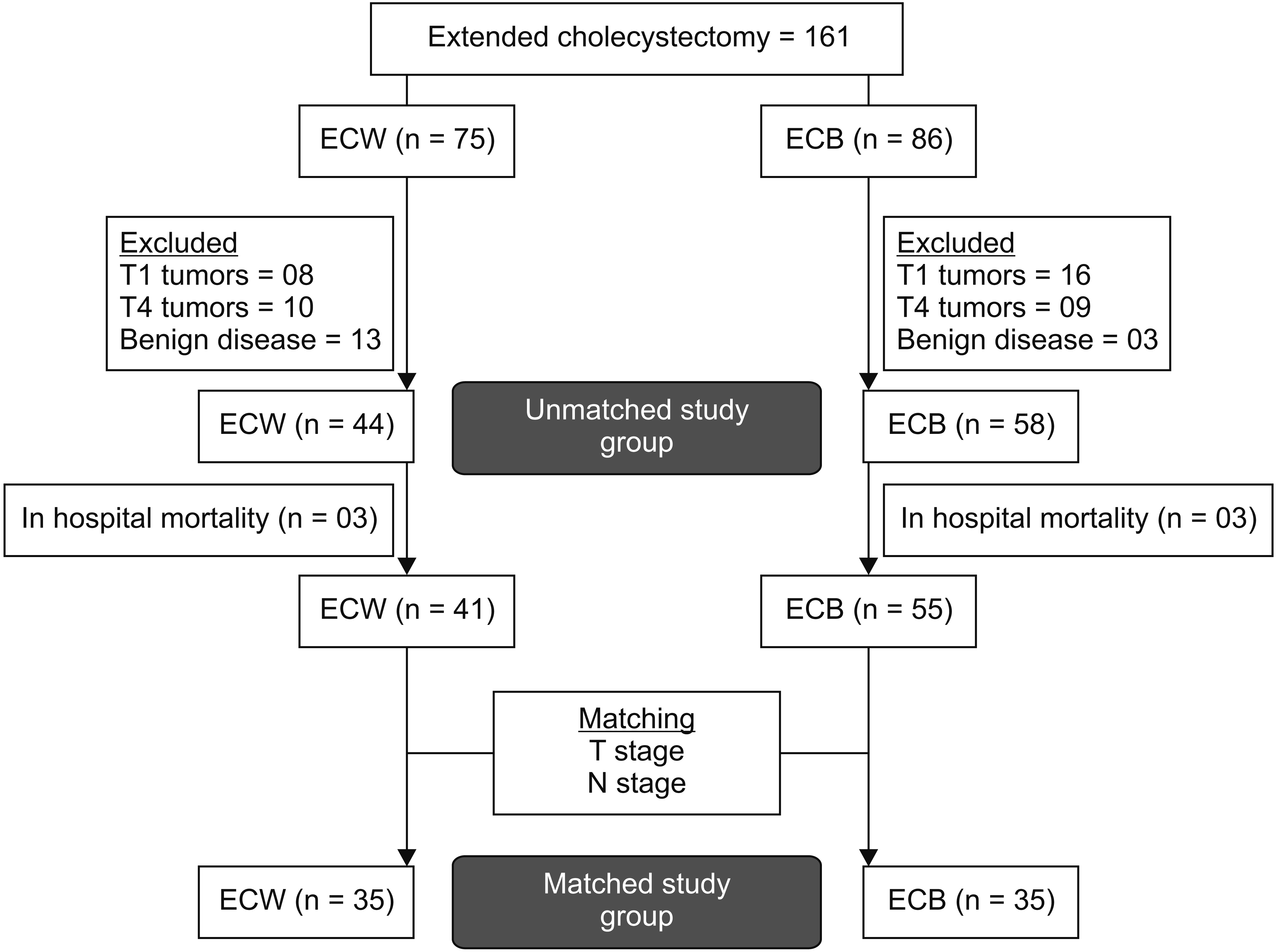
This matched case-control study was conducted at Govind Ballabh Pant Institute of Postgraduate Medical Education and Research, New Delhi, India. Hospital records of patients who were offered EC by single unit from May 2009 to February 2019 were reviewed. Follow-up data were collected up to February 2019. Written informed consent from patient was waived by the institutional committee of ethics [11]. Based on the type of hepatic resection, patients were divided into ECB and ECW groups. Patients with T1 GBC, T4 GBC, or benign diseases were excluded. Post-exclusion, both groups were exactly (1 : 1) matched for T and N stages using MedCalc software 19.6.1 (MedCalc Software Ltd, Ostend, Belgium; https://www.medcalc.org; 2020) (Fig. 1). Demographic, clinical, operative, and postoperative parameters of the two groups were then compared.
The preoperative workup for patients with GBC included clinical history, clinical examination, complete blood counts, biochemistry (renal function tests, liver function tests, international normalized ratio), and tumour markers (carcino-embryonic-antigen and carbohydrate antigen 19.9), ultrasound of the abdomen, contrast-enhanced computed tomography (CECT) of the chest and the abdomen, and endoscopic ultrasound (EUS). Fine needle aspiration cytology was conducted under conventional/EUS guidance if an inoperable disease was suspected on radiological imaging. The American Joint Committee on Cancer (AJCC) 8th classification system was employed. Due to a high prevalence of patients with incidental GBC (IGBC), sub-classification of T2 stage into T2a and T2b was omitted [12]. The severity of postoperative complications was described per the classification proposed by Dindo et al. [13]. Hospital mortality was defined as mortality within 90 days from the operation. ECB was defined as excision of GB plus hepatic segments s4b and 5 plus hepatoduodenal ligament nodes (HDLNs). ECW was defined as en-bloc excision of GB plus two centimetres of tumor-uninvolved liver conforming GBF and HDLNs. Staging laparoscopy was performed through an infra-umbilical endoscopic port. EC was abandoned in patients with confirmed N2 and M1 metastasis. Laparoscopic ECB was performed as described by Nag et al. [14]. Any involvement of adjacent organs such as common bile duct (CBD), stomach, duodenum, and colon were dealt with CBD resection, distal gastrectomy, duodenal sleeve resection, and colonic resection, respectively. Adjuvant chemotherapy was given after attaining consensus of surgeon, oncologist, and patient willingness.
Statistical analysis
Statistical package MedCalc 19.6.1 (MedCalc Software Ltd) was used for analysis and case-control matching. Parametric numerical data are presented as mean ± standard deviation. Non-parametric numerical data are presented as median (interquartile range). Categorical and ordinal data are presented as percentages. Parametric numerical data were compared with Student’s t-test. Non-parametric numerical data were compared with a Mann–Whitney U-test. Chi-square test and Fisher’s test were used to compare categorical and ordinal data. Kaplan Meier method (log-rank test) and Cox regression method were used for survival analysis. p-value < 0.05 was considered significant.
Follow up
Patients visited the outpatient department every three months for the first two years and every six months thereafter. Clinical examination, liver function test, tumour markers, and ultrasound of the abdomen were advised at every visit. CECT and/or 18-fluorodeoxyglucose positron emission tomography-computed tomography were advised selectively.
Go to :
RESULTS
Out of a total of 161 patients who received EC during the study period, 75 patients were offered ECW and 86 patients were offered ECB. After excluding those with T1 tumors, T4 tumors, or benign pathologies, 102 patients (ECW = 44, ECB = 58) had T2 and T3 stage tumors. After matching, there were 35 patients in each group (Fig. 1). All patients had R0 resection. This section will focus on post-matching data. As depicted in Table 1, most of demographic, clinical, and laboratory parameters were comparable between the two groups except that for the use of the laparoscopic approach was more frequent in the ECB group (p = 0.000). For ECB vs. ECW, hospital stay was 6 days vs. 8 days (p = 0.216), operation time was 270 minutes vs. 300 minutes (p = 0.748), and surgical blood loss was 200 mL vs. 250 mL (p = 0.005). The overall complication rate was 17.1% in the ECB group and 40.0% in the ECW group (p = 0.035). The incidence of grade II & III complications and bile leak were significantly less in the ECB group (p = 0.002) (Table 2).
Table 1
Comparison of demographic and laboratory parameters
| Variable | Before matching | After matching | |||||
|---|---|---|---|---|---|---|---|
|
|
|
||||||
| ECW (n = 44) | ECB (n = 58) | p-value | ECW (n = 35) | ECB (n = 35) | p-value | ||
| Age (yr) | 51 (43.5–60.0) | 50 (44–60) | 0.794 | 52 (42.7–60.0) | 49 (45–60) | 0.737 | |
| Sex (female) | 29 (65.9) | 41 (70.7) | 0.608 | 25 (71.4) | 27 (77.1) | 0.587 | |
| ASA grade | 0.386 | ||||||
| 0 | 0 | 5 (8.6) | 0 | 3 (8.6) | |||
| 1 | 27 (61.4) | 32 (55.2) | 0.527 | 24 (68.6) | 23 (65.7) | ||
| 2 | 17 (38.6) | 19 (32.8) | 11 (31.4) | 8 (22.9) | |||
| 3 | 0 | 2 (3.4) | 0 | 1 (2.9) | |||
| Comorbidity | 6 (13.6) | 9 (15.5) | 0.791 | 4 (11.4) | 5 (14.3) | 0.723 | |
| IGBC | 11 (25.0) | 13 (22.4) | 0.761 | 9 (25.7) | 9 (25.7) | 1.000 | |
| Pre-operative hemoglobin (gm/dL) | 10.9 (10–12.1) | 11.1 (9.5–11.9) | 0.633 | 10.8 (10.0–11.9) | 11 (9.0–11.7) | 0.462 | |
| Pre-operative bilirubin (mg/dL) | 0.8 (0.6–1.5) | 0.6 (0.5–0.9) | 0.01* | 0.7 (0.6–1.3) | 0.6 (0.5–0.9) | 0.05 | |
| Pre-operative albumin (U/L) | 3.5 (3.3–4.0) | 3.8 (3.4–4.1) | 0.179 | 3.6 (3.3–3.9) | 3.8 (3.4–4.1) | 0.209 | |
| CA19.9 (IU/mL) | 16.7 (5.9–34.8) | 11.3 (6.0–22.0) | 0.607 | 7.6 (4.5–33.2) | 8.7 (5.6–17.7) | 0.789 | |
| Laparoscopic approach | 3 (6.8) | 22 (37.9) | 0.000* | 3 (8.6) | 16 (45.7) | 0.000* | |
![]()
Table 2
Comparison of operation details and postoperative outcome
| Variable | Before matching | After matching | |||||
|---|---|---|---|---|---|---|---|
|
|
|
||||||
| ECW (n = 44) | ECB (n = 58) | p-value | ECW (n = 35) | ECB (n = 35) | p-value | ||
| Postoperative stay (day) | 8 (5–12.5) | 6 (5–8) | 0.158 | 8 (5–12.5) | 6 (5–8) | 0.216 | |
| Duration of surgery (min) | 300 (240–360) | 240 (200–320) | 0.059 | 300 (225–357) | 270 (240–325) | 0.748 | |
| Blood loss (mL) | 250 (200–300) | 200 (120–200) | 0.000* | 250 (200–300) | 200 (100–200) | 0.005* | |
| Postoperative complications | 20 (45.5) | 18 (31.0) | 0.137 | 14 (40.0) | 6 (17.1) | 0.035* | |
| Clavien-Dindo Grade I/Grade II/Grade III/Grade IV | 5/9/2/0 | 11/1/1/1 | 0.032* | 3/8/2/0 | 4/0/1/1 | 0.046* | |
| Post-operative bile leak | 11 (25.0) | 1 (1.7) | 0.000* | 8 (22.9) | 0 | 0.002* | |
| T stage | 1.000 | ||||||
| T2 | 23 (52.3) | 31 (53.4) | 0.906 | 19 (54.3) | 19 (54.3) | ||
| T3 | 21 (47.7) | 27 (46.6) | 16 (45.7) | 16 (45.7) | |||
| N stage | 0.039 | 1.000 | |||||
| N0 | 18 (40.9) | 36 (62.1) | 18 (51.4) | 18 (51.4) | |||
| N1 | 9 (20.5) | 12 (20.7) | 8 (22.9) | 8 (22.9) | |||
| N2 | 17 (38.6) | 10 (17.2) | 9 (25.7) | 9 (25.7) | |||
| AJCC staging | 0.029* | 1.000 | |||||
| Stage 2 | 12 (27.3) | 22 (37.9) | 12 (34.3) | 12 (34.3) | |||
| Stage 3 | 15 (34.1) | 27 (46.6) | 14 (40.0) | 14 (40.0) | |||
| Stage 4 | 17 (38.6) | 9 (15.5) | 9 (25.7) | 9 (25.7) | |||
| PNI/LVI positive | 15 (34.1) | 12 (20.7) | 0.130 | 13 (37.1) | 8 (22.9) | 0.195 | |
| Minimum margin (mm) | 8 (3–20) | 14 (8.2–19.5) | 0.098 | 9 (5–20) | 12 (8–15) | 0.487 | |
| Adjuvant chemotherapy | 25 (56.8) | 34 (58.6) | 0.855 | 22 (62.9) | 23 (65.7) | 0.804 | |
| Recurrence | 22 (50.0) | 15 (25.9) | 0.013* | 16 (45.7) | 11 (31.4) | 0.259 | |
| Recurrence free survival (mon) | 37.5 ± 4.9 | 64.9 ± 5.1 | 0.014* | 42.3 ± 5.41 | 58.2 ± 6.2 | 0.264 | |
| Overall survival (mon) | 39.0 ± 4.5 | 67.0 ± 4.9 | 0.008* | 43.4 ± 5.05 | 61.5 ± 5.8 | 0.161 | |
![]()
Distributions of patients according to T, N, and AJCC stages were similar in both groups (Table 2). The minimum hepatic margin was 12 mm in the ECB group and 9 mm in the ECW group (p = 0.487). In ECB and ECW groups, 65.7% and 62.9% of patients received adjuvant chemotherapy, respectively (p = 0.804). The recurrence rate was 31.4% in the ECB group and 45.7% in the ECW group (Table 2). Isolated hepatic recurrence was observed in four patients (3 in ECW group and 1 in ECB group). In the rest of patients, recurrence involved the liver, lymph nodes, and other organs simultaneously. Mean recurrence-free survival (RFS) was 58.2 months for the ECB group and 42.3 months for the ECW group (hazard ratio [HR]: 0.64, 95% confidence interval [CI]: 0.30–1.39, p = 0.264) (Table 2, Fig. 2). Mean overall survival (OS) was 61.5 months for the ECB group and 43.4 months for the ECW group (HR: 0.57, 95% CI: 0.26–1.25; p = 0.161) (Table 2, Fig. 3). The probability of OS was > 50% for the ECB group. Therefore, median OS could not be estimated for ECB group. The median OS for the ECW group was 40 months (Table 2). One-year, 3-year, and 5-year RFS rates for ECB vs. ECW groups were 81.6% vs. 64.9%, 63.9% vs. 51.2%, and 63.9% vs. 51.2%, respectively. One-year, 3-year, and 5-year OS rates for ECB vs. ECW groups were 90.8% vs. 88.3%, 65.6% vs. 50.8%, and 65.6% vs. 44.4%, respectively. The involvement of surrounding organs and the occurrence of various types of malignancies were comparable in both groups (Table 3). On univariate analysis, T stage and N stage were associated with poor prognosis. On multivariate analysis, T stage, N stage, and ASA grade were associated with poor RFS and OS (Table 3–5).
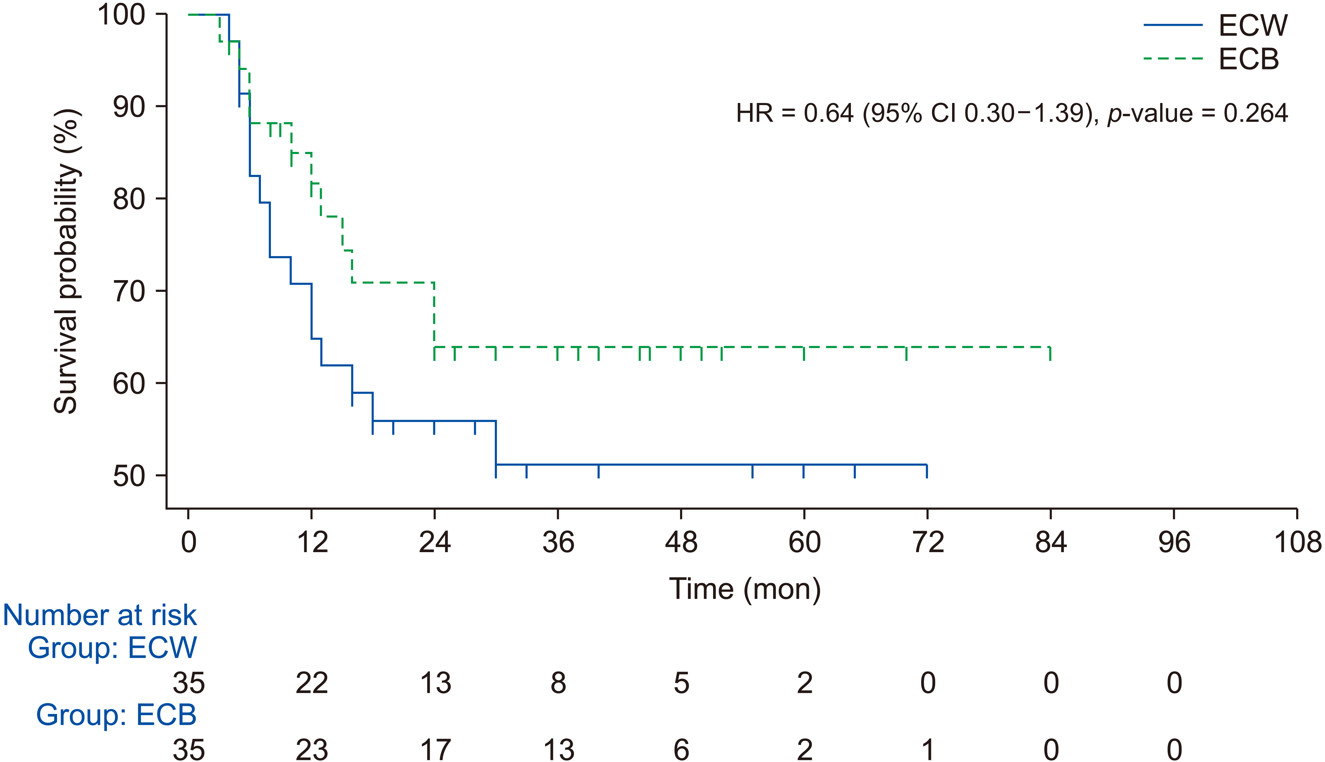 | Fig. 2Kaplan–Meier recurrence-free survival curves for ECB and ECW groups. ECB, extended cholecystectomy involving bi-segmentectomy s4b&5; ECW, extended cholecystectomy involving wedge hepatic resection; HR, hazard ratio; CI, confidence interval.
|
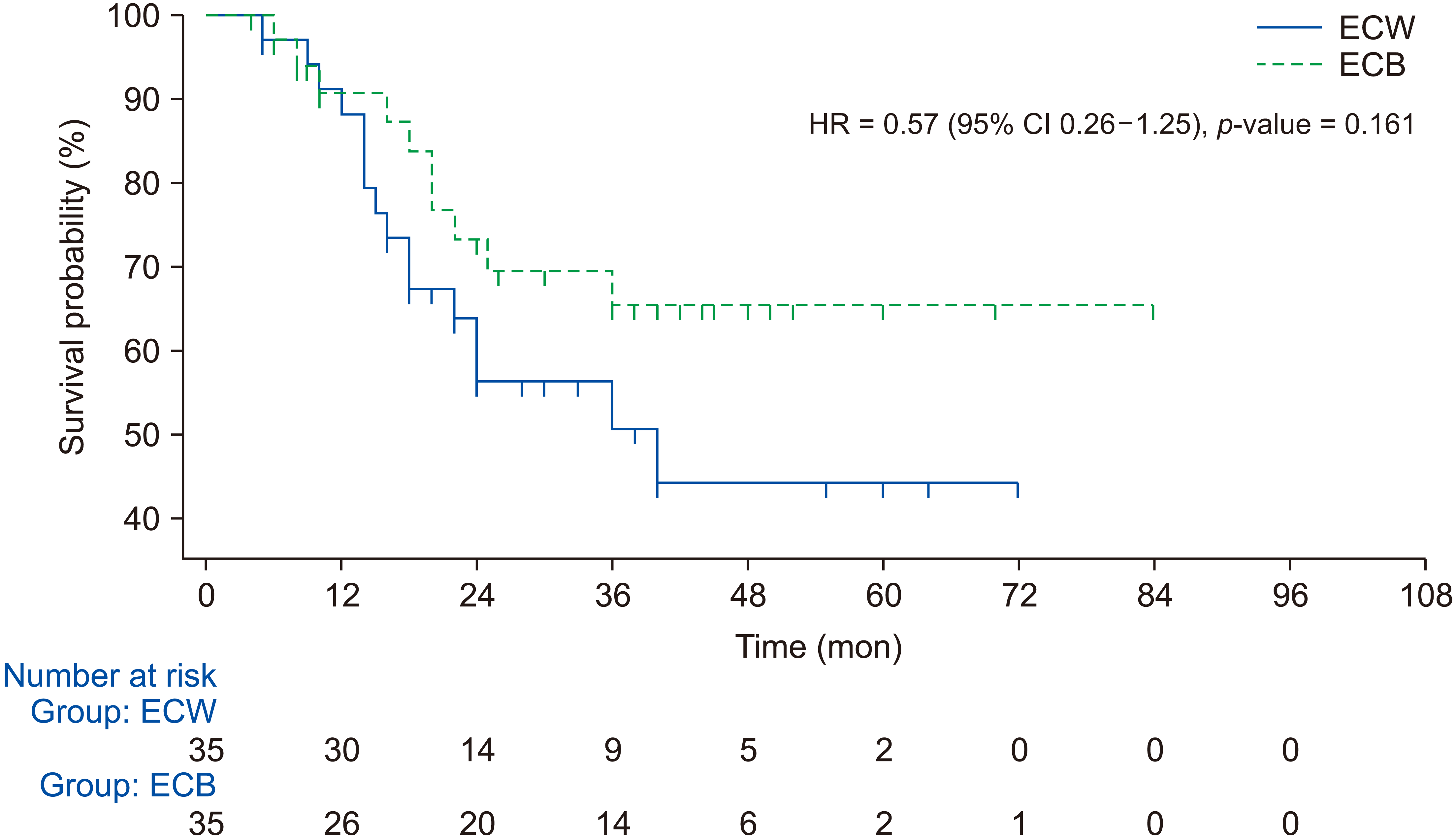 | Fig. 3Kaplan–Meier overall survival curves for ECB and ECW groups. ECB, extended cholecystectomy involving bi-segmentectomy s4b&5; ECW, extended cholecystectomy involving wedge hepatic resection; HR, hazard ratio; CI, confidence interval.
|
Table 3
Comparison of tumor involvement and histological types
![]()
Table 4
Cox proportional hazards analysis of prognostic factors for recurrence-free survival (matched population)
![]()
Table 5
Cox proportional hazards analysis of prognostic factors for overall survival (matched population)
![]()
Go to :
DISCUSSION
Anatomical location and drainage channels of the GB make segment 4b&5 of the liver most vulnerable to involvement by the GBC [1,15-18]. Previous studies have demonstrated that hepatic segment 4b&5 is the most common site of metastasis in early stage of GBC [7,17,18]. Shirai et al. [19,20] have demonstrated a correlation between the extent of microscopic angiolymphatic portal tract invasion and gross depth of hepatic invasion by the GBC. It has been established that patients with positive resection margins have poorer outcomes than patients with negative resection margins [20]. An adequate extent of hepatic resection to clear microscopic metastatic foci and to achieve R0 resection is necessary to improve the survival of patients with resectable GBC [20-23]. In the present study, liver margins from the tumor were larger in the ECB group than in the ECW group. The minimum macroscopic liver margin aimed for the ECW group was about 2 cm (20 mm). However, all patients had R0 resection.
In the present study, tumor locations were similarly distributed between the two groups. However, patient selection for either ECB or ECW was not preferably decided on the basis of tumor location. It was mainly based on the extent of hepatic infiltration and the possibility of R0 resection with offered resection. Either extended right hepatectomy or segment 4, 5, & 8 resection (whichever was appropriate to achieve R0 resection) was offered to patients with extensive hepatic infiltration or patients with tumor in close relation to the right anterior portal pedicle on CECT and such patients were excluded from this study.
In the present study, both disease-free survival and OS were longer for the ECB group, although differences between the two groups were not statistically significant. Similar results have been reported by two questionnaire surveys comparing ECB and ECW in patients with T2 GBC [9,10]. There were less surgical blood loss and postoperative complications in the ECB group than in the ECW group, reflecting more perfect control of portal pedicle in ECB [1]. However, other authors have reported more blood loss and bile leak with ECB [10]. The possible reason for significantly lower amount of blood loss in the ECB group might be a higher number of patients with laparoscopic approach in the ECB group, better portal pedicle control in the ECB group, and our practice of minimum use of Pringle’s manoeuvre during hepatic transection. Recurrence rate was similar in both groups. Sole hepatic involvement was not common. Therefore, other factors such as AJCC stage are more likely to be the cause of recurrence other than the type of hepatic recurrence if R0 resection has been achieved [1,9,10,17,24].
Adjuvant chemotherapy might have yielded a positive impact on our results, although the percentage of patients with adjuvant chemotherapy was similar in both groups [25,26]. Our study was prone to biases due to its retrospective nature. Therefore, we matched our patients for T and N stages. Laparoscopic control of intra-operative bleeding is a tedious and tiring process. A subconscious thought that an early control of portal pedicle could have better outcome for controlling bleeding might have biased us to perform ECB while attempting laparoscopic EC. The prevalence of laparoscopic procedures in the ECB group might have some effects on our results. However, we could not match our patients for the laparoscopic approach because very few patients in the ECW group were managed with this approach due to more frequent use of ECB with a laparoscopic approach. Similar results of laparoscopic and open surgical management of patients with GBC have been reported previously [27,28].
Our study may be criticised for not doing a subgroup analysis for patients with T2a and T2b GBC. As discussed earlier, we avoided this due to a high prevalence of IGBC in both groups as most of these patients with IGBC did not undergo a preoperative (pre-cholecystectomy) CECT necessary for this sub-classification.
In conclusion, the overall or recurrence free survival after ECB for T2 and T3 GBC was not significantly superior to that after ECW, although surgical blood loss and postoperative complications were lower following ECB.
Go to :
 XML Download