This article has been
cited by other articles in ScienceCentral.
Abstract
Unlike other aneurysms, blood blister-like aneurysms(BBA) occur at non-branching sites, are commonly located at the dorsal wall of the supraclinoid internal carotid artery (ICA), and are suspected to arise from dissecting aneurysms. They are typically diagnosed after a bleed because of their small size and unusual site. Therefore, lesions exist after brain computed tomography or magnetic resonance imaging, but they are often not found. In the case of BBA, which have a high risk of recurrence, the treatment is difficult, and selecting the treatment method is also challenging. This paper describes the treatment of a BBA with internal trapping of the ICA that eventually recurred despite performing coil embolization twice.
Go to :

Keywords: Blister, False aneurysm, Endovascular procedures
Blood blister-like aneurysms(BBAs)are found on the dorsal wall of the internal carotid artery (ICA) and occur uniquely where there are no branches.
1,
2 Although explanations for BBA located at different sites within intracranial circulation have been proposed, the initial and most classical definitions of BBA are limited to the ICA.
3,
4 They are particularly rare, comprising about 0.9–6.5% of all ICA aneurysms, 1% of all intracranial aneurysms, and 0.5 – 2% of ruptured aneurysms.
5–
7 They are usually diagnosed after bleeding because the first computed tomography (CT) angiography may miss them due to their small size. Since BBA assuming a saccular shape can be seen up to a few days after bleeding, special attention should be paid to radiological evaluation at different times, such as after rupture.
8
Nevertheless, the important point is the difficulty of diagnosis as well as the complexity of treatment. This is always challenging regardless of the treatment method. The extremely fragile wall of BBA makes management complex and dangerous. Here, we present a case of a BBA that had two recurrences despite endovascular coiling.
CASE
A 57-year-old female with a history of hypertension presented to the hospital emergency room with loss of consciousness. Upon arrival, the patient underwent a neurological examination, and the level of consciousness was stupor. As a result of brain CT, a distributed subarachnoid hemorrhage (SAH) was found in the basal cistern and sylvian fissure on both sides (
Fig. 1A). Upon taking cerebrovascular CT, a lesion showing outpouching in the right distal ICA was identified (
Fig. 1B).
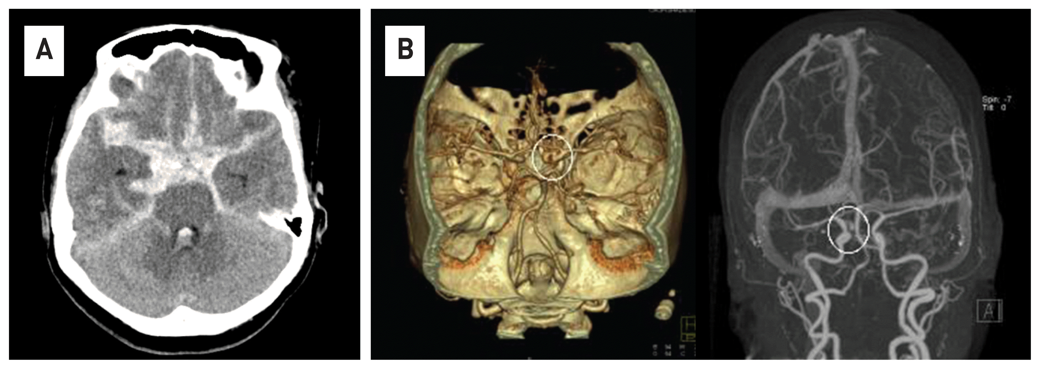 | Fig. 1Brain computed tomography(CT) scans show diffuse and thick subarachnoid hemorrhage in the basal cistern and both sylvian fissure (A). Contrast-enhanced CT angiography shows a cerebral aneurysm in the right distal ICA(white circle) (B). 
|
Immediately after diagnosis, transfemoral angiography was performed in the angiography room, and a BBA was identified from the right distal ICA toward the superior direction (
Fig. 2A). After checking the location and shape of the cerebral aneurysm, it was determined that endovascular coiling was possible, and the procedure was performed (
Fig. 2B, C). Coiling was performed with single open cell stent (Target® 360 detachable coils was used for coil, and a total of 5 coils were packed.) because no stenosis or dilation was found in other parts other than the dorsal wall of the right distal ICA. For the safety of vessel wall, the multiple stent technique was also considered, but it was not performed due to the high possibility of the thrombo-embolic complications during the procedure. Dual antiplatelet agents were used immediately after the procedure. In the case of BBA, the possibility of recurrence is very high, so a follow-up examination of the patient was performed 2 weeks later, and aneurysm regrowth was observed along with coil compaction (
Fig. 3A). It was determined that additional endovascular treatment was necessary, and the recurrence of the aneurysm sac was treated in the same way as before (
Fig. 3B). Additional Target® 360 detachable coils were packaged in the recurrent aneurysm sac, and additional open stents were deployed to prevent coil herniation. Then, 2 weeks later, an additional transfemoral angiography procedure was performed, and it was found that the aneurysm had recurred again (
Fig. 4A). It was determined that stent-assisted coiling was no longer effective, and other treatment methods were considered. Considering the endovascular trapping treatment method, the lesion-side ICA was blocked through balloon dilatation using a balloon catheter, and as a result of angiography of the opposite ICA, it was confirmed that the blood flow of the right side of the brain was sufficiently supplied through the anterior communicating artery, and the ICA on the lesion side was trapped. After the ICA was completely trapped with multiple Target® 360 detachable coils, follow-up angiography was performed on the ICA on the opposite side, and the procedure was terminated after confirming that sufficient blood flow was supplied to the brain on the side of the trapped ICA (
Fig. 4B, 4C). After the balloon test, the patient’s neurological symptoms should be checked, but the patient was unconscious at the time of this test. Therefore, inevitably, the procedure decision was determined only by 4-vesselimaging. And from this point on, the dual antiplatelet agent was stopped. The patient was discharged from the hospital about 2 months after visiting the emergency room, and there were no other neurological symptoms other than a slight decrease in muscle strength in the left upper limb at the time of discharge. Suspected cerebral infarction near the right motor cortex was observed on Brain CT taken 2 weeks after the second procedure, and the result of thrombo-embolic complications that occurred during three procedures is suspected (
Fig. 5).
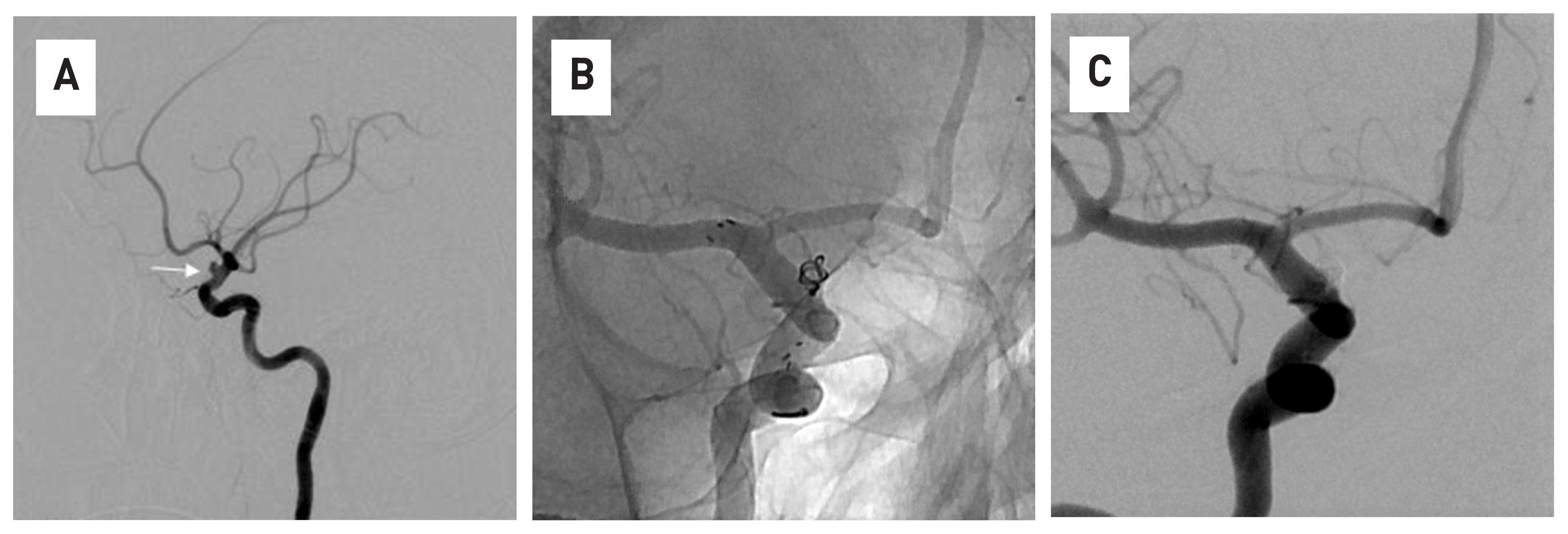 | Fig. 2In the 4-vesselangiography, a ruptured cerebral aneurysm of a saccular type is observed on the dorsal wall of distal Internal cerebral artery(ICA)(white arrow)(A). The stent was deployed from the proximal ICA to the proximal middle cerebral artery, and the microcatheter was placed in the sac of aneurysm, and then coiling was performed (B). Endovascular coiling was densely packed into the aneurysm sac, and the procedure was terminated after confirming that there was no contrast filling with an aneurysm sac anymore 
|
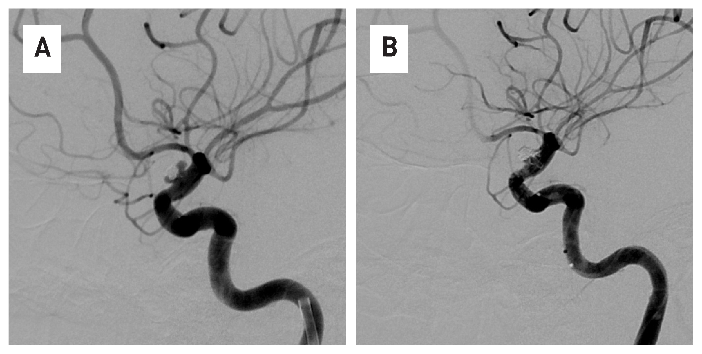 | Fig. 3The regrowth and coil compaction of aneurysm were observed in 4-vesselangiography two weeks after the first coil embolization (A). It was determined that additional treatment was necessary, endovascular coiling was performed on the recurrent aneurysm sac, and the procedure was terminated after checking the complete occlusion of the sac (B). 
|
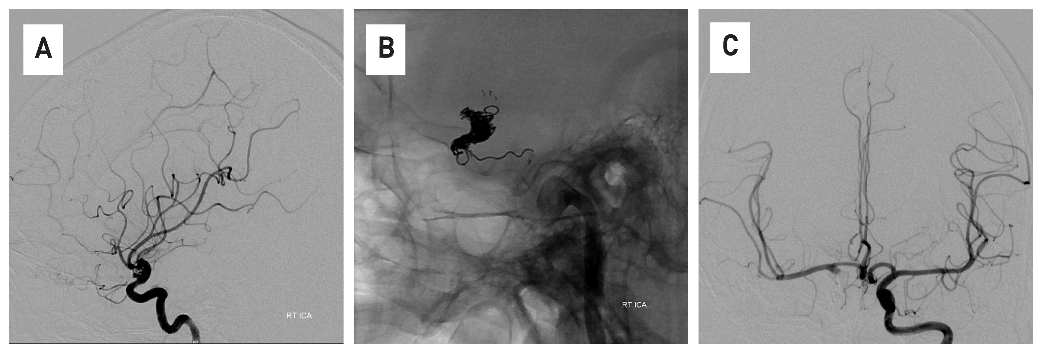 | Fig. 4The regrowth of cerebral aneurysm is observed again in 4-vesselangiography, which was performed two weeks after the additional procedure (A). Internal trapping treatment of distal internal cerebral artery was performed using coils (B). In the 4-vessel angiography conducted by the opposite internal cerebral artery, the flow of blood toward the lesion could be confirmed through anterior communicating artery (C). 
|
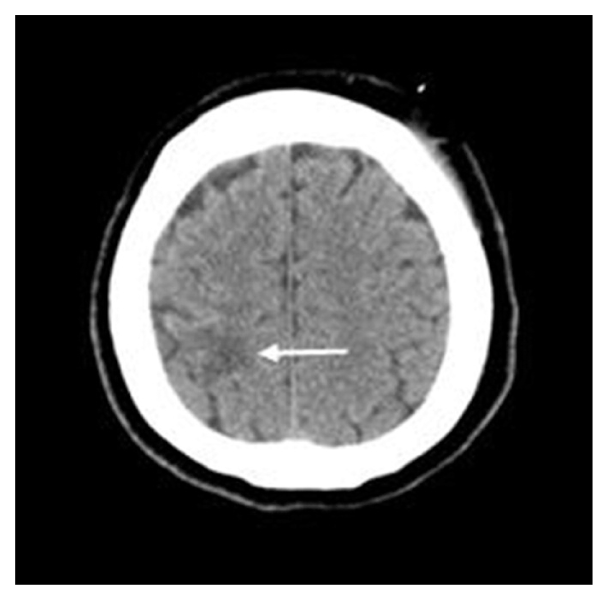 | Fig. 5After the second procedure, low density near the right motor cortex is observed and is thought to be a suspected cerebral infarction (white arrow). 
|
Go to :

DISCUSSION
An aneurysm located on the non-branched area on the dorsal wall of the supraclinoid portion of the ICA was first reported by Sundt et al.
9 in 1969. The extremely fragile wall of BBA makes the management complicated and risky. Various surgeries and endovascular treatments have been proposed for the treatment of BBA. As in this case, there is coil embolization using a stent, and a way to coil using several stents for the stability of the blood vessel wall. Surgical approach include a method of directly identifying a lesion and then clipping, and a method of bypass surgery after trapping the parent artery.
1,
2,
4 However, despite these various treatment efforts, BBA are very difficult to treat both surgically and endovascularly.
10 Pathological tests were performed on BBA, and a focal wall defect covered by thin layers of fibrous tissue and adventitia and a lack of collagenous layers were reported.
9 These unique characteristics of BBA lead to remarkable weaknesses in the walls and a high risk of premature rupture during surgery and intravascular procedures.
11 Moreover, the rapid increase in size at short intervals is probably related to the hemodynamic stress from the fragile walls and nonbranching site of BBA. These remarkable characteristics of BBA are associated with differences in treatment and prognosis from usual saccular aneurysms. These characteristics were likely the cause of the repeated recurrence in our case. The reason why we chose stent assisted coiling as the treatment of this patient was because of the low mobility and mortality of the procedure.
12 Difficulty in surgery increases the mortality rate of BBA. The mortality rate of SAH with a ruptured BBA is 57%.
1 As a result, in this case, the endovascular method of internal trapping was considered an effective choice of treatment, but in the case of internal trapping, the risk is higher than that of general endovascular techniques.
3–
6 It is difficult to choose the method of BBA treatment because none of the predictors of prognosis have been accurately identified. Stent assistance is generally recommended for the treatment of BBA with coil embolization.
3–
8 However, if recurrence occurs repeatedly as in this case, internal trapping is also considered a good treatment method.
Various endovascular treatments for aneurysms such as BBA have been documented. However, coil compaction with regrowth is very common after endovascular coiling of blister aneurysms. Therefore, it is thought that transfemoral angiography is necessary as a follow-up within a short period of time.
Go to :


 PDF
PDF Citation
 Print
Print


 XML Download