Journal List > Korean Circ J > v.51(12) > 1148451

Kim and Hwang: Vascular Calcification in Chronic Kidney Disease: Distinct Features of Pathogenesis and Clinical Implication

Author's summary

Vascular calcification (VC) is highly prevalent in patients with chronic kidney disease (CKD) and leads to increased cardiovascular morbidity and mortality. In patients with CKD, traditional factors do not fully explain the high prevalence of VC. This suggests that a CKD-specific pathobiology is involved in the development of VC and mounting evidence indicates that VC in CKD patients has distinct features of clinical presentation and that clinical implications are changed compared to those in the general population. In this review, we discuss the mechanism, diagnostic imaging modalities, clinical features and implications, and management of VC in patients with CKD.

Abstract

Chronic kidney disease (CKD) is associated with a higher prevalence of vascular calcification (VC) and cardiovascular disease. VC in CKD patients showed different pathophysiological features from those of the general population. The pathogenesis of VC in CKD is a highly organized process, and prior studies have suggested that patients with CKD have their own specific contributors to the phenotypic change of vascular smooth muscle cells (VSMCs), including uremic toxins, CKD-mineral and bone disease (CKD-MBD), inflammation, and oxidative stress. For the diagnosis and monitoring of VC in CKD, several imaging modalities, including plain radiography, ultrasound, and computed tomography have been utilized. VC in CKD patients has distinct clinical features and implications. CKD patients revealed a more intense and more prevalent calcification on the intimal and medial layers, whereas intimal calcification is predominantly observed in the general population. While a higher VC score is clearly associated with a higher risk of all-cause mortality and cardiovascular events, a greater VC score in CKD patients does not fully reflect the burden of atherosclerosis, because they have more calcification at equal volumes of atheromatous plaques. The primary goal of VC treatment in CKD is the prevention of VC progression, and the main management is to control the biochemical components of CKD-MBD. Cinacalcet and non-calcium-containing phosphate binders are the mainstay of VC prevention in CKD-MBD management. VC in patients with CKD is an ongoing area of research and is expected to advance soon.

INTRODUCTION

Vascular calcification (VC) is defined as mineral deposition in the vasculature in the form of calcium-phosphate complexes. Although VC is regarded as part of the normal aging process, certain pathological processes, such as diabetes, hypertension, chronic kidney disease (CKD), and rare hereditary disorders may precipitate the condition.1) VC causes a loss of arterial elasticity, increased pulse pressure and systolic blood pressure, and left ventricular hypertrophy, and the severity of VC is closely associated with an increased risk of cardiovascular morbidity and mortality.2)
VC has been observed more frequently in adults with CKD than in age-matched general population,3) and a similar trend is observed in younger CKD patients.4) VC in CKD patients was initially considered to occur only in end-stage renal disease; however, recent studies have shown that VC is also observed in the early stage of CKD.5) Considering that cardiovascular disease is the most common cause of death in CKD patients, it is important to understand the pathophysiology of VC in CKD patients to detect early signs of VC and prepare for appropriate management.6) In this review, we present a review of the mechanism, diagnostic imaging modalities, clinical manifestations and implications, and the available preventive management of VC in patients with CKD.

BIOLOGICAL MECHANISM OF VASCULAR CALCIFICATION IN CHRONIC KIDNEY DISEASE

Difference between chronic kidney disease and general population

CKD patients have a higher prevalence of VC than the general population, which contributes to the high risk of cardiovascular morbidity and mortality.7) In CKD patients, the traditional factors alone do not fully explain the high prevalence of VC. Although the precise underlying mechanisms of VC in CKD are not fully understood, previous studies have suggested that the CKD patients have specific contributors to the development of VC.8)9) Emerging evidence indicates that non-traditional risk factors including uremic toxins, CKD-mineral and bone disease (CKD-MBD), oxidative stress, and inflammation may contribute to the development of VC and cardiovascular disease in CKD patients.4)10)
It is well known that VC is a highly controlled process mainly regulated by the vascular smooth muscle cells (VSMCs).11) In the physiological conditions, VSMCs have a contractile phenotype and maintain the structural and functional homeostasis of the vessel wall. However, under pathological conditions, VSMCs transform into osteoblast-like cells and secrete collagenous extracellular matrix.12) In vitro studies have reported that serum derived from CKD subjects could induce calcification in human aortic smooth muscle cells. Under uremic conditions, the expression of markers of a contractile VSMC phenotype is downregulated, while those of a synthetic phenotype is upregulated.13)14) In patients with CKD, the VSMCs change their phenotype and proceed to VC through cellular proliferation, migration, and apoptosis.15)16)17) Other studies suggested that several factors relevant to CKD including uremic toxins, inflammatory molecules are associated with a phenotype switch and physiological function of VSMCs.16)18)

Burden of oxidative stress and inflammation

Oxidative stress is known to increase in correlation with declining renal function and increased oxidative stress may contribute to cardiovascular events in CKD patients.19) Since the kidney is one of the main sources of antioxidant enzymes, an impairment of the renal function is associated with decreased levels of these enzymes, and CKD patients have an impairment of antioxidant systems.20)21) Yamada et al.22) demonstrated that oxidative stress induced by uremic conditions influences the development of VC in CKD using an animal model. They also reported that antioxidant treatment ameliorated phenotypic changes in VSMCs, providing evidence for the clinical application of antioxidants to prevent VC in CKD patients. Huang et al.23) observed an elevation of oxidative stress in CKD patients and demonstrated a significant association between oxidative stress and VC. Their findings provide evidence supporting the pivotal role of oxidative stress in the phenotypic change of VSMCs in early CKD.
Inflammation also plays a critical role in the development of VC in CKD patients by inducing VSMC osteochondrogenic conversion.24)25) Previous studies have shown both systemic and local inflammation related to VC in CKD patients. Uremic toxins promote the development of an inflammatory state, and elevated plasma levels of inflammatory markers are observed in CKD patients. The levels of inflammatory markers are significantly associated with the prevalence of VC and cardiovascular events in CKD patients.26)27)28)29) Benz et al.30) demonstrated that the vessels obtained from early CKD patients showed an upregulation of inflammatory molecules. Other researchers also reported a more pronounced macrophage infiltration in coronary artery lesions of CKD.31) Mounting evidence indicates that inflammation triggers changes in the VSMCs and promotes the VC process by releasing calcified extracellular vesicles. It has been proposed that inflammation may activate the endoplasmic reticulum stress pathway, which is associated with increasing inorganic phosphate uptake and causes VSMC phenotypic transformation and mineral accumulation.32)

Chronic kidney disease-mineral and bone disease

CKD-MBD is a dysregulation of mineral and bone metabolism caused by CKD, which includes biochemical abnormalities of calcium, phosphate, vitamin D, parathyroid hormone (PTH), bone disorders, and calcification.33) Increased serum levels of calcium and phosphate are considered as the main risk factors for the development of VC in patients with CKD.34) Hypercalcemia was suggested to engage in VC by increasing Ca × P product and enhancing sodium-dependent phosphate cotransporters.35) Hyperphosphatemia is a common finding in CKD patients, which is caused by the reduction of renal excretion. It is assumed that the effects of hyperphosphatemia during VC development are primarily associated with sodium-dependent phosphate cotransporters.36) Previous studies have reported that elevated phosphate levels induce the expression of osteoblastic markers which stimulate the dysregulation of VSMCs. Within the vessel wall, increased levels of calcium and phosphate synergistically induce phenotypic changes in VSMCs.37)

Active process, not passive deposition of mineral

VC in CKD was initially considered to be a passive process by which minerals were deposited on the vessel wall by the dysregulation of calcium and phosphorus.38) However, recent evidence indicates that VC is an active and highly controlled process that involves the dysregulation and phenotypic transformation of VSMCs.10) In CKD patients, an increase in the calcification promoter level and a simultaneous decrease in the inhibitor level are associated with a high prevalence of VC. We previously investigated 90 iliac arteries obtained from the dialysis recipients at the time of transplantation to identify the characteristics of calcification.39) VC was observed in 77.8% of the participants using microscopic examination. Fetuin-A, an active calcific inhibitor, and C-reactive protein (CRP) stain was positive in 60.0% and 97.8% of patients, respectively (Figure 1). These findings indicate that the arteries in CKD patients significantly experience active calcification and a high rate of inflammation. We also identified a high correlation between a positive CRP stain score and calcification score, which suggests that local inflammation is closely related to calcification in the vessels of CKD patients.
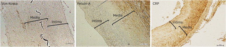
Figure 1

Immunohistochemistry of von Kossa, fetuin-A, and CRP in the iliac artery of kidney transplant recipients. Scale bar 100 μm. Original magnification×100.39)

CRP = C-reactive protein.
kcj-51-961-g001

DISCOVERY OF MULTI-FACTORS INVOLVED IN VASCULAR CALCIFICATION

Fibroblast growth factor-23–Klotho axis

Fibroblast growth factor (FGF)-23–Klotho axis is a recently identified new player in the field of CKD-MBD and is one of the most notable substances in the pathogenesis of VC in CKD patients. FGF-23 is a bone-derived hormone that regulates phosphate and vitamin D homeostasis and the expression of klotho.40) An excess of FGF-23 and klotho deficiency are closely associated with VC in CKD.10)41) In animal studies, despite the induction of CKD, transgenic mice with klotho overexpression showed better renal function and lesser calcification compared with wild-type uremic mice.42) Krishnasamy et al. 43) reported that elevated serum FGF-23 levels are associated with the progression of arterial stiffness and aortic calcification in patients with advanced CKD. Other studies also demonstrated that an increased serum FGF-23 concentration was an independent predictor of VC in CKD patients, and suggested that FGF-23 is an independent biomarker of VC in CKD patients.41)44)45)46) Some studies have reported the protective effect of FGF-23. Zhu et al. 47) showed the expression profiles of FGF-23 during VC and demonstrated that FGF-23 prevents VC through the activation of the ERK1/2 pathway. They suggested that FGF-23 could be a potential therapeutic strategy. However, the treatment effect of FGF-23 on VC remains controversial and further studies are required to clarify this.

Osteoprotegerin

Osteoprotegerin (OPG) is a key regulator of bone remodeling and is involved in the vascular system. It acts as a decoy receptor binding to the receptor activator of nuclear factor-kappa B ligand and inhibits its interaction with the receptor activator of nuclear factor-kappa B.48) Recently, the interest in the role of OPG in the pathogenesis of VC has increased in the general population. OPG knockout mice showed early onset osteoporosis and extensive aortic medial calcification, suggesting the role of OPG as an inhibitor of VC.49) However, paradoxically, OPG expression was upregulated in VC lesions, and several clinical studies reported a positive correlation between OPG concentrations and the severity of VC.50) An increase in the OPG levels may be a result of compensatory response, for the protection of further vascular damage.51)52) Morena et al.53) showed that the serum levels of OPG were significantly associated with coronary artery calcification in the CKD patients and increased along with decreased renal function. Other studies with CKD patients also reported significant correlations between serum OPG concentrations and VC.54)55) Based on prior studies, OPG has been suggested as a potential biomarker of VC, and related studies are ongoing.

Fetuin-A

Fetuin-A is a circulating inhibitor of calcification and has anti-inflammatory properties thus, it exhibits a protective role against atherosclerosis.56) It binds early calcium-phosphate crystals and reduces the crystal growth and deposition.57) The levels of circulating fetuin-A showed an inverse relationship with CRP, a representative marker of inflammation.56) Mice deficient in fetuin-A develop prominent soft tissue calcification, indicating its role as a systemic inhibitor of ectopic calcification.58) In dialysis patients, the serum fetuin-A levels were decreased and negatively correlated with aortic and valvular calcification, calciphylaxis, and cardiovascular mortality.59)60) Recently, fetuin-mineral complex (FMC) has received attention in the pathogenesis of VC in CKD. The centrifugation of serum from rats with renal failure yielded a complex of fetuin-A and minerals; this complex apparently reflects the extra-osseous calcification stress.61) Hamano et al.62) investigated the clinical relevance of FMC in hemodialysis patients and suggested that the quantitative measurement of FMC, not supernatant or serum total fetuin-A, reflects extraosseous calcification stress. Further studies are required to validate these findings.

DIAGNOSTIC IMAGING OF VASCULAR CALCIFICATION IN CHRONIC KIDNEY DISEASE

Comparison of multiple imaging modality

A variety of imaging modalities have been utilized in the diagnosis and monitoring of VC in CKD patients.63) Plain radiographs are mainly used to assess the calcification of large vessels. Atherosclerosis and calcification of the abdominal aorta can be evaluated using lateral abdominal radiography. Kauppila et al.64) suggested a simple scoring method to quantify VC in the abdominal aorta, which showed a significant correlation with arterial stiffness measured by the pulse wave velocity and coronary artery calcification measured by computed tomography (CT).65)66) The effectiveness of this scoring method has been validated not only in patients with mild to moderate renal dysfunction but also in patients with end-stage renal disease on dialysis.66) However, in our prior study using the iliac artery, no significant correlation was observed between radiologic and microscopic calcification. In addition, microscopic calcification was observed in 77.9% of patients without radiologic calcification. These findings raise the question of how sensitively plain radiographs can detect calcification.39) Despite these disadvantages, the information obtained from plain radiographs has the advantage that it is relatively easy to provide clinical relevance, considering that calcification observed on plain radiographs is significantly associated with cardiovascular events and mortality.67)68) Furthermore, plain radiography is widely used because it is inexpensive and easily available.
Ultrasound is suitable for evaluating large superficial vessels, such as the carotid and femoral arteries.69) Previous studies reported that calcified carotid plaques detected by ultrasound were significantly associated with the traditional risk factors of cardiovascular disease 70)71)72) and was an independent risk factor for cardiovascular events in CKD patients.73) Ultrasound has limitations that are operator dependent, subjective, and it is difficult to recognize the difference between intimal and medial calcifications. However, ultrasound could be a convenient alternative for VC evaluation because it can examine each vessel in detail without radiation exposure, detect the presence and extent of VC, and measure the pulse wave velocity, which is an index of arterial compliance and stiffness.63)69)74) Furthermore, in patients with CKD, ultrasound is often performed before dialysis vascular access creation. It provides information on the vascular status and calcification and assists preoperative planning and vascular mapping.75)76)
CT is more sensitive and objective for VC evaluation than other imaging modalities. Several studies have shown that the quantification of VC by CT has a high sensitivity and specificity and has a good prognostic value for cardiovascular events.63)77) The clinical significance of coronary artery and aortic calcification detected by CT is well established. Agatston et al.78) suggested a scoring system to quantify coronary artery calcification, which predicts cardiovascular events with high sensitivity and specificity. CT is considered as a reference standard for assessing VC because it accurately detects and quantifies VC. Furthermore, CT has the advantage of monitoring the progression of VC over a period of time and obtaining additional information such as valvular calcification and bone density.79) The limitations of CT lie in the high risk of radiation exposure, high cost, and low accessibility.80)81) In addition, because it is difficult to distinguish between intimal and medial calcification, its use for risk stratification is limited in CKD patients. Some studies have suggested reducing this limitation through a combination of biomarkers and other risk factors. Further studies are needed in this field.4)82)83)

Screening of vascular calcification

The Kidney Disease Improving Global Outcome (KDIGO) work group recommends screening for VC in CKD patients using either lateral abdominal radiography or CT.84) At the time of this review, there were several controversies regarding the benefit of screening for VC in CKD patients. Although the association between the VC and poor cardiovascular outcomes in CKD patients is well established, the impact of risk factor modifications through screening is not clear and there is no proper treatment for improving the clinical outcomes. Therefore, the KDIGO work group weakly recommends VC screening in patients with CKD.

CLINICAL PRESENTATION OF VASCULAR CALCIFICATION IN CHRONIC KIDNEY DISEASE

Intimal vascular calcification

Intimal VC is closely associated with atherosclerotic plaque formation.85) Traditional cardiovascular risk factors, including older age, smoking, hypertension, diabetes, obesity and hyperlipidemia provokes the formation of atherosclerotic plaques, and intimal calcification occurs in the plaque as a late event in atherosclerosis (Figure 2A). CKD patients already have a number of traditional risk factors for cardiovascular disease, and atherosclerotic arterial lesions are more prevalent in CKD patients.86)87)88) In addition, the progressive decline of renal function leads to the further progression of atherosclerosis.89)90) Consequently, the burden of atherosclerosis is considerably increased in CKD patients, and excess calcium and phosphate precipitates facilitate the process of intimal calcification and a higher frequency of intimal calcification.91)92)
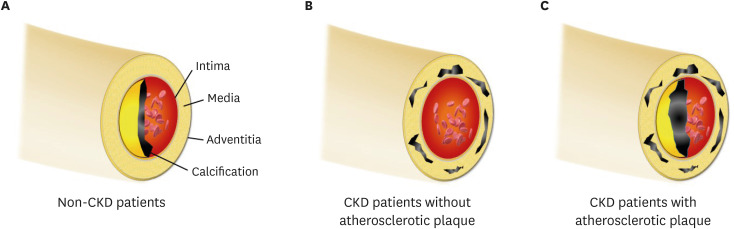
Figure 2

(A) In patients without renal dysfunction, calcification is deposited in the intimal layer; (B) CKD patients obtain the medial calcification in the absence of the atherosclerotic factor, because mineral bone disorder accelerates the calcific process; (C) In clinical practice, intimal and medial calcification frequently co-exists in CKD patients, because they already had several traditional risk factors. Notably, the intimal or medial calcific burden is greater in CKD patients than in non-CKD patients.

CKD = chronic kidney disease.
kcj-51-961-g002

Medial vascular calcification

Medial calcification theoretically appears to be more specific to CKD patients than intimal calcification, considering the pathogenic similarity between bone ossification and medial calcification.93)94) Hypercalcemia, hyperphosphatemia, secondary hyperparathyroidism and other CKD-MBD-related factors trigger mineral deposition on the medial layer of the vascular wall; also, the formation of micro-calcific foci accelerates the accumulation of additional calcification.18 Therefore, medial VC can frequently occur in CKD, despite the absence of traditional risk factors (Figure 2B).95) Arterial medial calcifications are not directly related to cholesterol deposits and do not induce occlusive lesions in the lumen. However, medial calcification is associated with an increased pulse pressure, arterial stiffness, poor vascular compliance and left ventricular hypertrophy.85)96) These changes lead to an increased rate of future cardiovascular events and mortality risk in the CKD population.97)

Which type of vascular calcification plays leading roles in chronic kidney disease?

The intimal and medial VCs represent different clinical entities with distinct pathogenic signatures and it would also appear that the basic pathogenic principles remain relevant in the CKD population (intimal calcification - atherosclerosis and medial calcification - loss of the cushioning function). However, the CKD patients retain common risk profiles and signaling pathways, which overlap in intimal and medial calcifications.30) In particular, oxidative and inflammatory signaling are the common pathways for both types of VC, and uremic conditions considerably activate these two pathways.98)99) Therefore, intimal and medial VC in CKD patients can frequently occur at the same time and several studies have reported the co-existence of the two types of VC (Figure 2C).100)

CLINICAL IMPLICATION OF VASCULAR CALCIFICATION IN CHRONIC KIDNEY DISEASE

Different presentation of vascular calcification in different chronic kidney disease setting

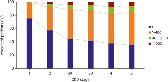
Lower renal function increases the burden of VC, and its prevalence is substantially increased in CKD patients compared to patients without renal impairment.101)102)103) In non-dialysis-dependent CKD, VC is observed with a variable prevalence range around 30% to 60% patients.104) There is an inverse relationship between estimated glomerular filtration rate (eGFR) and VC score independent of traditional risk factors (Figure 3).105)106) In addition, CKD patients with a greater severity of coronary calcification score are expected to show a more rapid progression of calcification.107)108)
Figure 3

The prevalence of coronary artery calcification in different CKD stages. KSN News Factsheet Vol. 15 (Sep. 2019).

CKD = chronic kidney disease; KSN = Korean Society of Nephrology.
kcj-51-961-g003
The prevalence and progression of VC increase dramatically, if CKD progression arrives at the latest stages. It has been reported that VC is detected in more than 50% of patients receiving dialysis treatment, and they have a 15–30% annual increase in coronary calcification.109)110)111) In our previous studies, it was also found that 40–45% of hemodialysis patients showed VC, which was detected in the aortic area using plain chest radiography.96)112)113) Notably, we found a strikingly increased prevalence of VC in the microscopic examination of the iliac artery compared to those on plain radiography of the aorta.39) The detected intimal calcification using von Kossa stain was observed in 62% of dialysis patients and medial calcification in 74% of the patients, while radiologic calcification was detected in 24% of the patients. These findings suggest that the actual prevalence of VC may be higher than previously reported.
Renal transplant recipients gain recovery of renal function from the successful engraftment of donor kidney. The restoration of the renal function allows for the elimination of multi-factors and toxins that play a role in promoting VCs. In addition, it is expected that CKD-MBD resolves spontaneously, because secondary hyperparathyroidism and impaired phosphate excretion disappear. Several studies have investigated whether VC progresses or regresses after successful renal transplantation and found that medial arterial calcification appeared to slow the progression. However, most renal transplant recipients take over the calcification trait from the dialysis days and these are not spontaneously regressed.114) Indeed, the transferred calcification from dialysis acts as strong predictor for the progression of calcification after transplantation and coronary arterial calcification progresses as approximately 10% increase/year after transplantation.115)116)117) In addition, secondary hyperparathyroidism continues in 40% of renal transplant recipients and persistent CKD-MBD is a significant determinant of progression of VC after transplantation.118)119)120) The current data support that all types of VC (intimal and medial) is gradually deteriorated after renal transplantation.

Cardiovascular consideration of vascular calcification

Cardiovascular events are the most common cause of death in CKD patients and VC is currently established as an uncontroversial risk marker of incident cardiovascular event.121)122)123) The presence of VC irrespective of the site is associated with a several-fold increase in the risk of cardiovascular morbidity and mortality in the entire spectrum of CKD patients and renal transplant recipients.124)125) The KDIGO international clinical practice guideline suggests that CKD patients with VC need to be considered as having the highest risk of incident cardiovascular events.126) In addition, VC is closely related to functional disorders of the cardiovascular system in CKD patients. Intradialytic hypotension is the most common hemodynamic dysfunction in hemodialysis patients and we found that VC is independently associated with a higher risk of intradialytic hypotension.113) The significant synergistic interaction between VC and intradialytic hypotension was also observed in the prediction of all-cause mortality and cardiovascular events, suggesting that the prognostic significance of VC is linked to the functional impairment in the hemodynamic system.
In the general population, the coronary arterial calcification score correlates well with the presence of atherosclerotic plaques. Nonetheless, the association between the severity of calcification and impaired perfusion is not strong in CKD patients.127) A recent report showed that the coronary artery calcification showed a weak relationship with myocardial ischemia, suggesting that coronary artery calcification may not be a good reflection of the atherosclerotic burden in CKD patients.128) As the calcification deposits in CKD patients are more active in the medial layer than those in patients with a normal renal function, the coupling power between the calcification score and ischemia becomes weaker. The anatomic pathology of atheromatous plaques provides additional constructive evidence. Previous studies on direct pathologic examination found that the volume of atheromatous plaque was not the main distinguishing factor of atherosclerosis between CKD and non-CKD patients. The differences originate in the composition of the plaque and calcific content (Figure 2A and C).31)129)130) CKD patients have more intense calcification on the intimal layer with additional medial calcification, which may not allow the VC score to fully reflect the atherosclerotic burden.
We reasonably question how reliable calcification is a predictor of coronary atherosclerotic events, not the overall cardiovascular mortality and survival rate. Undoubtedly, the dialysis patients with higher coronary artery calcification scores are significantly associated with coronary atheroma.131)132) In addition, higher calcification scores clearly predict the incidence of atherosclerotic coronary disease in CKD patients. However, the cutoff value of calcification score seems to be higher than non-CKD patients.133) Further studies are needed to determine the extent of calcification for the risk stratification of atherosclerotic events in CKD patients. The development of a new diagnostic modality to differentiate intimal and medial calcifications would be highly useful for atherosclerotic risk assessment of VC in CKD patients.

Non-cardiovascular consideration of vascular calcification

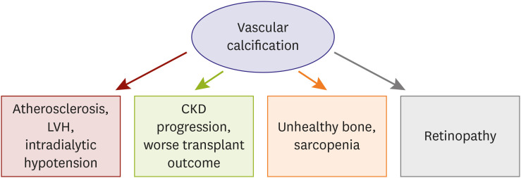
CKD patients are considerably exposed to calcification-prone conditions and any arterial system in our body presents calcific deposition. VC also evokes functional impairment in large and small arteries and causes problems in the non-cardiovascular system (Figure 4). We previously evaluated the relationship between VC and retinopathy, based on this plausible hypothesis.134) We found that retinopathy was significantly associated with the presence of aortic calcification, suggesting that large vessel calcification is linked with microvascular dysfunction. Furthermore, the combination of retinopathy and VC was stronger in predicting renal function decline than retinopathy alone. These findings support the concept that VC has prognostic implications in non-cardiovascular organs. The effect of VC on the renal outcomes was also associated with renal transplant recipients. Intimal calcification was significantly associated with a faster decline in the renal allograft function and a lower graft survival rate.135) The deposition of fetuin-A, a key regulator of calcific process, further reduces the allograft survival rate and is an independent predictor of renal graft function decline.
Figure 4

Clinical connection of vascular calcification to cardiovascular or non-cardiovascular organs in CKD.

CKD = chronic kidney disease; LVH = left ventricular hypertrophy.
kcj-51-961-g004
Secondary hyperparathyroidism and CKD-MBD are powerful inducers of VC in patients with CKD and it serves as a connecting link to bone health status. Several clinical studies have demonstrated that VC is associated with bone histomorphometry, low bone activity, low bone volume, and abnormal bone turnover in dialysis patients, suggesting an interplay between the bone health and VC.136)137)138) Notably, these studies consistently show that the mineralization in bone and vessel counterworks, while VC and bone mineralization share common pathophysiologic processes.139)140) CKD-MBD leads to bone resorption, decreased bone mineralization, and increased calcium efflux from bones promoting a positive calcium balance to vascular mineralization and calcification. This inverse relationship has been extended to recent research, which shows that VC is independently associated with incident fractures in dialysis patients.141)142)
It has been reported that increased adipose tissue volume and low muscle mass are positively associated with prevalent aortic and coronary calcification.143)144) Studies on CKD patients showed similar trend. Lee et al. 145) analyzed the nationwide prospective cohort of CKD in Korea and reported that normal body mass index with central obesity has increased risk of coronary artery calcification, and adipose tissue volume has a significant correlation with VC. 146)147) In addition, the prevalence of low muscle mass is increased as CKD is more advanced 148) and sarcopenia in hemodialysis patients shows an independent relationship with VC.149) These results suggest that pathogenesis of obesity, sarcopenia and VC may involve some shared mechanism, and inflammation is potential common pathway of them.

MANAGEMENT OF VASCULAR CALCIFICATION IN CHRONIC KIDNEY DISEASE

Significance of chronic kidney disease-mineral and bone disease management in vascular calcification

VC is rarely observed to regress, and the primary goals of management are the prevention and stabilization of the existing calcifications. The optimal management of CKD-MBD is a fundamental key in determining the progression of VC, and the presence of an effective prevention method is the distinct differences in CKD compared to those of other metabolic conditions. The biochemical components of CKD-MBD are serum calcium, phosphate, and PTH levels, and nephrologists attempt to control all of these components using dialysis treatment and medications.150)151) The achievement of CKD-MBD parameters in the target range significantly reduced the risk of VC, cardiovascular mortality and all-cause death (Figure 5).92)
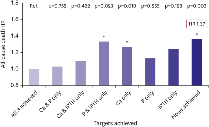
Figure 5

Target achievement of the CKD-MBD parameters and mortality risk in dialysis patients. KSN News Factsheet Vol. 18 (May 2020).

Ca = calcium; CKD-MBD = chronic kidney disease-mineral and bone disease; HR = hazard ratio; KSN = Korean Society of Nephrology; P = phosphate; iPTH = intact parathyroid hormone.
kcj-51-961-g005

PTH suppressing agents

Increased PTH levels are associated with deteriorated clinical outcomes in CKD patients, and treatment strategies focus primarily on the reduction of PTH levels. However, lower levels of PTH are also associated with an increased risk of mortality, and PTH should be controlled in the target range, which is different between CKD stages.126) Calcimimetics, paricalcitol, and active vitamin D are the three major agents for PTH reduction. Active vitamin D suppresses the proliferation of parathyroid cells; however, it also increases the absorption of calcium and phosphate from vitamin D receptors in the gastrointestinal tract. Therefore, the administration of vitamin D at therapeutic doses increased the serum calcium and phosphate levels, despite of PTH reduction. This phenomenon promotes the precipitation of calcium and phosphate products and enhances the progression of VC. The K/DOQI clinical practice guidelines recommend that serum calcium and phosphate should be monitored regularly with the use of active vitamin D compounds. Some experimental studies demonstrated that the therapeutic doses of vitamin D supplementation had protective effects against CKD-stimulated VC and low circulating levels of vitamin D was associated with greater calcification score.152)153) However, patients with higher level of vitamin D also revealed advanced VC and there is a lack of clinical studies on the bimodal association between vitamin D supplementation and VC, particularly in a prospective randomized design. Therefore, additional evidence is required to translate the bimodal effect of vitamin D into clinical practice.
Paricalcitol is a synthetic vitamin D analogue that works on the parathyroid gland more specifically than on non-parathyroid organs and showed greater activity in PTH reduction than other PTH suppressing agents.154) The higher selectivity of paricalcitol on the parathyroid gland reduces hypercalcemia and hyperphosphatemia. In addition, the dialysis patients receiving paricalcitol have a significant survival advantage compared to those receiving standard forms of active vitamin D compounds.155) Nevertheless, paricalcitol retains the mineral absorption effects on the gastrointestinal tract and is less effective in preventing the progression of VC.156) In contrast, calcimimetics act only on the calcium sensing receptor of the parathyroid gland and the serum calcium level is moderately decreased through PTH suppression. Therefore, of all the PTH suppressing agents, cinacalcet showed the best effectiveness in preventing VC and can reduce the risk of major adverse cardiovascular events and bone fracture.157)158)159)

Phosphate binder

CKD patients exhibit an impaired renal excretion of phosphate, and dialysis treatment does not fully eliminate the phosphate to the extent that it maintains normal the serum phosphate levels. Therefore, phosphate binders are usually administered to manage hyperphosphatemia in the advanced stages of CKD. The phosphate binders are classified into calcium-or non-calcium-containing regimens and these two types of phosphate binders show different results in the prevention of VC. Non-calcium-containing phosphate binders are more effective in avoiding a positive calcium balance in blood vessels; additionally, this benefit is translated into clinical practice. In dialysis patients, the non-calcium-containing phosphate binders interrupt the progression of VC and the protective effect against VC is more prominent in the patients with preexisting VC.160)161) In addition, there is some evidence that non-calcium-containing phosphate binders improved the survival rate in non-dialysis-dependent CKD and dialysis patients.162)163) Consequently, recent guidelines suggest that calcium-containing phosphate binders should be restricted and that non-calcium-containing regimens are preferred in the presence of VC.84)

Warfarin

Warfarin, a vitamin K antagonist, is used clinically as an oral anticoagulant for the management of atherothrombotic cardiovascular disorders. In CKD patients, the prevalence of atrial fibrillation is higher than in non-CKD patients, and the risk of major adverse cardiovascular outcomes is synergistically increased in the coexistence of CKD and atrial fibrillation.135) Therefore, the necessity of anticoagulation is increased in CKD patients. However, warfarin promotes calcification because it suppresses γ-carboxylation of the matrix Gla protein, an inhibitor of calcification. A number of studies implicate warfarin treatment in the development of VCs, and the high prevalence of vitamin K deficiency in CKD patients may potentiate the calcification-inducible effect of warfarin.164)165)166) Notably, the clinical benefit of warfarin is also disputed in CKD patients. It has been report that the incidence of stroke was even higher in dialysis patients on warfarin than in those on aspirin, clopidogrel, or no treatment.167) In non-dialysis-dependent CKD, warfarin treatment was observed to increase the risk of bleeding.168)
The adverse effects of warfarin on VC question whether the attractive options of new oral anticoagulants are valid in CKD patients to minimize VC and other adverse effects. In an experimental model of atherosclerosis, rivaroxaban showed a greater effect on the prevention of VC when compared to warfarin.169) Several human studies demonstrated that new oral anticoagulants lead to slow down the progression of calcification compared to warfarin, suggesting the benefit of new oral anticoagulants in clinical practice.170)171)172)173) In a recently published study on hemodialysis patients, switching from a vitamin K antagonist to rivaroxaban also revealed favorable effects in reducing the risk of life-threatening and major bleeding. However, rivaroxaban with vitamin K2 supplementation showed no significant protection against VC progression in hemodialysis patients.174) The authors suggest that VC already progressed in studied participants to the extent that intervention treatment is ineffective and that the use of rivaroxaban at earlier stages of CKD might be beneficial in preventing VC. Therefore, we need additional evidence to opt for the newly developed anticoagulants over warfarin in patients with CKD.

CONCLUSION

In this review, we showed that VC in CKD is a highly organized process, and that uremic toxins, CKD-MBD, inflammation, and oxidative stress have contributed to the development of VC. Several factors involved in the pathogenesis of VC in CKD have been discovered and suggested as biomarkers, but further studies are needed. The clinical presentation of VC in patients with CKD also differs from that in the general population. The prevalence of VC and rate of VC progression change with different CKD status. The association between a higher VC score and greater burden of atherosclerosis is modified in CKD patients, and the prognostic significance of VC is linked to non-cardiovascular organs. In addition, the management of VC is more focused on CKD-MBD, not atherosclerosis, indicating that the clinical implications of VC are different. We anticipate that this updated review, ranging from the pathophysiology to clinical implications, will help to improve understanding VC in CKD patients.

Notes

Funding: This work was supported by grants from Kyung Hee University in 2020 (KHU-20201235) and was supported by grants from the Patient-Centered Clinical Research Coordinating Center, which is funded by the Ministry of Health & Welfare, Republic of Korea (H19C0481, HC19C0041).

Conflict of Interest: The authors have no financial conflicts of interest.

Data Sharing Statement: The data generated in this study is available from the corresponding author upon reasonable request.

Author Contributions:

  • Conceptualization: Kim JS, Hwang HS.

  • Methodology: Hwang HS.

  • Supervision: Hwang HS.

  • Visualization: Kim JS, Hwang HS.

  • Writing - original draft: Kim JS, Hwang HS.

  • Writing - review & editing: Kim JS, Hwang HS.

References

1. Leopold JA. Vascular calcification: an age-old problem of old age. Circulation. 2013; 127:2380–2382. PMID: 23690467.
2. Demer LL, Tintut Y. Vascular calcification: pathobiology of a multifaceted disease. Circulation. 2008; 117:2938–2948. PMID: 18519861.
3. Braun J, Oldendorf M, Moshage W, Heidler R, Zeitler E, Luft FC. Electron beam computed tomography in the evaluation of cardiac calcification in chronic dialysis patients. Am J Kidney Dis. 1996; 27:394–401. PMID: 8604709.
4. Goodman WG, Goldin J, Kuizon BD, et al. Coronary-artery calcification in young adults with end-stage renal disease who are undergoing dialysis. N Engl J Med. 2000; 342:1478–1483. PMID: 10816185.
crossref
5. Fang Y, Ginsberg C, Sugatani T, Monier-Faugere MC, Malluche H, Hruska KA. Early chronic kidney disease-mineral bone disorder stimulates vascular calcification. Kidney Int. 2014; 85:142–150. PMID: 23884339.
crossref
6. Mizobuchi M, Towler D, Slatopolsky E. Vascular calcification: the killer of patients with chronic kidney disease. J Am Soc Nephrol. 2009; 20:1453–1464. PMID: 19478096.
crossref
7. Rennenberg RJ, Kessels AG, Schurgers LJ, van Engelshoven JM, de Leeuw PW, Kroon AA. Vascular calcifications as a marker of increased cardiovascular risk: a meta-analysis. Vasc Health Risk Manag. 2009; 5:185–197. PMID: 19436645.
crossref
8. Fang Y, Ginsberg C, Seifert M, et al. CKD-induced wingless/integration1 inhibitors and phosphorus cause the CKD-mineral and bone disorder. J Am Soc Nephrol. 2014; 25:1760–1773. PMID: 24578135.
crossref
9. Zhan JK, Wang YJ, Wang Y, et al. The protective effect of GLP-1 analogue in arterial calcification through attenuating osteoblastic differentiation of human VSMCs. Int J Cardiol. 2015; 189:188–193. PMID: 25897902.
crossref
10. Hénaut L, Chillon JM, Kamel S, Massy ZA. Updates on the mechanisms and the care of cardiovascular calcification in chronic kidney disease. Semin Nephrol. 2018; 38:233–250. PMID: 29753400.
crossref
11. Metz RP, Patterson JL, Wilson E. Vascular smooth muscle cells: isolation, culture, and characterization. Methods Mol Biol. 2012; 843:169–176. PMID: 22222531.
crossref
12. Durham AL, Speer MY, Scatena M, Giachelli CM, Shanahan CM. Role of smooth muscle cells in vascular calcification: implications in atherosclerosis and arterial stiffness. Cardiovasc Res. 2018; 114:590–600. PMID: 29514202.
crossref
13. Monroy MA, Fang J, Li S, et al. Chronic kidney disease alters vascular smooth muscle cell phenotype. Front Biosci (Landmark Ed). 2015; 20:784–795. PMID: 25553479.
crossref
14. Madsen M, Aarup A, Albinsson S, et al. Uremia modulates the phenotype of aortic smooth muscle cells. Atherosclerosis. 2017; 257:64–70. PMID: 28107707.
crossref
15. Patidar A, Singh DK, Thakur S, Farrington K, Baydoun AR. Uremic serum-induced calcification of human aortic smooth muscle cells is a regulated process involving Klotho and RUNX2. Biosci Rep. 2019; 39:BSR20190599. PMID: 31519772.
crossref
16. Cazaña-Pérez V, Cidad P, Donate-Correa J, et al. Phenotypic modulation of cultured primary human aortic vascular smooth muscle cells by uremic serum. Front Physiol. 2018; 9:89. PMID: 29483881.
crossref
17. Patidar A, Singh DK, Winocour P, Farrington K, Baydoun AR. Human uraemic serum displays calcific potential in vitro that increases with advancing chronic kidney disease. Clin Sci (Lond). 2013; 125:237–245. PMID: 23464884.
crossref
18. Moradi H, Sica DA, Kalantar-Zadeh K. Cardiovascular burden associated with uremic toxins in patients with chronic kidney disease. Am J Nephrol. 2013; 38:136–148. PMID: 23941724.
crossref
19. Cottone S, Lorito MC, Riccobene R, et al. Oxidative stress, inflammation and cardiovascular disease in chronic renal failure. J Nephrol. 2008; 21:175–179. PMID: 18446711.
20. Kelly D, Rothwell PM. Disentangling the multiple links between renal dysfunction and cerebrovascular disease. J Neurol Neurosurg Psychiatry. 2020; 91:88–97. PMID: 31511306.
crossref
21. Johnson-Davis KL, Fernelius C, Eliason NB, Wilson A, Beddhu S, Roberts WL. Blood enzymes and oxidative stress in chronic kidney disease: a cross sectional study. Ann Clin Lab Sci. 2011; 41:331–339. PMID: 22166502.
22. Yamada S, Taniguchi M, Tokumoto M, et al. The antioxidant tempol ameliorates arterial medial calcification in uremic rats: important role of oxidative stress in the pathogenesis of vascular calcification in chronic kidney disease. J Bone Miner Res. 2012; 27:474–485. PMID: 21987400.
crossref
23. Huang M, Zheng L, Xu H, et al. Oxidative stress contributes to vascular calcification in patients with chronic kidney disease. J Mol Cell Cardiol. 2020; 138:256–268. PMID: 31866376.
crossref
24. Podkowińska A, Formanowicz D. Chronic kidney disease as oxidative stress- and inflammatory-mediated cardiovascular disease. Antioxidants. 2020; 9:752.
crossref
25. Bover J, Evenepoel P, Ureña-Torres P, et al. Pro: cardiovascular calcifications are clinically relevant. Nephrol Dial Transplant. 2015; 30:345–351. PMID: 25712934.
crossref
26. Kiu Weber CI, Duchateau-Nguyen G, Solier C, et al. Cardiovascular risk markers associated with arterial calcification in patients with chronic kidney disease Stages 3 and 4. Clin Kidney J. 2014; 7:167–173. PMID: 24683472.
crossref
27. Benz K, Hilgers KF, Daniel C, Amann K. Vascular calcification in chronic kidney disease: the role of inflammation. Int J Nephrol. 2018; 2018:4310379. PMID: 30186632.
crossref
28. Choi SR, Lee YK, Cho AJ, et al. Malnutrition, inflammation, progression of vascular calcification and survival: Inter-relationships in hemodialysis patients. PLoS One. 2019; 14:e0216415. PMID: 31048884.
crossref
29. Hénaut L, Massy ZA. New insights into the key role of interleukin 6 in vascular calcification of chronic kidney disease. Nephrol Dial Transplant. 2018; 33:543–548. PMID: 29420799.
crossref
30. Benz K, Varga I, Neureiter D, et al. Vascular inflammation and media calcification are already present in early stages of chronic kidney disease. Cardiovasc Pathol. 2017; 27:57–67. PMID: 28171827.
crossref
31. Gross ML, Meyer HP, Ziebart H, et al. Calcification of coronary intima and media: immunohistochemistry, backscatter imaging, and x-ray analysis in renal and nonrenal patients. Clin J Am Soc Nephrol. 2007; 2:121–134. PMID: 17699396.
crossref
32. Masuda M, Miyazaki-Anzai S, Levi M, Ting TC, Miyazaki M. PERK-eIF2α-ATF4-CHOP signaling contributes to TNFα-induced vascular calcification. J Am Heart Assoc. 2013; 2:e000238. PMID: 24008080.
crossref
33. Moe SM, Drüeke T, Lameire N, Eknoyan G. Chronic kidney disease-mineral-bone disorder: a new paradigm. Adv Chronic Kidney Dis. 2007; 14:3–12. PMID: 17200038.
crossref
34. Block GA, Klassen PS, Lazarus JM, Ofsthun N, Lowrie EG, Chertow GM. Mineral metabolism, mortality, and morbidity in maintenance hemodialysis. J Am Soc Nephrol. 2004; 15:2208–2218. PMID: 15284307.
crossref
35. Yang H, Curinga G, Giachelli CM. Elevated extracellular calcium levels induce smooth muscle cell matrix mineralization in vitro. Kidney Int. 2004; 66:2293–2299. PMID: 15569318.
36. Giachelli CM. The emerging role of phosphate in vascular calcification. Kidney Int. 2009; 75:890–897. PMID: 19145240.
crossref
37. Jono S, McKee MD, Murry CE, et al. Phosphate regulation of vascular smooth muscle cell calcification. Circ Res. 2000; 87:E10–E17. PMID: 11009570.
crossref
38. Jono S, Shioi A, Ikari Y, Nishizawa Y. Vascular calcification in chronic kidney disease. J Bone Miner Metab. 2006; 24:176–181. PMID: 16502129.
crossref
39. Hwang HS, Lim SW, Sun IO, et al. Clinical significance of preexisting microcalcification in the iliac artery in renal transplant recipients. Transplantation. 2015; 99:811–817. PMID: 25211516.
crossref
40. Quarles LD. Role of FGF23 in vitamin D and phosphate metabolism: implications in chronic kidney disease. Exp Cell Res. 2012; 318:1040–1048. PMID: 22421513.
crossref
41. Scialla JJ, Xie H, Rahman M, et al. Fibroblast growth factor-23 and cardiovascular events in CKD. J Am Soc Nephrol. 2014; 25:349–360. PMID: 24158986.
crossref
42. Hu MC, Shi M, Zhang J, et al. Klotho deficiency causes vascular calcification in chronic kidney disease. J Am Soc Nephrol. 2011; 22:124–136. PMID: 21115613.
crossref
43. Krishnasamy R, Tan SJ, Hawley CM, et al. Progression of arterial stiffness is associated with changes in bone mineral markers in advanced CKD. BMC Nephrol. 2017; 18:281. PMID: 28870151.
crossref
44. Baralić M, Brković V, Stojanov V, et al. Dual roles of the mineral metabolism disorders biomarkers in prevalent hemodilysis patients: in renal bone disease and in vascular calcification. J Med Biochem. 2019; 38:134–144. PMID: 30867641.
crossref
45. Desjardins L, Liabeuf S, Renard C, et al. FGF23 is independently associated with vascular calcification but not bone mineral density in patients at various CKD stages. Osteoporos Int. 2012; 23:2017–2025. PMID: 22109743.
crossref
46. Morena M, Jaussent I, Halkovich A, et al. Bone biomarkers help grading severity of coronary calcifications in non dialysis chronic kidney disease patients. PLoS One. 2012; 7:e36175. PMID: 22567137.
crossref
47. Zhu D, Mackenzie NC, Millan JL, Farquharson C, MacRae VE. A protective role for FGF-23 in local defence against disrupted arterial wall integrity? Mol Cell Endocrinol. 2013; 372:1–11. PMID: 23523568.
crossref
48. Boyce BF, Xing L. Functions of RANKL/RANK/OPG in bone modeling and remodeling. Arch Biochem Biophys. 2008; 473:139–146. PMID: 18395508.
crossref
49. Bucay N, Sarosi I, Dunstan CR, et al. Osteoprotegerin-deficient mice develop early onset osteoporosis and arterial calcification. Genes Dev. 1998; 12:1260–1268. PMID: 9573043.
50. Kiechl S, Werner P, Knoflach M, Furtner M, Willeit J, Schett G. The osteoprotegerin/RANK/RANKL system: a bone key to vascular disease. Expert Rev Cardiovasc Ther. 2006; 4:801–811. PMID: 17173497.
crossref
51. Kaden JJ, Bickelhaupt S, Grobholz R, et al. Receptor activator of nuclear factor kappaB ligand and osteoprotegerin regulate aortic valve calcification. J Mol Cell Cardiol. 2004; 36:57–66. PMID: 14734048.
52. Van Campenhout A, Golledge J. Osteoprotegerin, vascular calcification and atherosclerosis. Atherosclerosis. 2009; 204:321–329. PMID: 19007931.
crossref
53. Morena M, Jaussent I, Dupuy AM, et al. Osteoprotegerin and sclerostin in chronic kidney disease prior to dialysis: potential partners in vascular calcifications. Nephrol Dial Transplant. 2015; 30:1345–1356. PMID: 25854266.
crossref
54. Mesquita M, Demulder A, Damry N, et al. Plasma osteoprotegerin is an independent risk factor for mortality and an early biomarker of coronary vascular calcification in chronic kidney disease. Clin Chem Lab Med. 2009; 47:339–346. PMID: 19676147.
crossref
55. Mikami S, Hamano T, Fujii N, et al. Serum osteoprotegerin as a screening tool for coronary artery calcification score in diabetic pre-dialysis patients. Hypertens Res. 2008; 31:1163–1170. PMID: 18716364.
crossref
56. Heiss A, DuChesne A, Denecke B, et al. Structural basis of calcification inhibition by alpha 2-HS glycoprotein/fetuin-A. Formation of colloidal calciprotein particles. J Biol Chem. 2003; 278:13333–13341. PMID: 12556469.
57. Cai MM, Smith ER, Holt SG. The role of fetuin-A in mineral trafficking and deposition. Bonekey Rep. 2015; 4:672. PMID: 25987986.
crossref
58. Schäfer C, Heiss A, Schwarz A, et al. The serum protein alpha 2-Heremans-Schmid glycoprotein/fetuin-A is a systemically acting inhibitor of ectopic calcification. J Clin Invest. 2003; 112:357–366. PMID: 12897203.
59. Ketteler M, Bongartz P, Westenfeld R, et al. Association of low fetuin-A (AHSG) concentrations in serum with cardiovascular mortality in patients on dialysis: a cross-sectional study. Lancet. 2003; 361:827–833. PMID: 12642050.
crossref
60. Reynolds JL, Skepper JN, McNair R, et al. Multifunctional roles for serum protein fetuin-a in inhibition of human vascular smooth muscle cell calcification. J Am Soc Nephrol. 2005; 16:2920–2930. PMID: 16093453.
crossref
61. Matsui I, Hamano T, Mikami S, et al. Fully phosphorylated fetuin-A forms a mineral complex in the serum of rats with adenine-induced renal failure. Kidney Int. 2009; 75:915–928. PMID: 19190677.
crossref
62. Hamano T, Matsui I, Mikami S, et al. Fetuin-mineral complex reflects extraosseous calcification stress in CKD. J Am Soc Nephrol. 2010; 21:1998–2007. PMID: 20947626.
crossref
63. Disthabanchong S, Boongird S. Role of different imaging modalities of vascular calcification in predicting outcomes in chronic kidney disease. World J Nephrol. 2017; 6:100–110. PMID: 28540199.
crossref
64. Kauppila LI, Polak JF, Cupples LA, Hannan MT, Kiel DP, Wilson PW. New indices to classify location, severity and progression of calcific lesions in the abdominal aorta: a 25-year follow-up study. Atherosclerosis. 1997; 132:245–250. PMID: 9242971.
crossref
65. Raggi P, Bellasi A, Ferramosca E, Islam T, Muntner P, Block GA. Association of pulse wave velocity with vascular and valvular calcification in hemodialysis patients. Kidney Int. 2007; 71:802–807. PMID: 17311068.
crossref
66. Bellasi A, Ferramosca E, Muntner P, et al. Correlation of simple imaging tests and coronary artery calcium measured by computed tomography in hemodialysis patients. Kidney Int. 2006; 70:1623–1628. PMID: 16955104.
crossref
67. Okuno S, Ishimura E, Kitatani K, et al. Presence of abdominal aortic calcification is significantly associated with all-cause and cardiovascular mortality in maintenance hemodialysis patients. Am J Kidney Dis. 2007; 49:417–425. PMID: 17336703.
crossref
68. Verbeke F, Van Biesen W, Honkanen E, et al. CORD Study Investigators. Prognostic value of aortic stiffness and calcification for cardiovascular events and mortality in dialysis patients: outcome of the calcification outcome in renal disease (CORD) study. Clin J Am Soc Nephrol. 2011; 6:153–159. PMID: 20829424.
crossref
69. Bellasi A, Raggi P. Techniques and technologies to assess vascular calcification. Semin Dial. 2007; 20:129–133. PMID: 17374086.
70. Guérin AP, London GM, Marchais SJ, Metivier F. Arterial stiffening and vascular calcifications in end-stage renal disease. Nephrol Dial Transplant. 2000; 15:1014–1021. PMID: 10862640.
crossref
71. Oh DJ. Continuous ambulatory peritoneal dialysis patients show high prevalence of carotid artery calcification which is associated with a higher left ventricular mass index. J Korean Med Sci. 2005; 20:848–852. PMID: 16224161.
crossref
72. Sumida Y, Nakayama M, Nagata M, et al. Carotid artery calcification and atherosclerosis at the initiation of hemodialysis in patients with end-stage renal disease. Clin Nephrol. 2010; 73:360–369. PMID: 20420796.
crossref
73. Nakayama M, Ura Y, Nagata M, et al. Carotid artery calcification at the initiation of hemodialysis is a risk factor for cardiovascular events in patients with end-stage renal disease: a cohort study. BMC Nephrol. 2011; 12:56. PMID: 21999942.
crossref
74. Disthabanchong S. Vascular calcification in chronic kidney disease: Pathogenesis and clinical implication. World J Nephrol. 2012; 1:43–53. PMID: 24175241.
crossref
75. Lima A, Carrilho P, Germano A. Clinical and ultrasound evaluation for hemodialysis access creation. Nefrologia (Engl Ed). 2021.
crossref
76. Shenoy S, Darcy M. Ultrasound as a tool for preoperative planning, monitoring, and interventions in dialysis arteriovenous access. AJR Am J Roentgenol. 2013; 201:W539–W543. PMID: 24059390.
crossref
77. Dube P, DeRiso A, Patel M, et al. Vascular calcification in chronic kidney disease: diversity in the vessel wall. Biomedicines. 2021; 9:404. PMID: 33917965.
crossref
78. Agatston AS, Janowitz WR, Hildner FJ, Zusmer NR, Viamonte M Jr, Detrano R. Quantification of coronary artery calcium using ultrafast computed tomography. J Am Coll Cardiol. 1990; 15:827–832. PMID: 2407762.
crossref
79. Karohl C, D'Marco Gascón L, Raggi P. Noninvasive imaging for assessment of calcification in chronic kidney disease. Nat Rev Nephrol. 2011; 7:567–577. PMID: 21862991.
crossref
80. De Mauri A, Brambilla M, Chiarinotti D, Matheoud R, Carriero A, De Leo M. Estimated radiation exposure from medical imaging in hemodialysis patients. J Am Soc Nephrol. 2011; 22:571–578. PMID: 21355057.
crossref
81. Marinelli A, Pistolesi V, Pasquale L, et al. Diagnosis of arterial media calcification in chronic kidney disease. Cardiorenal Med. 2013; 3:89–95. PMID: 23922548.
crossref
82. Mehrotra R, Budoff M, Hokanson JE, Ipp E, Takasu J, Adler S. Progression of coronary artery calcification in diabetics with and without chronic kidney disease. Kidney Int. 2005; 68:1258–1266. PMID: 16105059.
crossref
83. Poli FE, Gulsin GS, McCann GP, Burton JO, Graham-Brown MP. The assessment of coronary artery disease in patients with end-stage renal disease. Clin Kidney J. 2019; 12:721–734. PMID: 31583096.
crossref
84. Kidney Disease: Improving Global Outcomes (KDIGO) CKD-MBD Update Work Group. KDIGO 2017 clinical practice guideline update for the diagnosis, evaluation, prevention, and treatment of chronic kidney disease-mineral and bone disorder (CKD-MBD). Kidney Int Suppl (2011). 2017; 7:1–59. PMID: 30675420.
85. Reiss AB, Miyawaki N, Moon J, et al. CKD, arterial calcification, atherosclerosis and bone health: inter-relationships and controversies. Atherosclerosis. 2018; 278:49–59. PMID: 30253289.
crossref
86. Zoccali C. Traditional and emerging cardiovascular and renal risk factors: an epidemiologic perspective. Kidney Int. 2006; 70:26–33. PMID: 16723985.
crossref
87. Kim Y, Cho JS, Cho WK, et al. Retinopathy and left ventricular hypertrophy in patients with chronic kidney disease: Interrelationship and impact on clinical outcomes. Int J Cardiol. 2017; 249:372–376. PMID: 29121742.
crossref
88. Hwang HS, Choi YA, Kim SY, et al. Diabetes retinopathy is a poor predictor for renal and cardiovascular outcomes in comparison with hypertensive retinopathy in patients with chronic kidney disease. Diabetes Res Clin Pract. 2015; 109:312–318. PMID: 26008724.
crossref
89. Drüeke TB, Massy ZA. Atherosclerosis in CKD: differences from the general population. Nat Rev Nephrol. 2010; 6:723–735. PMID: 20978469.
crossref
90. Nakano T, Ninomiya T, Sumiyoshi S, et al. Association of kidney function with coronary atherosclerosis and calcification in autopsy samples from Japanese elders: the Hisayama study. Am J Kidney Dis. 2010; 55:21–30. PMID: 19765871.
crossref
91. Nakamura S, Ishibashi-Ueda H, Niizuma S, Yoshihara F, Horio T, Kawano Y. Coronary calcification in patients with chronic kidney disease and coronary artery disease. Clin J Am Soc Nephrol. 2009; 4:1892–1900. PMID: 19833908.
crossref
92. Oh KH, Kang M, Kang E, et al. The KNOW-CKD Study: What we have learned about chronic kidney diseases. Kidney Res Clin Pract. 2020; 39:121–135. PMID: 32550711.
crossref
93. Goodman WG, London G, Amann K, et al. Vascular calcification in chronic kidney disease. Am J Kidney Dis. 2004; 43:572–579. PMID: 14981617.
crossref
94. Moe SM, O'Neill KD, Duan D, et al. Medial artery calcification in ESRD patients is associated with deposition of bone matrix proteins. Kidney Int. 2002; 61:638–647. PMID: 11849407.
crossref
95. Milliner DS, Zinsmeister AR, Lieberman E, Landing B. Soft tissue calcification in pediatric patients with end-stage renal disease. Kidney Int. 1990; 38:931–936. PMID: 2266678.
crossref
96. Hwang HS, Cho JS, Hong YA, et al. Vascular calcification and left ventricular hypertrophy in hemodialysis patients: interrelationship and clinical impacts. Int J Med Sci. 2018; 15:557–563. PMID: 29725245.
crossref
97. London GM, Marchais SJ, Guérin AP, Métivier F. Arteriosclerosis, vascular calcifications and cardiovascular disease in uremia. Curr Opin Nephrol Hypertens. 2005; 14:525–531. PMID: 16205470.
crossref
98. Lee HY, Lim S, Park S. Role of inflammation in arterial calcification. Korean Circ J. 2021; 51:114–125. PMID: 33525066.
crossref
99. Mori D, Matsui I, Shimomura A, et al. Protein carbamylation exacerbates vascular calcification. Kidney Int. 2018; 94:72–90. PMID: 29716796.
crossref
100. Voelkl J, Cejka D, Alesutan I. An overview of the mechanisms in vascular calcification during chronic kidney disease. Curr Opin Nephrol Hypertens. 2019; 28:289–296. PMID: 30985336.
crossref
101. Merjanian R, Budoff M, Adler S, Berman N, Mehrotra R. Coronary artery, aortic wall, and valvular calcification in nondialyzed individuals with type 2 diabetes and renal disease. Kidney Int. 2003; 64:263–271. PMID: 12787418.
crossref
102. Kramer H, Toto R, Peshock R, Cooper R, Victor R. Association between chronic kidney disease and coronary artery calcification: the Dallas Heart Study. J Am Soc Nephrol. 2005; 16:507–513. PMID: 15601745.
crossref
103. Lee W, Yoon YE, Kwon O, et al. Evaluation of coronary artery calcium progression in asymptomatic individuals with an initial score of zero. Korean Circ J. 2019; 49:448–457. PMID: 30808082.
crossref
104. Bover J, Aguilar A, Arana C, et al. Clinical approach to vascular calcification in patients with non-dialysis dependent chronic kidney disease: mineral-bone disorder-related aspects. Front Med (Lausanne). 2021; 8:642718. PMID: 34095165.
crossref
105. Budoff MJ, Rader DJ, Reilly MP, et al. CRIC Study Investigators. Relationship of estimated GFR and coronary artery calcification in the CRIC (Chronic Renal Insufficiency Cohort) Study. Am J Kidney Dis. 2011; 58:519–526. PMID: 21783289.
crossref
106. Górriz JL, Molina P, Cerverón MJ, et al. Vascular calcification in patients with nondialysis CKD over 3 years. Clin J Am Soc Nephrol. 2015; 10:654–666. PMID: 25770175.
crossref
107. Kestenbaum BR, Adeney KL, de Boer IH, Ix JH, Shlipak MG, Siscovick DS. Incidence and progression of coronary calcification in chronic kidney disease: the Multi-Ethnic Study of Atherosclerosis. Kidney Int. 2009; 76:991–998. PMID: 19692998.
crossref
108. Kronmal RA, McClelland RL, Detrano R, et al. Risk factors for the progression of coronary artery calcification in asymptomatic subjects: results from the Multi-Ethnic Study of Atherosclerosis (MESA). Circulation. 2007; 115:2722–2730. PMID: 17502571.
109. Reynolds JL, Joannides AJ, Skepper JN, et al. Human vascular smooth muscle cells undergo vesicle-mediated calcification in response to changes in extracellular calcium and phosphate concentrations: a potential mechanism for accelerated vascular calcification in ESRD. J Am Soc Nephrol. 2004; 15:2857–2867. PMID: 15504939.
crossref
110. Daniel WT, Weber C, Bailey JA, Raggi P, Sharma J. Prospective analysis of coronary calcium in patients on dialysis undergoing a near-total parathyroidectomy. Surgery. 2013; 154:1315–1321. PMID: 24238050.
crossref
111. Savage T, Clarke AL, Giles M, Tomson CR, Raine AE. Calcified plaque is common in the carotid and femoral arteries of dialysis patients without clinical vascular disease. Nephrol Dial Transplant. 1998; 13:2004–2012. PMID: 9719155.
crossref
112. Schlieper G, Aretz A, Verberckmoes SC, et al. Ultrastructural analysis of vascular calcifications in uremia. J Am Soc Nephrol. 2010; 21:689–696. PMID: 20203159.
crossref
113. Kim SY, Hong YA, Yoon HE, et al. Vascular calcification and intradialytic hypotension in hemodialysis patients: Clinical relevance and impact on morbidity and mortality. Int J Cardiol. 2016; 217:156–160. PMID: 27183451.
crossref
114. Alappan HR, Vasanth P, Manzoor S, O'Neill WC. Vascular calcification slows but does not regress after kidney transplantation. Kidney Int Rep. 2020; 5:2212–2217. PMID: 33305114.
crossref
115. Schankel K, Robinson J, Bloom RD, et al. Determinants of coronary artery calcification progression in renal transplant recipients. Am J Transplant. 2007; 7:2158–2164. PMID: 17640315.
crossref
116. Bargnoux AS, Dupuy AM, Garrigue V, et al. Evolution of coronary artery calcifications following kidney transplantation: relationship with osteoprotegerin levels. Am J Transplant. 2009; 9:2571–2579. PMID: 19775319.
crossref
117. Mazzaferro S, Pasquali M, Taggi F, et al. Progression of coronary artery calcification in renal transplantation and the role of secondary hyperparathyroidism and inflammation. Clin J Am Soc Nephrol. 2009; 4:685–690. PMID: 19211668.
crossref
118. Messa P, Sindici C, Cannella G, et al. Persistent secondary hyperparathyroidism after renal transplantation. Kidney Int. 1998; 54:1704–1713. PMID: 9844148.
crossref
119. Lou I, Foley D, Odorico SK, et al. How well does renal transplantation cure hyperparathyroidism? Ann Surg. 2015; 262:653–659. PMID: 26366545.
crossref
120. Mathur A, Sutton W, Ahn JB, et al. Association between treatment of secondary hyperparathyroidism and posttransplant outcomes. Transplantation. 2021.
crossref
121. Hwang HS, Kim JS, Kim YG, et al. Circulating PCSK9 level and risk of cardiovascular events and death in hemodialysis patients. J Clin Med. 2020; 9:9.
crossref
122. Hwang HS, Kim JS, Kim YG, et al. Circulating neprilysin level predicts the risk of cardiovascular events in hemodialysis patients. Front Cardiovasc Med. 2021; 8:684297. PMID: 34212014.
crossref
123. Cho IJ, Chang HJ, Lee SE, Shim CY, Hong GR, Chung N. Prognostic application of thoracic aortic calcium scoring for adverse clinical outcome risk in elderly patients with left ventricular hypertrophy. Korean Circ J. 2017; 47:918–928. PMID: 29035431.
crossref
124. Chen J, Budoff MJ, Reilly MP, et al. Coronary artery calcification and risk of cardiovascular disease and death among patients with chronic kidney disease. JAMA Cardiol. 2017; 2:635–643. PMID: 28329057.
crossref
125. Nelson AJ, Raggi P, Wolf M, Gold AM, Chertow GM, Roe MT. Targeting vascular calcification in chronic kidney disease. JACC Basic Transl Sci. 2020; 5:398–412. PMID: 32368697.
crossref
126. Kidney Disease: Improving Global Outcomes (KDIGO) CKD-MBD Work Group. KDIGO clinical practice guideline for the diagnosis, evaluation, prevention, and treatment of Chronic Kidney Disease-Mineral and Bone Disorder (CKD-MBD). Kidney Int Suppl. 2009; S1–S130.
127. Dilsizian V, Gewirtz H, Marwick TH, et al. Cardiac imaging for coronary heart disease risk stratification in chronic kidney disease. JACC Cardiovasc Imaging. 2021; 14:669–682. PMID: 32828780.
128. Herzog CA, Natwick T, Li S, Charytan DM. Comparative utilization and temporal trends in cardiac stress testing in U.S. Medicare beneficiaries with and without chronic kidney disease. JACC Cardiovasc Imaging. 2019; 12:1420–1426. PMID: 29909107.
129. Mathew RO, Bangalore S, Lavelle MP, et al. Diagnosis and management of atherosclerotic cardiovascular disease in chronic kidney disease: a review. Kidney Int. 2017; 91:797–807. PMID: 28040264.
130. Schwarz U, Buzello M, Ritz E, et al. Morphology of coronary atherosclerotic lesions in patients with end-stage renal failure. Nephrol Dial Transplant. 2000; 15:218–223. PMID: 10648668.
crossref
131. Sharples EJ, Pereira D, Summers S, et al. Coronary artery calcification measured with electron-beam computerized tomography correlates poorly with coronary artery angiography in dialysis patients. Am J Kidney Dis. 2004; 43:313–319. PMID: 14750097.
crossref
132. Haydar AA, Hujairi NM, Covic AA, Pereira D, Rubens M, Goldsmith DJ. Coronary artery calcification is related to coronary atherosclerosis in chronic renal disease patients: a study comparing EBCT-generated coronary artery calcium scores and coronary angiography. Nephrol Dial Transplant. 2004; 19:2307–2312. PMID: 15213315.
crossref
133. Yiu KH, de Graaf FR, van Velzen JE, et al. Different value of coronary calcium score to predict obstructive coronary artery disease in patients with and without moderate chronic kidney disease. Neth Heart J. 2013; 21:347–353. PMID: 23579986.
crossref
134. Hwang HS, Kim SY, Hong YA, et al. Clinical impact of coexisting retinopathy and vascular calcification on chronic kidney disease progression and cardiovascular events. Nutr Metab Cardiovasc Dis. 2016; 26:590–596. PMID: 27089976.
crossref
135. Hwang HS, Park MW, Yoon HE, et al. Clinical significance of chronic kidney disease and atrial fibrillation on morbidity and mortality in patients with acute myocardial infarction. Am J Nephrol. 2014; 40:345–352. PMID: 25358406.
crossref
136. Naves M, Rodríguez-García M, Díaz-López JB, Gómez-Alonso C, Cannata-Andía JB. Progression of vascular calcifications is associated with greater bone loss and increased bone fractures. Osteoporos Int. 2008; 19:1161–1166. PMID: 18180973.
crossref
137. London GM, Marty C, Marchais SJ, Guerin AP, Metivier F, de Vernejoul MC. Arterial calcifications and bone histomorphometry in end-stage renal disease. J Am Soc Nephrol. 2004; 15:1943–1951. PMID: 15213285.
crossref
138. Lee SM, Kim SE, Lee JY, Jeong HJ, Son YK, An WS. Serum myostatin levels are associated with abdominal aortic calcification in dialysis patients. Kidney Res Clin Pract. 2019; 38:481–489. PMID: 31537054.
crossref
139. Osako MK, Nakagami H, Koibuchi N, et al. Estrogen inhibits vascular calcification via vascular RANKL system: common mechanism of osteoporosis and vascular calcification. Circ Res. 2010; 107:466–475. PMID: 20595654.
140. Viegas C, Araújo N, Marreiros C, Simes D. The interplay between mineral metabolism, vascular calcification and inflammation in Chronic Kidney Disease (CKD): challenging old concepts with new facts. Aging (Albany NY). 2019; 11:4274–4299. PMID: 31241466.
crossref
141. Nam YJ, Hwang SY, Kim DW, Kim D, Shin SJ, Yoon HE. Sex-specific relationship between vascular calcification and incident fracture in patients with end-stage renal disease. Kidney Res Clin Pract. 2020; 39:344–355. PMID: 32522894.
crossref
142. An WS. Sex hormones impact vascular calcification and fracture in dialysis patients. Kidney Res Clin Pract. 2020; 39:236–238. PMID: 32958722.
crossref
143. Jensky NE, Criqui MH, Wright CM, Wassel CL, Alcaraz JE, Allison MA. The association between abdominal body composition and vascular calcification. Obesity (Silver Spring). 2011; 19:2418–2424. PMID: 21475146.
crossref
144. Ko BJ, Chang Y, Jung HS, et al. Relationship between low relative muscle mass and coronary artery calcification in healthy adults. Arterioscler Thromb Vasc Biol. 2016; 36:1016–1021. PMID: 27034471.
crossref
145. Lee MJ, Park JT, Park KS, et al. Normal body mass index with central obesity has increased risk of coronary artery calcification in Korean patients with chronic kidney disease. Kidney Int. 2016; 90:1368–1376. PMID: 27884313.
crossref
146. Paoli S, Mitsnefes MM. Coronary artery calcification and cardiovascular disease in children with chronic kidney disease. Curr Opin Pediatr. 2014; 26:193–197. PMID: 24632542.
crossref
147. Cordeiro AC, Qureshi AR, Lindholm B, et al. Visceral fat and coronary artery calcification in patients with chronic kidney disease. Nephrol Dial Transplant. 2013; 28(Suppl 4):iv152–iv159. PMID: 23832273.
crossref
148. Moreno-Gonzalez R, Corbella X, Mattace-Raso F, et al. Prevalence of sarcopenia in community-dwelling older adults using the updated EWGSOP2 definition according to kidney function and albuminuria: the Screening for CKD among Older People across Europe (SCOPE) study. BMC Geriatr. 2020; 20(Suppl 1):327. PMID: 33008317.
crossref
149. Kim SH, Choi G, Song Y, et al. Low muscle mass in patients receiving hemodialysis: correlations with vascular calcification and vascular access failure. J Clin Med. 2021; 10:10.
crossref
150. Pérez-Ricart A, Galicia-Basart M, Comas-Sugrañes D, Cruzado-Garrit JM, Segarra-Medrano A, Montoro-Ronsano JB. Long-term effectiveness of cinacalcet in non-dialysis patients with chronic kidney disease and secondary hyperparathyroidism. Kidney Res Clin Pract. 2019; 38:229–238. PMID: 31022778.
crossref
151. Park EJ, Jung SW, Kim DR, et al. Conversion from acetate dialysate to citrate dialysate in a central delivery system for maintenance hemodialysis patients. Kidney Res Clin Pract. 2019; 38:100–107. PMID: 30754934.
crossref
152. Shroff R, Egerton M, Bridel M, et al. A bimodal association of vitamin D levels and vascular disease in children on dialysis. J Am Soc Nephrol. 2008; 19:1239–1246. PMID: 18337484.
crossref
153. Mathew S, Lund RJ, Chaudhary LR, Geurs T, Hruska KA. Vitamin D receptor activators can protect against vascular calcification. J Am Soc Nephrol. 2008; 19:1509–1519. PMID: 18448587.
crossref
154. Ketteler M, Martin KJ, Wolf M, et al. Paricalcitol versus cinacalcet plus low-dose vitamin D therapy for the treatment of secondary hyperparathyroidism in patients receiving haemodialysis: results of the IMPACT SHPT study. Nephrol Dial Transplant. 2012; 27:3270–3278. PMID: 22387567.
crossref
155. Teng M, Wolf M, Lowrie E, Ofsthun N, Lazarus JM, Thadhani R. Survival of patients undergoing hemodialysis with paricalcitol or calcitriol therapy. N Engl J Med. 2003; 349:446–456. PMID: 12890843.
crossref
156. Anis KH, Pober D, Rosas SE. Vitamin D analogues and coronary calcification in CKD Stages 3 and 4: a randomized controlled trial of calcitriol versus paricalcitol. Kidney Med. 2020; 2:450–458. PMID: 32775985.
crossref
157. Raggi P, Chertow GM, Torres PU, et al. The ADVANCE study: a randomized study to evaluate the effects of cinacalcet plus low-dose vitamin D on vascular calcification in patients on hemodialysis. Nephrol Dial Transplant. 2011; 26:1327–1339. PMID: 21148030.
crossref
158. Moe SM, Abdalla S, Chertow GM, et al. Effects of cinacalcet on fracture events in patients receiving hemodialysis: the EVOLVE trial. J Am Soc Nephrol. 2015; 26:1466–1475. PMID: 25505257.
crossref
159. Chertow GM, Block GA, Correa-Rotter R, et al. Effect of cinacalcet on cardiovascular disease in patients undergoing dialysis. N Engl J Med. 2012; 367:2482–2494. PMID: 23121374.
crossref
160. Komaba H, Wang M, Taniguchi M, et al. Initiation of sevelamer and mortality among hemodialysis patients treated with calcium-based phosphate binders. Clin J Am Soc Nephrol. 2017; 12:1489–1497. PMID: 28724618.
crossref
161. Kakuta T, Tanaka R, Hyodo T, et al. Effect of sevelamer and calcium-based phosphate binders on coronary artery calcification and accumulation of circulating advanced glycation end products in hemodialysis patients. Am J Kidney Dis. 2011; 57:422–431. PMID: 21239096.
crossref
162. Patel L, Bernard LM, Elder GJ. Sevelamer versus calcium-based binders for treatment of hyperphosphatemia in CKD: a meta-analysis of randomized controlled trials. Clin J Am Soc Nephrol. 2016; 11:232–244. PMID: 26668024.
crossref
163. Block GA, Raggi P, Bellasi A, Kooienga L, Spiegel DM. Mortality effect of coronary calcification and phosphate binder choice in incident hemodialysis patients. Kidney Int. 2007; 71:438–441. PMID: 17200680.
crossref
164. Krueger T, Schlieper G, Schurgers L, et al. Vitamin K1 to slow vascular calcification in haemodialysis patients (VitaVasK trial): a rationale and study protocol. Nephrol Dial Transplant. 2014; 29:1633–1638. PMID: 24285427.
crossref
165. Caluwé R, Pyfferoen L, De Boeck K, De Vriese AS. The effects of vitamin K supplementation and vitamin K antagonists on progression of vascular calcification: ongoing randomized controlled trials. Clin Kidney J. 2016; 9:273–279. PMID: 26985380.
crossref
166. Caluwé R, Verbeke F, De Vriese AS. Evaluation of vitamin K status and rationale for vitamin K supplementation in dialysis patients. Nephrol Dial Transplant. 2020; 35:23–33. PMID: 30590803.
crossref
167. Chan KE, Lazarus JM, Thadhani R, Hakim RM. Warfarin use associates with increased risk for stroke in hemodialysis patients with atrial fibrillation. J Am Soc Nephrol. 2009; 20:2223–2233. PMID: 19713308.
crossref
168. Olesen JB, Lip GY, Kamper AL, et al. Stroke and bleeding in atrial fibrillation with chronic kidney disease. N Engl J Med. 2012; 367:625–635. PMID: 22894575.
crossref
169. Rattazzi M, Faggin E, Bertacco E, et al. Warfarin, but not rivaroxaban, promotes the calcification of the aortic valve in ApoE-/- mice. Cardiovasc Ther. 2018; 36:e12438. PMID: 29847020.
crossref
170. Hasific S, Øvrehus KA, Gerke O, et al. Extent of arterial calcification by conventional vitamin K antagonist treatment. PLoS One. 2020; 15:e0241450. PMID: 33119722.
crossref
171. Plank F, Beyer C, Friedrich G, et al. Influence of vitamin K antagonists and direct oral anticoagulation on coronary artery disease: a CTA analysis. Int J Cardiol. 2018; 260:11–15. PMID: 29530620.
crossref
172. Lee J, Nakanishi R, Li D, et al. Randomized trial of rivaroxaban versus warfarin in the evaluation of progression of coronary atherosclerosis. Am Heart J. 2018; 206:127–130. PMID: 30227941.
crossref
173. Win TT, Nakanishi R, Osawa K, et al. Apixaban versus warfarin in evaluation of progression of atherosclerotic and calcified plaques (prospective randomized trial). Am Heart J. 2019; 212:129–133. PMID: 31002997.
crossref
174. De Vriese AS, Caluwé R, Pyfferoen L, et al. Multicenter randomized controlled trial of vitamin K antagonist replacement by rivaroxaban with or without vitamin K2 in hemodialysis patients with atrial fibrillation: the Valkyrie study. J Am Soc Nephrol. 2020; 31:186–196. PMID: 31704740.
crossref
TOOLS
Similar articles