

 PDF
PDF Citation
 Print
Print


I. Introduction
The knowledge of anatomy is integrated with many other disciplines of medical and dental education. Students in dentistry, medicine, and other health professions need to develop a clear spatial understanding of a physiological system to comprehend its composition, orientation, and interaction with surrounding structures. The three-dimensional (3D) conception of anatomical structures forms the foundation of understanding physiology and pathology [1]. Students in the Dentistry and Dental Hygiene Programs at the University of Alberta extensively study dental and oral anatomy, pathology, and oral microbiology. To understand the pathology of a disease or the effect of a localized lesion, a student must have a spatial understanding of the anatomical structures of the mouth. Knowledge of oral anatomy is also required to study oral microbiology. Oral health professionals need a detailed understanding of the diversity and function of the oral microbiota in different parts of the mouth. The oral microbiota varies and changes dynamically based on age, sex, genetics, and nutritional habits. Understanding the oral microbiome’s localization, function, and gradual transformation is often challenging for students who are only presented with 2D textbook images.
Wax or ceramic 3D models are traditionally used to teach dental anatomy; however, these static models fail to represent dynamic cellular and physiological mechanisms. Moreover, the structural details of the oral cavity, which are critical for dental education, are often omitted or are presented in insufficient detail in existing models. Educators are now more frequently using Internet-based animations and videos to explain physiological mechanisms. However, a significant drawback of these videos and animations is that learners cannot interact with these modalities, and instructors cannot manipulate the pre-made animations to meet their specific teaching needs.
The recent curricular transformation in dental and medical schools centers around integrating knowledge among disciplines and pedagogical improvement to enhance students’ learning experiences [2–4]. Approaches to facilitate student-centered learning include introducing innovative teaching techniques and blending traditional face-to-face teaching with technology-enhanced learning [5]. In an integrated medical curriculum, vertical integration connects basic and clinical science, while horizontal integration blurs the line between traditionally separated subject areas [6,7].
We developed a computer program called Jawnatomy that displays a human head model. In this model, we aimed to incorporate details of the oral and dental anatomical structures needed for dental education. The program also illustrates the gradual progression of some oral pathologies and the localization of the oral microbiome in different parts of the mouth. Jawnatomy can be used as a teaching and learning tool for anatomy, pathology, and the microbiology of oral structures in an integrated manner.
Go to : 
II. Description
Jawnatomy was built as a 3D graphic representation of the human head and oral cavity, including hard and soft tissues. The primary goal of this computer program was to create an integrated platform to teach anatomy, pathology, and microbiology to dental students using a single model. Along with the normal, physiological view of the oral cavity and tooth, Jawnatomy has two modules: pathology and microbiology (Figures 1, 2). Users can choose to examine the internal and external anatomy of a healthy tooth. Jawnatomy displays the gradual progression of tooth decay when the pathology module is selected, starting from a healthy tooth to severe periodontal disease. Learners can visualize disease progression externally, on the surface of a tooth model, and internally from a cross-sectional view of the same tooth (Figure 3). The pathological changes that occur during tooth decay are overlaid on the model of a healthy tooth. Users can toggle and visualize the impact of the accumulation of subgingival plaque, gingiva inflammation, and gradual consequential loss of the alveolar bone surrounding the affected tooth (Figure 3). Jawnatomy also enables learners to learn oral and facial anatomy by removing certain facets of the face to visualize internal oral structures (Figure 1D). After introducing the program in-class, lessons and projects will be designed to enhance students’ confidence in succeeding if they exert effort. At the end of the course, surveys and focus groups will be conducted to evaluate the effectiveness of Jawnatomy.
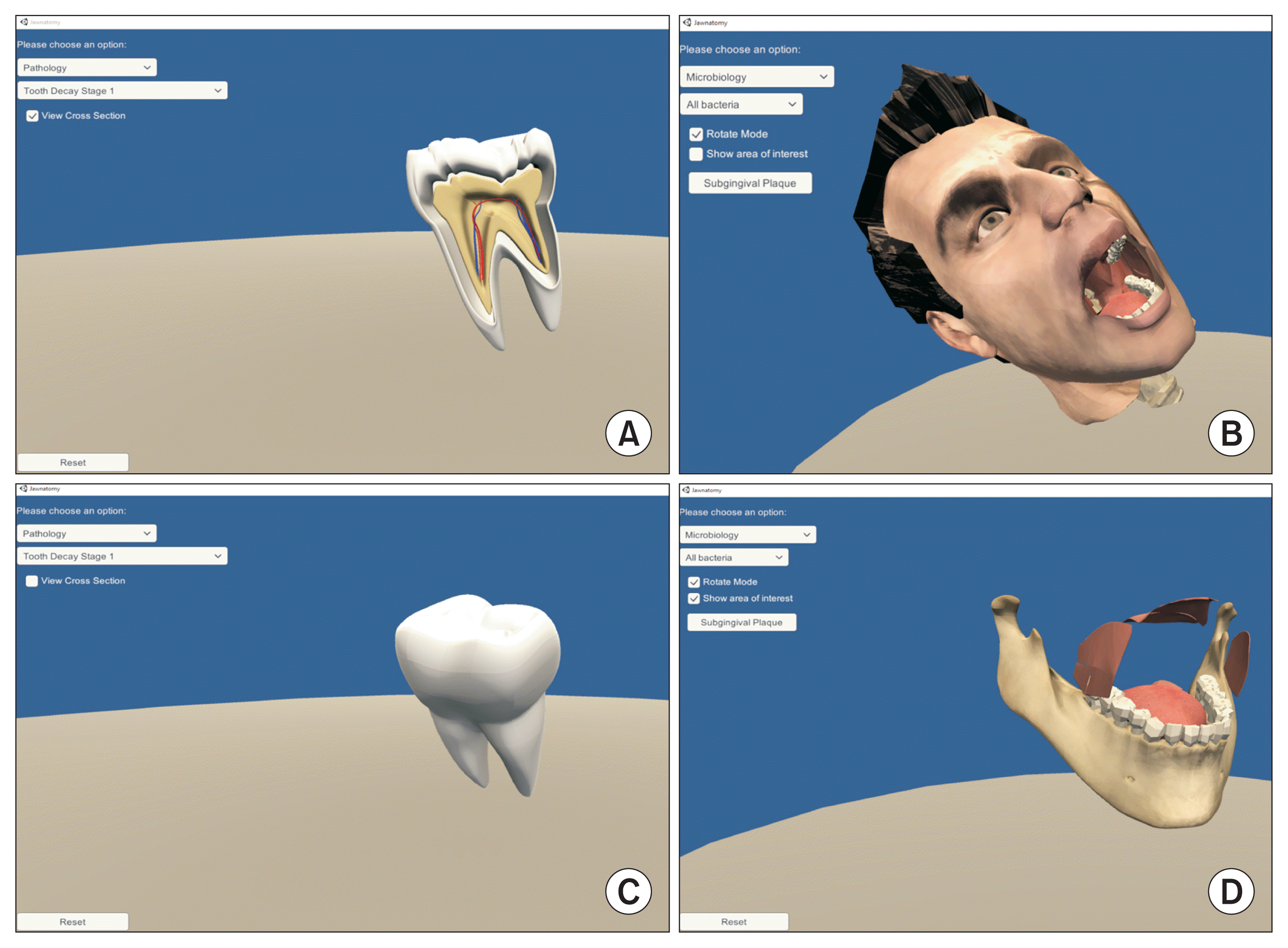 | Figure 1Learning modules of Jawnatomy. Along with a model of the physiological condition, Jawnatomy offers two other learning modules: (A) pathology and (B) microbiology. Jawnatomy allows learners to view the anatomical structure of a healthy tooth in both its surface view (C) and cross-sectional view (A). (D) Students can also remove external facial structures to visualize internal oral structures to learn oral anatomy.
|
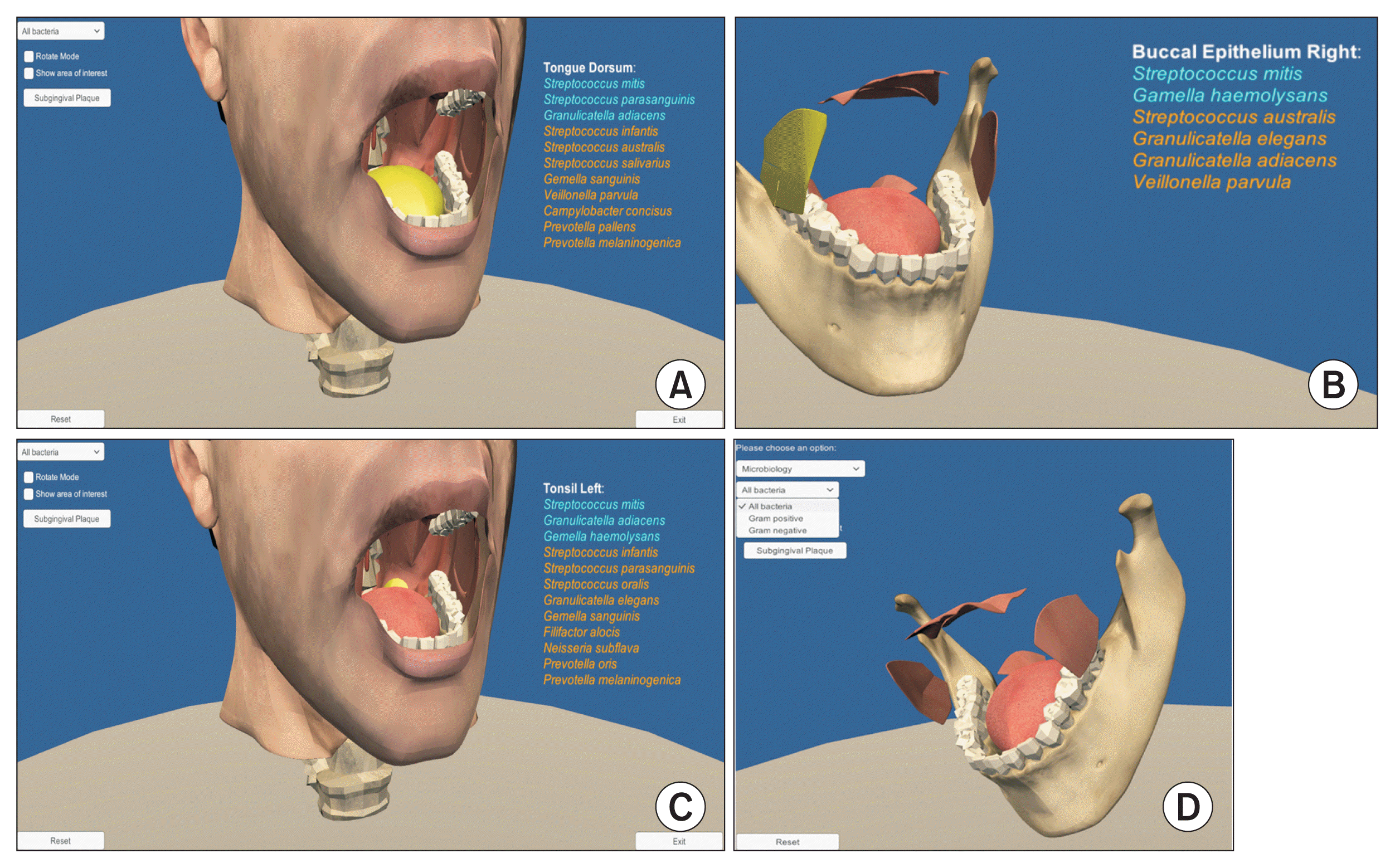 | Figure 2Microbiology module of Jawnatomy. Jawnatomy allows the learner the explore the normal microbial flora of the oral cavity. When selected, each oral cavity region shows the list of bacterial species found in that area. The lists of bacteria colonizing the tongue dorsum, buccal epithelium, and tonsil are shown in (A), (B), and (C), respectively. The list of bacteria is color-coded according to their prevalence, with those in blue being more abundant in that region. (D) Students can also select and group bacteria according to their cell wall properties (Gram-positive and Gram-negative).
|
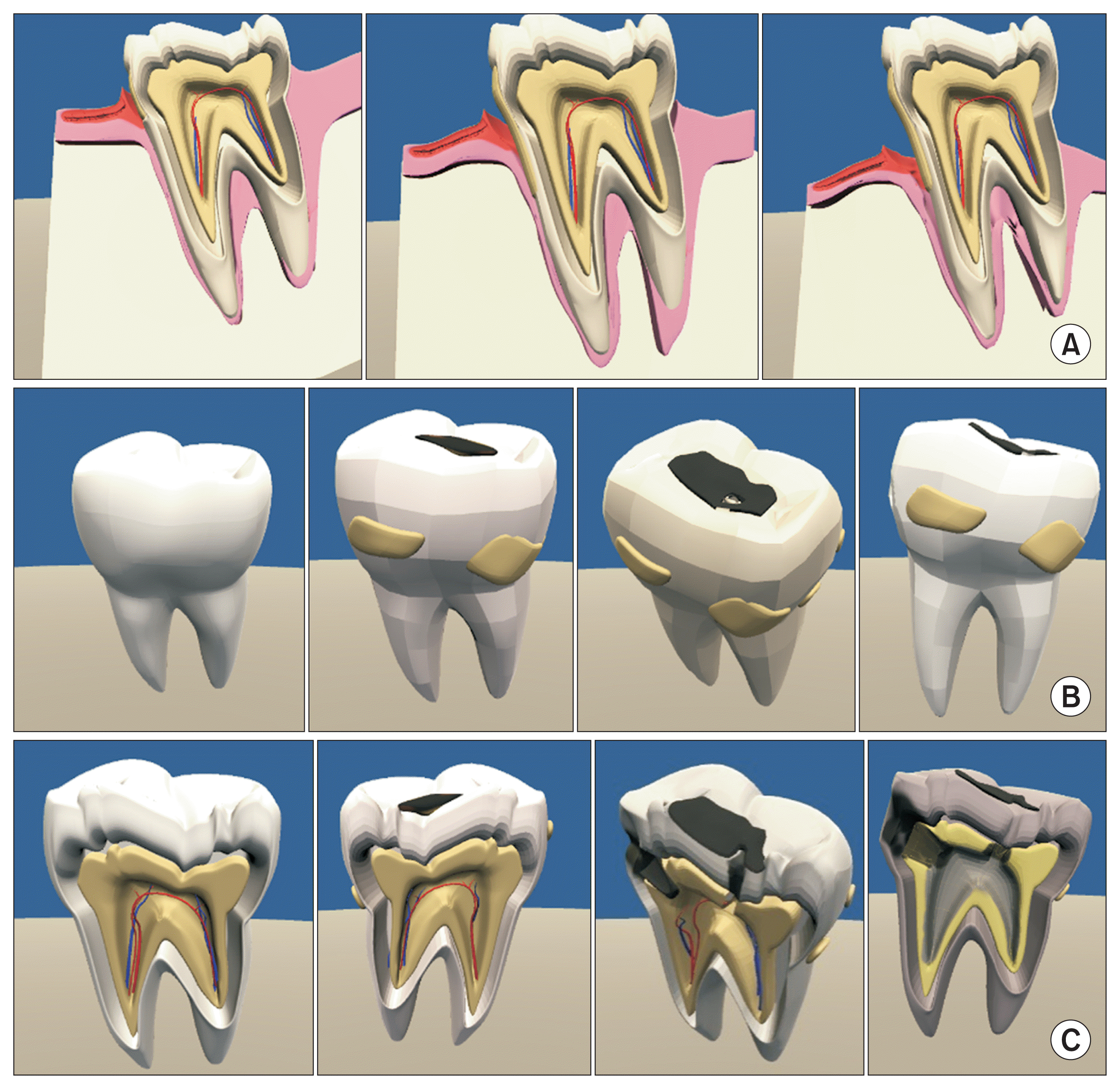 | Figure 3Pathology module of Jawnatomy. The pathology module of Jawnatomy allows learners to visualize the pathological processes observed in (A) periodontal disease and (B, C) dental caries. Periodontal disease is a chronic inflammatory disease that starts with bacterial plaque accumulation in the subgingival region. (A) Students can observe the progression of gingival inflammation and gradual loss of underlying alveolar bone to understand the development of periodontal disease. (B) Learners can also visualize plaque accumulation and dental caries formation on a molar tooth. They can rotate the tooth model to different angles and magnify areas to observe structural details. (C) Students can switch to the cross-sectional mode to analyze the effect of dental caries on the tooth’s internal structure. This module allows students to understand the gradual progression of the disease and compare it with healthy teeth and oral structures.
|
In the microbiology module of Jawnatomy, learners can explore the normal microbial flora in different locations of the healthy oral cavity. Students can interact with various parts of the oral cavity to investigate bacterial colonization. When selected, each region shows the list of bacterial species typically found in that region. The bacterial list is color-coded according to their prevalence and grouped into Gram-positive and Gram-negative bacteria (Figure 2). The authors prepared the location-specific list of bacteria based on the published literature [8,9].
Blender [10], a 3D computer graphics software, was used to create the tooth, surrounding oral structures, and facial model for Jawnatomy. The same software was used to develop the cross-sections and the different pathological states of the tooth, gingiva (gum), and alveolar bone. The authors agreed upon the anatomical details needed for teaching in dentistry and dental hygiene programs. The oral anatomy and pathology graphics were built under the supervision of content experts (AC and NS). Regular meetings were held between content experts and graphic designers to review and modify the anatomical structures and disease states for accuracy. Unity 3D [11] was used to incorporate interactivity, enabling users to magnify and rotate the models, toggle between cross-sections, stages of periodontitis, and investigate the localized microbial communities within the open mouth of the 3D head model. Jawnatomy is packaged as an executable file, which instructors supply to students. To run Jawnatomy in Windows or Mac OS, the user needs to unzip the file and start the executable file named “Jawnatomy.”
Several educational theories guided the development of Jawnatomy. Learning anatomy requires comprehension of the 3D anatomical structures and the structure-function relationships in a biological system [12]. Recognizing that students often struggle with the formation of complex mental models that require the integration of 3D structures and the changes that occur in pathological states, Jawnatomy was developed to reduce the cognitive load experienced by students during the learning process. Cognitive load theory is based on the premise that learning becomes difficult when the brain’s limited resources are taxed due to intrinsic and extrinsic loads [13]. The inherent complexity and the nature of the content contribute to the intrinsic load, which is not easily altered. The extrinsic load, however, comes from the way the material is presented [13], and Jawnatomy reduces this extrinsic load by helping students form 3D mental models and visualize the interactions that can occur between the oral anatomy and microbiota.
Motivation is an essential component of learning behavior. Studies have found that students with high motivation show more exploratory learning behaviors [14]. Using Keller’s attention, relevance, confidence, and satisfaction (ARCS) model [15], Jawnatomy was developed to increase student motivation and interaction with the material. According to Keller’s ARCS model, students are motivated to learn when if the material (1) captures and sustains students’ attention; (2) explains the subject’s relevance; (3) increases students’ confidence that they can succeed if they exert effort; and (4) ensures overall student satisfaction [16]. Attention can be earned by perceptual arousal, inquiry arousal, and variability [15,17]. Jawnatomy includes interactive 3D computer graphics to capture students’ visual attention.
Go to :
III. Discussion
Jawnatomy was designed to (1) facilitate the horizontal integration of oral anatomy, pathology, and microbiology in dental education, (2) increase student motivation, and (3) reduce the extrinsic load of learning and integrating different disciplines. This program will be introduced to first-year students in the dentistry and dental hygiene programs. In addition to regular classroom teaching, Jawnatomy will enable in-class project- and team-based learning by facilitating peer discussions and interactions, supported by Bandura’s social learning theory [18]. Jawnatomy will also be made available to students as a self-directed learning tool to help them strengthen their anatomical knowledge at their own pace. Students in dentistry and dental hygiene programs are emerging adult learners. Knowles’ theory of andragogy emphasizes that adults are self-directed in their learning [19]. Jawnatomy gives learners control over their own learning by allowing them to access the material when they are most ready to learn.
Computer-aided programs are valuable and have great potential in medical and dental education [20,21]. The effectiveness of several computer-based teaching tools, games, and simulations has been demonstrated through improvements in student satisfaction, in-class participation [22], and academic performance [23,24]. Jawnatomy enables students to visualize 3D oral structures and the dynamic process of some oral pathologies, thereby improving their understanding of anatomy and pathology and facilitating the retention of visually learned information [12].
Although Jawnatomy and other technologies can improve learning experiences, they can also cause restrictions for students who are less advantaged. The students at the School of Dentistry must have a computer or similar personal device. However, not all students have the privilege to access the up-to-date computers necessary to run programs like Jawnatomy. Despite this limitation, we believe that Jawnatomy will become a valuable addition to dental education. We will continue to update and expand this computer program based on students’ feedback.
Go to :
 XML Download