INTRODUCTION: DEFINING SKELETAL MUSCLE FAT INFILTRATION
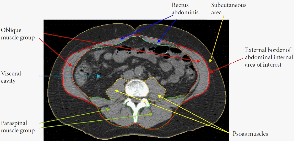
 | Fig. 1.Representation of areas of interest (adipose tissue and muscle measures) derived from abdominal computed tomography (CT) scans in the Multi-Ethnic Study of Atherosclerosis (MESA) cohort. The morphologically discrete tissues measured during CT image processing are: subcutaneous area which lies between the dermis and the fascia of the abdominal muscles; visceral cavity area with the internal organs; and abdominal skeletal muscles. The specific muscle groups denoted by psoas muscles (yellow arrows), paraspinal muscles (green arrows), oblique muscles (red arrows), and rectus abdominis muscles (blue arrows). Adipose tissue is identified as being between –190 and –30 Hounsfield units (HU). Lean tissue is identified as being between 0 and 100 HU. Inter-muscular adipose tissue is the sum of the pixels within a given muscle fascia. Intramuscular fat is computed as the mean of the pixel HU values between 0 and 99 for a given muscle group, with a higher mean density value for muscle indicating less intramuscular fat. |
MYOSTEATOSIS AND INSULIN RESISTANCE AND TYPE 2 DIABETES MELLITUS
Table 1.
| Reference | Design | Sample | Exposure(s) | Relevant outcome(s) | Significant associations (+) positive; (–) inverse; (n) null | |
|---|---|---|---|---|---|---|
| Insulin resistance and T2DM | ||||||
| Goodpaster et al. (2003) [13] | Cross-sectional | 1,447 Men /1,517 women | Thigh IMAT | Glucose | Glucose (n) | |
| Mean age 74 years | Insulin | Insulin (+) in normal weight | ||||
| Black and White | T2DM | Insulin (n) in overweight and obese | ||||
| Health ABC Study | T2DM (+) | |||||
| Kim et al. (2003) [14] | Cross-sectional | 23 Men/52 women | Thigh low-density muscle | HOMA-IR | HOMA-IR (+) | |
| Mean age 41.9 years | ||||||
| Obese, Korean | ||||||
| Miljkovic-Gacic et al. (2008) [15] | Cross-sectional | 1249 African-Caribbean men | Calf IMAT | Prevalent T2DM | Prevalent T2DM (+) | |
| Mean age 59 years | ||||||
| Age range 40–91 years | ||||||
| Tobago Health Study | ||||||
| Miljkovic et al. (2013) [17] | Cross-sectional | 393 Men | Abdominal IMAT (total abdominal, psoas, paraspinal) | Glucose | Total abdominal and psoas IMAT associated with: | |
| Mean age 74 years | Insulin | Glucose (+) | ||||
| White | HOMA-IR | Insulin (+) | ||||
| Osteoporotic Fractures in Men Study | HOMA-IR (+) | |||||
| Granados et al. (2019) [18] | Cross-sectional | 3,170 Men and women | Abdominal IMAT | Prediabetes | Prediabetes (+) | |
| Age range 43–55 years | T2DM | T2DM (+) | ||||
| Black and White | ||||||
| CARDIA Study | ||||||
| Miljkovic et al. (2020) [20] | Cross-sectional | 718 African-Caribbean men | Calf muscle density | T2DM | T2DM (–) in non-obese men only | |
| Mean age 64 years | ||||||
| Tobago Health Study | ||||||
| Flowers et al. (2019) [22] | Cross-sectional | 2,615 Men and women | Abdominal IMAT | Impaired fasting glucose | Impaired fasting glucose (+) in White and HispanicImpaired fasting glucose (n) in South Asian-, | |
| Mean age 61 years | T2DM | Chinese- and African-American | ||||
| South Asian, White, Chinese | T2DM (+) in White and Hispanic | |||||
| American, Black, and Hispanic | T2DM (–) in African American | |||||
| MASALA and MESA | T2DM (n) in South Asian- and Chinese-American | |||||
| Therkelsen et al. (2013) [19] | Cross-sectional | 1,466 Men/1,479 women | Paraspinous muscle density | Glucose | Glucose (–) | |
| Mean age 51 years | HOMA-IR | HOMA-IR (–) | ||||
| White | Impaired fasting glucose | Impaired fasting glucose (–) in women | ||||
| Framingham Heart Study | T2DM | Impaired fasting glucose (n) in men | ||||
| T2DM (–) | ||||||
| Miljkovic et al. (2016) [23] | Longitudinal | 1,515 African-Caribbean men | Calf IMAT | Incident T2DM | Incident T2DM (+) | |
| Mean age 57 years | ||||||
| Tanaka et al. (2020) [24] | Longitudinal | 621 Japanese men and women without diabetes | Abdominal muscle density | Incident T2DM | Muscle density associated with incident T2DM (–) | |
| Middle-aged | Abdominal low-attenuation muscle | Low attenuation muscle associated with incident T2DM (+) | ||||
| Nishimura Health Survey | ||||||
| Lipids | ||||||
| Miljkovic et al. (2013) [34] | Cross-sectional | 1,821 African-Caribbean men | Calf IMAT | Triglycerides | Triglycerides (n) and HDL-C (n) | |
| Mean age 59 years | Calf muscle density | LDL-C | Muscle density associated with LDL-C (+) | |||
| Tobago Health Study | HDL-C | |||||
| Durheim et al. (2008) [35] | Longitudinal | 40 Men/33 women | Thigh IMAT | LDL-C particle size | Reduction in thigh IMAT associated with shifts in HDL-C and LDL-C to larger particles in men only | |
| Age range 50–65 years | HDL-C particle size | |||||
| White and Black | ||||||
| Vella et al. (2020) [36] | Cross-sectional | 928 Men/940 women | Abdominal muscle density (total abdominal, stability and locomotor) | Triglycerides | Triglycerides (n) | |
| Mean age 65 years | LDL-C | LDL-C (n) | ||||
| White, Chinese, Black, Hispanic | VLDL cholesterol | Total abdominal muscle density associated with total cholesterol (+) | ||||
| MESA | HDL-C | Locomotor muscle density associated with VLDL (+), HDL (–), and total (+) cholesterol | ||||
| Total cholesterol | ||||||
| Mesinovic et al. (2019) [37] | Cross-sectional | 84 Overweight and obese men and women | Forearm muscle density | Triglycerides | Triglycerides (n) | |
| Age ≥50 years | Calf muscle density | HDL-C | HDL-C (n) | |||
| Inflammation | ||||||
| Miljkovic et al. (2011) [38] | Cross-sectional | 187 Men/284 women | Calf muscle density | CRP | CRP (–) | |
| Mean age 43 years | IL-6 | IL6 (n) | ||||
| Age range 18–103 years African-Caribbean | TNF-α | TNF (n) | ||||
| Tobago Family Health Study | ||||||
| Van Hollebeke et al. (2018) [39] | Cross-sectional | 1,000 Men/961 women | Abdominal muscle density (total abdominal, stability, and locomotor) | CRP | CRP (–) | |
| Mean age 65 years | IL-6 | IL-6 (–) | ||||
| Range 46–88 years | TNF-α | TNF (n) | ||||
| White, Chinese, Black, Hispanic | Resistin | Resistin (–) | ||||
| MESA | ||||||
| Vella et al. (2018) [40] | Cross-sectional | 983 Men/961 women | Abdominal muscle density (total abdominal, stability, and locomotor) | Adiponectin | Adiponectin (–) | |
| Mean age 65 years | Leptin | Leptin (+) | ||||
| White, Chinese, Black, Hispanic | ||||||
| MESA | ||||||
| Vella et al. (2018) [41] | Cross-sectional | 947 Men/950 women | Total abdominal muscle IMAT | CRP | CRP (+) | |
| Mean age 65 years | IL-6 | IL-6 (+) | ||||
| White, Chinese, Black, Hispanic | Adiponectin | Leptin (+) | ||||
| MESA | Leptin | Adiponectin (–) | ||||
| Resistin | Resistin (n) | |||||
| Beasley et al. (2009) [42] | Cross-sectional | 1,302 Men/1,349 women | Thigh IMAT | CRP | CRP (+) | |
| Age range 70–79 years | IL-6 | IL-6 (+) | ||||
| Black and White | TNF-α | TNF-α (+) in women | ||||
| Health ABC Study | TNF-α (n) in men | |||||
T2DM, type 2 diabetes mellitus; Health ABC, Health Aging and Body Composition; IMAT, intermuscular adipose tissue; HOMA-IR, homeostasis model assessment for insulin resistance; CARDIA, Coronary Artery Risk Development in Young Adults; MASALA, Mediators of Atherosclorisis in South Asians Living in America; MESA, Multi-Ethnic Study of Atherosclerosis; LDL-C, low-density lipoprotein cholesterol; VLDL, very low-density lipoprotein; HDL-C, high-density lipoprotein cholesterol; CRP, C-reactive protein; IL-6, interleukin-6; TNF-α, tumor necrosis factor alpha.


 PDF
PDF Citation
 Print
Print


 XML Download