

 PDF
PDF Citation
 Print
Print


INTRODUCTION
Ultrasonography is noninvasive, safe, and can be used to examine the morphology and functionality of the human body. Furthermore, it can be performed relatively quickly and repeated at a low cost. Carotid duplex ultrasound can be used to determine the presence and shape of atherosclerotic plaques. Since it is a relatively inexpensive and safe noninvasive examination method, it is widely used to diagnose and monitor carotid artery disease. However, despite its ease of access, inexperienced examiners may find the procedure difficult to perform. The purpose of this review is to present the standard method of carotid duplex ultrasound, and to interpret its findings and clinical applications.
Go to : 
HOW TO EXPLORE
Position
The examiner can observe the carotid artery from either an overhead or a lateral sitting position.1 In the overhead position, the examiner sits behind the head of the patient beside the end of the bed and uses both hands in the test. This position provides an expanded sonic window with a clear view of the carotid artery and allows many ultrasound probing positions. However, the examiner should be familiar with the practice of using both hands. A lateral sitting position is used for most ultrasonography examinations. The examiner generally uses their right hand to evaluate both carotid arteries. This position makes it easy to control the ultrasound probe. However, obtaining right-posterior projections is difficult. The optimal position for tilting the head of the patient is approximately 45° away from the relevant artery. Tilting the face too far from the test site can distort the anatomy or compress blood vessels, especially veins. The neck of the patient should be relaxed and the chin should be slightly raised. Placing a pillow induces a poor evaluation window for the carotid artery and therefore normally should not be used.
Ultrasound device settings
The standard equipment used in this technique includes a high-resolution grayscale B-mode system, with linear ultrasound transducers operating at > 7 MHz.2 Optimal image quality can be obtained when appropriate depths of focus (e.g., 30-40 mm) and optimal frame rates (25 Hz) are applied.3,4 Log gain compensation (dynamic range) is recommended to be around 60 dB.2 Gain settings are adjusted to achieve symmetrical brightness on the near and far walls or in the midfield to avoid intraluminal artifacts.4
Go to :
EXAMINATION PROCEDURE
The arterial wall should be assessed longitudinally, exactly perpendicular to the ultrasound projection. Both walls should be distinctly visualized to accurately measure the diameter. The diameter should be measured during diastole using automatic cine-loop detection or by measuring the minimum diameter during the cardiac cycle.3 A lateral probe position is recommended as it provides the best midfield resolution. Imaging of the carotid bifurcation is essential for serial imaging. Longitudinal and cross-sectional views of the carotid tree are required to examine focal atherosclerosis. Color flow Doppler imaging helps to identify hypoechogenic boundaries.
An adequate acoustic angle is important in obtaining an accurate color Doppler image. The ultrasound beam should be perpendicular to the skin, with a linear probe generating a grayscale image. However, to accurately record velocity using color Doppler ultrasonography, the angle must be between 30° and 60°.1 In most patients, the probe surface runs parallel to the common carotid artery (CCA) in its usual position, which allows scanning of the carotid artery without applying pressure. Pushing the head- or foot-side edge creates an angle between the probe surface and the vessel, which can achieve the optimal Doppler angle. This is called the heel-and-toe technique, and is a method for steering probes.1 Pulse-wave Doppler is needed to precisely measure flow velocity, and uses a small sample volume from the vessel center to assess the velocity at that segment. The peak velocity is measured to detect significant stenosis. Angle correction is crucial for measuring the actual flow velocity, which should be performed along the flow direction instead of the vessel wall.5 The flow direction is generally the same as the vessel direction, but it can differ from the vessel direction when eccentric atherosclerotic plaques are present.
Go to :
HOW TO DISTINGUISH BETWEEN ARTERIES
Carotid arteries
First, in the cross-sectional view, trace the carotid artery from the proximal CCA to the distal internal carotid artery (ICA) and then the external carotid artery (ECA), or vice versa. Then, observe the shape of the carotid artery in the longitudinal view. The CCA divides into the ICA and ECA. The ICA has a larger diameter, is located deeper than the ECA, and is directed toward the mastoid process. In contrast, the ECA is located superficially and is directed toward the face. The proximal ICA does not have branches, while the ECA does. The Doppler spectrums of the ICA indicated lower resistance patterns (Fig. 1). The velocity difference between the systolic and diastolic phases of the blood is smaller in the ICA than in the ECA. Another method for differentiating the ECA from the ICA involves placing the fingertips on the ipsilateral temporal artery when obtaining the ECA Doppler spectrums, which generates a serration-like artifact. This temporal artery tapping-induced artifact is generally not observed in the normal ICA Doppler spectrum. This so-called temporal tapping is a useful method for differentiating between the ICA and ECA (Fig. 1). However, waveform oscillations induced by temporal tapping can be found beyond the ECA in the CCA and ICA, especially in diseased ICAs.6
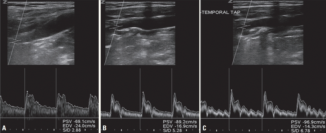 | Fig. 1.Examination of internal carotid artery (ICA) and external carotid artery (ECA). (A) Low-resistance waveform of the ICA. The waveform had a blunter systolic peak and greater diastolic flow for the ICA than for the ECA. (B) High-resistance waveform of the ECA. Sharp rise in flow velocity during systole, rapid decline toward baseline, and diminished diastolic flow. (C) ECA after temporal tapping. Repeated tapping of the superficial temporal artery caused waveform oscillation (small deflection) in the ECA spectrum.
|
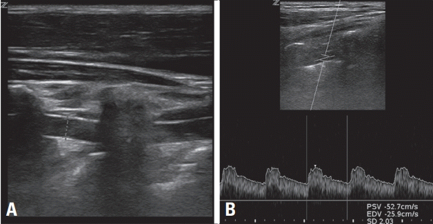
Vertebral arteries
Vertebral artery is a branch of the subclavian artery, and the V2 segment can be detected using carotid duplex ultrasound. The probe is placed parallel to the carotid artery and then moved posterolaterally to the neck. The vertebral artery can be distinguished by its acoustic shadow caused by the transverse processes (Fig. 2). The vertebral artery can be easily observed when the examinee pushes their shoulder down. Percussion of the C1 atlas may help differentiate between the vertebral artery, the thyrocervical artery, and the CCA.3
Go to :
EXAMINATION FINDINGS
The examination report should include the intima-media thickness (IMT), presence of plaques and their characteristics, flow velocities of each segment, diameter of each segment, and presence of stenosis and other vascular abnormalities. Furthermore, other abnormalities arising in the neck area, such as thyroid disease, should be noted and reported when they are observed.
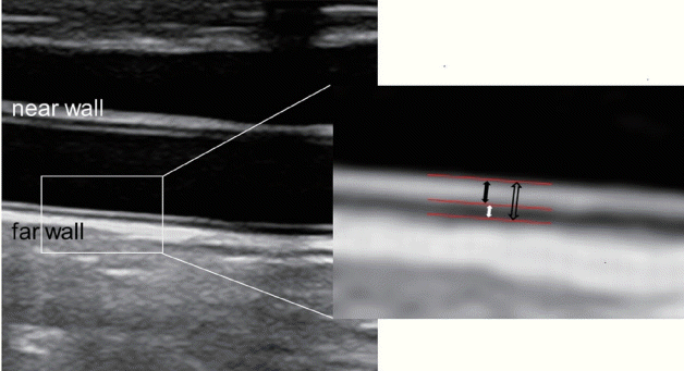
Intima-media thickness
The vascular wall consists of the intima, media, and adventitia. The IMT is observed in the longitudinal B-mode view of the CCA as two bright lines: the first line adjacent to the lumen is the intima, and the outside second line is the adventitia. The dark area between the two lines is the media, and the thickness from the interface between the lumen and the inner membrane to that between the media and the adventitia is the IMT (Fig. 3). It can be more clearly observed in the far than in the near wall. CCA IMT should be measured at the far wall without focal lesions over a 10-mm segment located approximately 5 mm proximal to the carotid bulb.2,4 Automatic or semiautomatic measurements are recommended, since manual measurements take longer and their values can vary between examinees. The average or maximum thickness is used in clinical practice.
Normal IMT values depend on age, sex, and race. They can also differ according to the type of measurement method used. Although normal IMT values among Koreans have been reported, large-scale studies are needed to predict cardio-cerebrovascular disease risk. Some studies have identified the normal IMT values in the Korean population. Two studies included healthy people who had no previous vascular diseases or other risk factors.7,8 One study used an average IMT value,7 and found mean IMTs in the overall population of 0.6 mm and 0.53 mm in males and females, respectively. The age was 56 ± 8 years (mean ± standard deviation) and 54 ± 8 years for males and females, respectively, and IMT increased with age. Another study used the mean maximum IMT value measured on the bilateral CCA.8 That study compared IMT according to the presence of metabolic syndrome. IMTs were larger in patients with metabolic syndrome (0.77 mm in males and 0.75 mm in females) than in the healthy population (0.76 mm and 0.71 mm, respectively). The difference in IMT between these two studies might be attributable to the older population included in the latter study (age of 62.1 ± 7.7 years in males and 61.0 ± 7.9 years in females).
Plaque
A plaque is defined as a focal structure protruding into the arterial lumen by at least 0.5 mm or 50% of the surrounding IMT, or having an IMT of > 1.5 mm.2 The maximum thickness should be measured at two different insonation angles in the longitudinal and cross-sectional views.
The size, morphology, and characteristics of plaques should be described, including echogenicity and intraplaque hemorrhage presence, neovascularization, ulceration, or its mobile portions. Plaque echogenicity can be described as echogenic, isoechoic, echolucent, or heterogeneous. The echogenicity of the intima-media complex is a criterion that determines an isoechoic plaque. The plaque surface can be described as smooth, irregular, or ulcerated.
Blood flow measurement
Blood flow velocity and endovascular diameter can be measured using two methods: 1) quantitative measurement using spectral Doppler ultrasound and 2) color M-mode ultrasound.3 The first method involves measuring the angle-corrected time-averaged mean flow velocity and inner diameter of blood vessels using B-mode images. The second is also called color velocity imaging quantification. When the intravessel luminal diameter or systolic velocity is at its maximum, the flow rate and inner diameter are measured simultaneously from a still image. Artifacts such as overlapping or color bleeding should be avoided when performing measurements in color M-mode imaging. In previous studies, the spectral Doppler method has been reported to show faster blood flow than the color M method, which could be attributed to the larger inner diameter measured by the spectral Doppler method. The following data should be included: peak systolic velocity and end-diastolic velocity for all vessel segments, Doppler spectral waveform morphologies from the CCA, ICA, and ECA, flow direction, and the peak systolic velocity in the vertebral arteries.
Internal carotid artery
To measure the blood flow rate and inner diameter of the ICA, the point where the blood vessel extends straight should be selected at least 1.5-2 cm above the ICA bifurcation, after the head is turned to the opposite side by 25-45°.3
Vertebral artery
The vertebral artery is normally measured between the C4 and C5 transverse processes when the head is turned approximately 10° to the side opposite to that being examined.3
Go to :
CLINICAL APPLICATIONS
Symptomatic carotid stenosis
Symptomatic carotid stenosis refers to atherosclerotic stenosis of the ICA, which induces ischemia symptoms. Ischemic events associated with carotid artery disease include ocular symptoms such as amaurosis fugax of the ipsilateral eye, central or branch retinal artery occlusion, ischemic optic neuropathy, cerebral infarction, and transient ischemic attack (TIA).
Diagnosis
The consensus criteria suggested by the Society of Radiologists in Ultrasound are widely used to evaluate the degree of carotid artery stenosis (Table 1).9 These criteria suggest that carotid artery stenosis can diagnosed as < 50%, 50-69%, ≥ 70%, or complete occlusion based on the maximum systolic ICA blood velocity combined with a comprehensive examination which determines atherosclerotic plaque presence, end-diastolic velocity, and the maximum systolic blood velocity ratio between the ICA and CCA. However, the specificity of carotid stenosis varies between reports. Therefore, to determine whether an operation or a procedure should be performed, magnetic resonance angiography or CT angiography or a subsequent examination of the carotid ultrasound by another examiner is recommended to minimize the probability of performing unnecessary procedures.10,11
Table 1.
Parameters used in carotid duplex ultrasound to estimate the degree of carotid artery stenosis
| Degree of stenosisa (%) |
Primary parameter |
Additional parameter |
||
|---|---|---|---|---|
| ICA PSV (cm/s) | Plaque estimate (%) | ICA/CCA PSV ratio | ICA EDV (cm/s) | |
| Normal | < 125 | None | < 2.0 | < 40 |
| < 50 | < 125 | < 50 | < 2.0 | < 40 |
| 50-69 | 125-230 | ≥ 50 | 2.0-4.0 | 40-100 |
| ≥ 70 | > 230 | ≥ 50 | > 4.0 | > 100 |
| Near occlusion | High, low, or undetectable | Visible | Variable | Variable |
| Total occlusion | Undetectable | Visible, no detectable lumen | Not applicable | Not applicable |
![]()
Predicting potential revascularization complications
Carotid duplex ultrasound can be used to predict complications that can occur during revascularization procedures such as carotid endarterectomy (CEA) or carotid artery angioplasty and stenting (CAS). Carotid ultrasound can provide important information for carotid artery stenting in determining the mechanical characteristics of an atherosclerotic plaque, such as its surface (smooth or irregular) and structure (homogeneous, nonhomogeneous, intraplaque hemorrhage, or calcification).12,13 However, carotid duplex sonography does not sufficiently assess the risk of surgical treatment for screening purposes because of a lack of standardized data.14 Another limitation is that it cannot be used to evaluate cerebral perfusions, intracranial collateral circulation, or other vascular abnormalities such as aneurysms.
Follow-up after revascularization
There are several reports of the restenosis incidence ranging from 5% to 22% and from 2.7% to 33% after CEA and CAS, respectively.15 After carotid revascularization, the blood flow velocity is observed to be greater than normal using carotid ultrasound, and so caution is needed when interpreting the observations.14 Appropriate criteria for interpreting carotid duplex sonography findings after carotid revascularization have not yet been established, and so it is necessary to establish and regularly verify laboratory standards.
Follow-up after carotid endarterectomy
CEA results in the blood vessels at the endarterectomy site becoming considerably wider, but the distal ones remain narrow, leading to higher measured blood flow velocities. This finding occurs particularly when artificial blood vessels are used in the stricture area during surgery. Reintervention consideration is suggested in postcarotid endarterectomies at 70-99% asymptomatic restenosis.10 There is no consensus on follow-up examinations using carotid ultrasound after CEA. Restenosis is known to occur less in cases of patch angioplasty than in those of primary closure.16 Therefore, a carotid ultrasonography follow-up is recommended for primary closure after 1 month, 6 months, and then annually.17 For patch angioplasty, further regular follow-ups are not required if findings are normal at the 6-month follow-up.18 The Society for Vascular Surgery in the United States recommend carotid duplex ultrasound at baseline and every 6 months for 2 years, and annually thereafter until it stabilizes (i.e., until no restenosis is observed in two consecutive annual scans).19
Follow-up after carotid angioplasty and stenting
In-stent restenosis after carotid artery stent placement is more common than restenosis after CEA. Restenosis occurrence should therefore be monitored. However, the degree of stenosis may be overestimated because of changes in blood flow and vascular compliance caused by the stent.19 In carotid artery stenting, since the degree of carotid artery stiffness increases due to the stent, the blood flow velocity through this area could increase. To reduce this, velocity thresholds need to be modified for follow-up observations of intrastent restenosis in the carotid artery (Table 2).20-22 To monitor for intrastent restenosis, it is suggested to perform carotid ultrasonography within 30 days of the procedure, then every 6 months for 2 years, and annually thereafter.10,23 In contrast to the relatively high restenosis incidence, the risk of stroke is minimal. It is therefore recommended that asymptomatic restenosis should be treated medically at > 70% after CAS.15
Table 2.
Suggested criteria for diagnosing in-stent restenosis after carotid artery stenting
| Stenosisa (%) | PSV (cm/s) | EDV (cm/s) | ICA/CCA ratio | |
|---|---|---|---|---|
| Lal et al.22 | 0-19 | < 150 | < 2.15 | |
| 20-49 | 150-219 | |||
| 50-79 | 220-339 | ≥ 2.7 | ||
| 80-99 | ≥ 340 | ≥ 4.15 | ||
| AbuRahma et al.21 | 30-49 | > 154 | > 42 | > 1.533 |
| 50-79 | > 224 | > 88 | > 3.439 | |
| 80-99 | > 325 | > 119 | > 4.533 | |
| Zhou et al.20 | > 70 | ≥ 350 | ≥ 4.75 |
![]()
Asymptomatic carotid stenosis
Asymptomatic carotid artery stenosis refers to stenosis that is accidentally revealed in a test and has not induced any neurological symptoms. It can be diagnosed if a patient has not experienced cerebral or transient ischemia symptoms in the area supplied by the stenotic carotid artery or has experienced cerebral ischemia symptoms for more than 6 months.24 The degree of stenosis and plaque characteristics can be used to estimate future ischemia risk. A study that assessed the future stroke risk in asymptomatic carotid stenosis estimated that the annual stroke risks was 1-2% in cases of 50-79% stenosis, 2-4% in cases of 80-89% stenosis, and 4-6% in cases of 90-99% stenosis.25 Typically 20-25% of patients with asymptomatic carotid artery stenosis have been reported as showing progression in their degree of stenosis during follow-up examinations.26,27 TIA or ischemic stroke risk has been reported to increase by approximately threefold when the degree of asymptomatic carotid artery stenosis progresses to ≥ 80% compared with cases without progression.26 As characteristics of atherosclerotic plaques, echolucent or hypoechoic plaques are associated with plaque instability,28 whereas echogenic or hyperechoic plaques are associated with plaque stability and are mostly observed in patients who have had asymptomatic carotid artery stenosis for a long period.25 A study involving an asymptomatic population found that hypoechoic plaques were significantly associated with ipsilateral stroke risk after adjusting for other factors including stenosis, grayscale median, presence of discrete white areas with no acoustic shadowing to indicate neovascularization, plaque area, and contralateral TIA or stroke history.29 Carotid ultrasound screening for detecting asymptomatic carotid artery stenosis is generally not recommended.30 However, when coronary artery disease, peripheral artery disease, or ≥ 2 risk factors for atherosclerosis are present, screening for carotid artery stenosis may be considered.17
Prediction of cardiovascular disease risk
Assessing carotid IMT and carotid atherosclerotic plaque measurements may be useful for predicting cardiovascular disease risk. The following patient characteristics can lead to consideration of carotid ultrasonography for evaluating the cardiovascular disease risk: early cardiovascular disease in immediate family members, aged < 60 years with at least one definite risk factor, and females aged < 60 years with two or more risk factors.4 However, carotid ultrasonography is not recommended when the patient has a diagnosed atherosclerotic disease, when the test results would not affect the treatment, or when the purpose is simply to identify worsening or improvement. The need for regular follow-up examinations is also contentious. A relationship between the progression (increase) of the carotid IMT over time and the risk of cardiovascular disease in a general population has not yet been established.31 It is also unclear whether treating increased carotid IMT or carotid atherosclerosis improves the cardiovascular disease prognoses.
Dissection
Cervical artery dissection is accompanied by various intravascular and extravascular changes, because blood vessels narrow after endovascular injury. The occurrence of an intimal tear results in the formation of an intramural hematoma between the intima and media. Intramural hematomas cause varying degrees of stenosis and occlusion. Duplex sonography can reveal hypoechogenic thickening of the blood vessel wall. Its other findings include irregular stenosis, dissection flap, double lumen, pseudoaneurysm, or gradually narrowed vessel with distal obstruction.32,33 However, the ultrasonographic findings of carotid artery dissections are mostly nonspecific stenosis or obstruction, and they rarely reveal characteristic intimal detachment, double lumen, or pseudoaneurysm.34
Carotid web
The carotid web is a thin membrane-like shape attached to the blood vessel wall that forms as the intima thickens and the media thins.35 Although conventional digital subtraction angiography is the gold standard for diagnosing a carotid web, ultrasound can also be helpful, especially in young patients with cryptogenic stroke.36,37 Because current sonographic findings are mostly nonspecific, consideration and awareness of the condition are necessary. Several sonographic features relate to the carotid web, including mild bulbar outgrowth, hypoechoic masses with a linear band extending into the lumen, focally arising artifacts with elevated peak systolic velocity, irregular echogenic plaque causing stenosis, and thin strands protruding into the lumen.38
Subclavian steal syndrome
The Doppler ultrasound effect can be used to assess blood flow reflux in the vertebral artery. An alternating flow pattern or prominent midsystolic deceleration is observed unless a complete blood flow reversal occurs.39,40 Hyperemia tests using a blood pressure cuff can induce blood flow reversal and diagnose the hidden steal phenomenon.39
Go to :
SUGGESTIONS
Based on the discussed findings, we now present a summary of standard guidelines for performing carotid ultrasound in clinical practice (Table 3). These suggestions are based on an article published in Journal of Neurosonology and Neuroimaging:14 1) Both the carotid and vertebral arteries should be examined, and B-mode and Doppler velocity measurements should be performed. 2) The necessity of carotid ultrasound is determined by the clinical symptoms, medical history, and other test results of a patient. Carotid ultrasound is not recommended for screening individuals with no related symptoms, medical history, or disease.
Table 3.
Carotid artery ultrasonography may be performed or considered in the following cases
![]()
Go to :
 XML Download