Journal List > Obstet Gynecol Sci > v.64(5) > 1147817

Nagandla, Lin, Chitra, and Jamli: Role of microRNAs as biomarkers of cervical carcinogenesis: a systematic review

Abstract

We performed a systematic review to identify the role of microRNAs (miRNAs) as biomarkers in the progression of cervical precancerous lesions. A comprehensive search of the Cochrane Controlled Register of Trials, PubMed, ScienceDirect, and Embase databases was performed for articles published between January 2010 and June 2020. The following Medical Subject Headings (MeSH) terms were searched: “microRNA” and “cervical” and “lesion.” All study designs that aimed to evaluate the correlation of miRNA expression with different precancerous cervical staging and/ or cervical cancer were included, except for case reports and case series. Approximately 82 individual miRNAs were found to be significant in differentiating the stages of cervical carcinogenesis. Among the miRNAs, miR-21 is the most prevalent, and it is consistently upregulated progressively from normal cervical to worsening cervical lesion stages in both cell and serum samples. miR-205 has been shown to have a higher specificity than human papilloma virus testing in predicting the absence of high-grade squamous intraepithelial lesions (HSILs) in exfoliated cell samples. The tumor suppressor miRNAs miR-34, let-7, miR-203 miR-29, and miR-375 were significantly downregulated in low-grade squamous intraepithelial lesions, HSILs, and cervical cancer. We found significant dysregulated miRNAs in cervical carcinogenesis with their dynamic expression changes and ability to detect viral persistency, risk prediction of low-grade lesions (cervical intraepithelial neoplasia [CIN] 2) to high-grade lesions (CIN 3), and progression of CIN 3 to cancer. Their ability to discriminate HSILs from non-dysplastic lesions has potential implications in early diagnosis and reducing overtreatment of otherwise regressive early preinvasive lesions.

Introduction

Cervical cancer continues to be one of the most common cancers among women globally [1]. The main causative agent of cervical cancer is persistent infection with oncogenic human papillomavirus (HPV) at the cervix, which results in intraepithelial lesions, defined histologically as cervical intraepithelial neoplasia (CIN) lesions. Low-grade lesions include CIN1, which is a morphological expression of HPV infection. High-grade lesions include CIN2, which is considered a mixture of CIN1 and CIN3 and has a higher frequency of regression, and CIN3, which progresses to invasion in 9 years [2,3]. Progression to intraepithelial pre-cancers involves the disruption of the HPV virus cell cycle control pathways mediated by overexpression of the viral E6 and E7 proteins that, among other actions, functionally inactivate the products of the retinoblastoma and p53 immunosuppressive genes. The overall process is complex and includes a number of genetic and epigenetic alterations [4,5]. The specificity of high-risk HPV DNA testing is too low to be useful for the diagnosis of low-grade lesions (23% for atypical squamous cells of undetermined significance and 17% for low-grade squamous intraepithelial lesions [LSILs]) [6,7]. A potential biomarker with high specificity values is required to avoid over-diagnosis and potential overtreatment in this category of patients.
MicroRNAs (miRNAs) are an abundant class of small, non-coding RNA genes that comprise short sequences of nucleotides (approximately 22 nucleotides) that control post-transcriptional regulation of gene expression [8]. Accumulating evidence on miRNA profiles in carcinogenesis has demonstrated their strong ability to differentiate various cancers and their tumor stages [9,10]. Analysis of miRNA functions has established that miRNAs play a crucial role in many physiological processes and developmental health [11]. In turn, miRNA expression levels are altered in many diseases, especially human cancers [12]. Various miRNAs have been identified in many cancers, including hepatocellular cancer, pancreatic cancer, stomach adenocarcinomas, ovarian cancer, breast cancer, and cervical cancer [12].
However, the exact role of miRNAs in cervical carcinogenesis is still largely unknown, especially in non-conventional sampling materials such as cervical mucus and plasma serum [13]. We systematically reviewed the literature to evaluate the role of miRNAs as biomarkers in the progression of cervical precancerous and cancerous lesions.

Methods

A systematic literature search was conducted using electronic databases for studies published between January 2010 and June 2020. The databases used included PubMed, Science-Direct, Embase, and Cochrane Controlled Register of Trials. Boolean operators and text words were combined to create the following keywords: “microRNA” and “cervical” and “lesion.” Reference lists of selected studies and review articles were examined to ensure that no relevant studies were omitted. Table 1 lists the search queries used and the number of articles obtained from each electronic database. To ensure that the studies included were eligible for subsequent review, specific criteria were used to distinguish relevant studies from all the initial studies found. The selected studies fulfilled the inclusion criteria detailed in the next sector.

Study selection

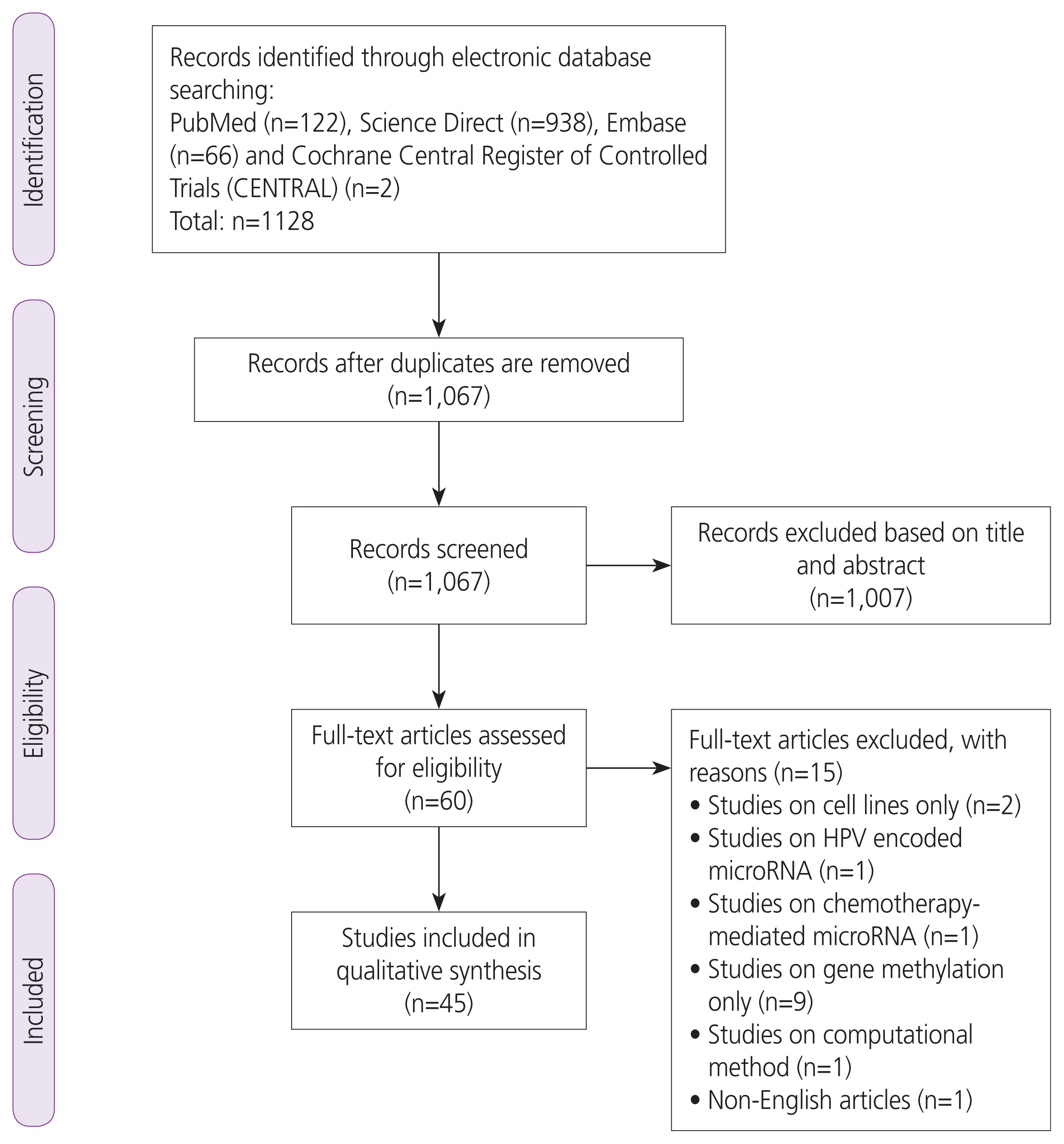
Two authors (K.N. and E.B.) independently screened the title and abstract of each record retrieved to identify potentially relevant articles for full-text review. If the two reviewers did not agree, a third reviewer arbitrated. Inclusion criteria included all study designs that aimed to evaluate correlations between miRNA expression and different precancerous cervical stages and/or cervical cancer were included except case reports and case series. All meta-analyses; systematic reviews; randomized and nonrandomized clinical trials; retrospective and prospective observational studies; phase I, II, and III clinical trials with study samples consisting of cervical cells, cervical tissues, serum, mucus, or a combination of these were included. The exclusion criteria included narrative reviews, letters, editorials, commentaries, case studies, case series, non-human studies, non-cervical cancer cases, studies experimenting only on cell lines, miRNAs that were investigated only in advanced cervical cancers or lymph node metastasis, studies on miRNA-related polymorphisms, and studies not published in English. The selected articles were discussed among the investigators during the screening phase. The selection process included three main processes: (1) identification of records, (2) screening of records, and (3) assessing eligibility of records and were presented accordingly in a Preferred Reporting Items for Systematic Reviews and Meta-Analyses (PRISMA) flow chart.

Data extraction and synthesis

Two authors (K.H. and M. F.) independently extracted the data from the included studies in a piloted data extraction form. The data extracted from individual studies included information on the title, study design, first author, publication year, publication journal, sample size, sample type, country of study, age group of participants, discovery and quantification methods of miRNAs, internal reference gene used for normalization, miRNAs significantly expressed in different types of samples, miRNA expression profiles (upregulated or downregulated) in various cervical disease states, HPV-related findings, and prognosis of patients. The piloted data form is provided in Appendix B.1.

Risk of bias assessment

For methodological quality assessment of observational studies, which included cross-sectional studies, cohort studies, and case-control studies, two different assessment tools were utilized to determine how well each individual study addressed the potential risk of bias.
All existing cohort studies and case-control studies were assessed using the Newcastle-Ottawa Scale by Wells et al. [14] and cross-sectional studies in the present review were analyzed using the The Joanna Briggs Institute (JBI) Critical Appraisal Checklist for Analytical Cross-Sectional Studies [15].

Results

The study selection process is summarized in Fig. 1 (PRISMA Flow Chart). Our initial search identified 1,128 potentially relevant articles. We included 62 articles after screening the titles and abstracts. Seventeen articles were excluded after further scrutiny. A complete list of the excluded studies is available from the authors.

Characteristics of selected studies

In total, 45 articles (22 case-control studies, 4 prospective cohort studies, and 19 cross-sectional studies) were included in this systematic review. The studies were carried out across several continents, namely Asia (n=30), Europe (n=11), South America (n=3), and Africa (n=1). Twenty-five studies were from China, four studies from the Netherlands [1619], three studies from Portugal [2022], two studies each from India [23,24] and Mexico [25,26], and one study each from Thailand, Japan, Russia, Italy, Hungary, Czech Republic, Sweden, Brazil, and Nigeria [2735]. The main characteristics of the selected studies are listed in Table 2.

Dysregulated miRNAs in cervical carcinogenesis

1. Upregulated miRNAs

Approximately 82 individual miRNAs were found to be significant in differentiating the stages of cervical carcinogenesis. The significantly upregulated miRNAs included miR-21 and miR-15 in LSIL, high-grade squamous intraepithelial lesions (HSILs), and cervical cancer in plasma serum, cervical exfoliated cells, and cervical tissue samples. miR-21 expression was found to be predominantly detected in the tumor-associated stroma of squamous cell carcinomas [27]. miR-21 was also found to be correlated with advanced cervical cancer lesions with progressively increased expression in more severe clinical manifestations [24].
Other miRNAs such as miR-196, miR-20, miR-155, miR-27, miR-200, miR-9, miR-146a, miR-193, miR-148, miR-10, miR-132, miR-141, and miR-93 were found to be upregulated in cervical cancer as compared to normal plasma serum, cervical mucus, and cervical tissue samples in studies conducted in Europe, Nigeria, Mexico and mainly in Asia [1621,3638]. miR-141 expression levels were found to be significantly higher in younger participants than in older cohorts [39].
In groups comparing expression levels of LSIL to HSIL, upregulated miRNAs include miR-20, miR-155, miR-27, and miR-16 in plasma serum and cervical tissue samples [31,40]. Interestingly, miR-200 was upregulated in cervical cell samples but downregulated in serum samples from patient groups comparing LSIL to HSIL [35]. In groups comparing HSIL to cervical cancer stages, miR-155, miR-27, miR-196, miR-200, miR-222, miR-9, miR-146a, miR-1, miR-17, miR-106, miR-148, miR-10, miR-132, and miR-205 were upregulated. miR-205 has also been shown to have a higher specificity than HPV testing in predicting the absence of HSIL in exfoliated cell samples [33].

2. Downregulated miRNAs

The significantly downregulated miRNAs included, let-7, miR-203, and miR-34 in LSIL, HSIL, and cervical cancer compared to controls. Furthermore, miR-34 is predominantly downregulated in HSIL compared to LSIL [31,41].
Other miRNAs, such as miR-29, miR-20, miR-125, miR-199, miR-23b, miR-99a, miR-126, miR-145, miR-149, miR-143, and miR-26a were downregulated in plasma serum, cervical exfoliated cells, and cervical tissue samples [16,19,21,29,31,42,43] miR-126 was found to be upregulated in cervical mucus, but downregulated in cervical tissues in two respective studies [28,29]. In particular, miR-20 was found to have a higher detection performance in LSIL and HSIL than in cervical cancer samples [39]. miR-146 was also found to have a higher expression in plasma serum than in cervical cells [35]. The expression of the five most common miRNAs and their dysregulation patterns are shown in Table 3.

miRNAs in non-conventional sample types

1. Cervical mucus samples

A total of four miRNAs, miR-126 miR-20, miR-451, and miR-144, were found to be significantly expressed in the cervical mucus samples. In particular, miR-451 and miR-144 were found to be significantly upregulated in cervical mucus samples [28]. Interestingly, miR-126 was upregulated in cervical mucus but downregulated in cell samples of cervical cancer patients compared to control patients [28,29]. Table 4 shows the miRNAs detected in the cervical mucus samples.

2. Plasma serum samples

A total of 11 miRNAs were investigated in serum samples from four studies [35,42,44,45]. Nine miRNAs, namely miR-21, miR-146, miR-155, miR-200, miR-34, miR-let-7, miR-182, miR-145, and miR-1266 were found to be differentially expressed in the cells, tissue, and serum samples of patients with abnormal cervical smears. Two miRNAs, namely miR-214 and miR-466, were found to be significantly expressed only in serum samples [44,46].

3. MiRNAs and HPV infection

MiR-21 was upregulated in HPV-positive samples compared to HPV-negative samples, while miR-3156-3p and miR-155-5p expression were lower in HPV-positive cervical cancer [27,47]. Several studies have found correlations between the expression of miRNAs and HPV E6 and E7 viral genes. A positive correlation was observed between miR-25 and miR-21 with HPV E7 and E6, respectively, indicating that these miRNAs are specific to these viral proteins [24,48]. A negative correlation was found between miR-129-5p, miR-409-3p, and let-7a with the level of HPV E6/E7 [24,32,41]. HPV-positive cervical mucus samples demonstrated upregulation of miR-126-3p, miR-20b-5p, miR-451a, and miR-144-3p, respectively [28].

4. Risk of bias assessment

The quality of the case-control and cohort studies was evaluated using the Newcastle-Ottawa Scale. The overall quality of the case-control studies was not outstanding, as 50% (n=11) of the studies reviewed were scored as good quality. For most case-control studies, the authors failed to provide or did not mention a suitable comparability of study design between the cases and controls. Most studies enrolled healthy patients (normal controls) through clinical settings that were convenient to investigators. Only three studies recruited normal and healthy participants through community screenings, which generally contributed to higher scoring [16,17,49]. Similar mediocre results were achieved during the assessment of cohort studies, with half of the studies scored as good quality [44,46]. For cohort studies, there was a loss of follow-up noted in one study conducted by Wang et al. [50]. High-quality studies were prioritized based on this scoring system.
For the 19 cross-sectional studies, the quality of the studies was analyzed using the JBI Critical Appraisal Checklist for Analytical Cross-Sectional Studies [15]. Most of the studies failed to identify any confounding factors that may influence the expression levels of miRNAs; only two studies analyzed sociodemographic factors [31,40]. A lack of clear definition was observed for the inclusion and exclusion criteria in a total of nine studies. Most studies utilized appropriate statistical analyses; however, four studies did not provide a clear explanation of the statistical tests used [41,5052]. All 19 studies were ultimately included in the present study. Supplementary Table 13, Fig. 1 show the quality assessment for case-control, cross-sectional, and cohort studies.

Discussion

1. Summary of main results

A systematic review was performed to identify the role of miRNAs as biomarkers in the progression of cervical precancerous lesions. The results of the systematic analysis identified the most prominent dysregulated miRNAs. Among the upregulated mRNAs, miR-21 has been shown to be consistently upregulated progressively from normal cervical to worsening cervical lesion stages in both cell and serum samples [18,24,27,35,43,51,53]. miR-21 is a key miRNA that is found to be increased in many diseased states, such as inflammation, cardiac injuries, and various tumors in mammalian tissues [36,54]. Apart from cervical cancer, miR-21 has been shown to have oncogenic effects in both solid tumors such as liver cancer, lung cancer, and non-solid tumors such as chronic lymphocytic leukemia [36]. It has been shown to be expressed in hematopoietic cells of the immune system, which respond to tumor progression, and is linked to greater immune cell activation, supporting our findings that miR-21 is an oncogenic miRNA [54]. Moreover, there is a strong correlation between upregulated miR-21 and gene hypomethylation in the regulation of cancer genes and poor survival rate in cancer patients [38]. It was also noted to be particularly high in advanced cancers with severe manifestations [24]. Evidently, miR-21 is a reliable, highly profiled miRNA that is commonly overexpressed in cervical cancer, making it a promising biomarker for tumor detection.
Among the downregulated miRNAs, miR-34, let-7, and miR-203 were proven to be significantly downregulated in LSIL, HSIL, and cervical cancer across multiple studies [2124,3134]. miR-34 and miR-21 were also found to be more specific than HPV testing in differentiating LSIL from HSIL and may provide more accurate detection results [53]. The dysregulation of these four miRNAs was observed to be an early onset event in the development of cervical cancer. It has also been reported that miR-34 has a synergistic effect with p53, a well-known tumor suppressor, further reinforcing its regulatory role in carcinogenesis [37]. Again, miR-34 was found to be upregulated in thyroid and liver cancer but downregulated in cervical, breast, and lung cancer, suggesting that the individual miRNAs may have different expression levels in various cell types [37].
It was discovered that miRNAs in cervical mucus have higher detection accuracies for detecting adenocarcinoma than conventional cytological methods [55]. However, significant results were only found at late stages of precancerous lesions (HSIL/CIN II-III), suggesting that miRNAs in cervical mucus could be a potential biomarker for HSIL and cervical cancer [28].
It was also shown that miRNAs maintain their stability despite being subjected to harsh conditions [56]. Wang et al. [42] confirmed that the expression levels of miRNAs were consistently similar in both plasma serum and cervical biopsy tissue, which further suggests that plasma serum reflects the true miRNA levels with less painful methods. Due to the low cost and relatively less invasive nature of phlebotomy, a blood-based test for cervical cancer would be an appealing option [57]. If proven successful, the detection of cervical-specific miRNAs will be a cost-effective and convenient option for patients who concurrently require blood tests for other health issues [57]. It would also allow for further community screening, which reduces the late detection rate of cervical cancer.
Currently, the available data on serum-based and cervical mucus-based tests are limited, despite their suitability for widespread use [58]. The comparability of the sensitivities and specificities of miRNAs in body fluids as compared to biopsy tissues is uncertain. Hence, more research is needed to directly compare these sampling methods and their efficacy.
The review found a correlation between miRNA expression and the presence of HPV infection in multiple studies. Many miRNAs exhibit dysregulated expression in HPV-positive patients. HPV 16 positivity was correlated with the expression of miR-27 and miR-34 from LSIL to HSIL [59]. Multiple HPV genotypes have been found to act synergistically in cervical carcinogenesis [31].
One study discovered that the viral genes E6 and E7 could both enhance the levels of miR-92a, but only viral E7 was able to upregulate the expression of miR-25 [48]. This demonstrated that certain oncoproteins have specific targeted miRNAs and different abilities to navigate HPV infection in cervical cells.

2. Study limitations

This systematic review has several limitations. First, the variation in sample sizes across selected studies may have affected the results of some miRNAs. Some studies have hugely disproportionate sample sizes in each category, which may have impacted the precision of the data. There was considerable heterogeneity, and hence, a meta-analysis was not possible. However, most of the included studies had sufficient sample sizes and data to draw valid conclusions.
The quality of certain included studies varied because the primary aim was not to investigate the correlation of miRNA expression with different cervical lesions. However, all included studies provided adequate data on miRNA expression levels in various cervical lesions. The quality of collected samples varied, as a few studies collected both cervical cancer cells and adjacent healthy cervical cells from the same patient. It is recommended that future studies obtain diseased and healthy cells from two different patient groups, respectively, for more accurate results.

3. Implications for clinical practice

The overwhelming amount of data available has validated that the ubiquitously aberrant expression of miRNAs is closely associated with the pathogenesis and progression of various human malignancies. This review presents significantly dysregulated miRNAs in cervical carcinogenesis with their dynamic expression changes and ability to detect viral persistency, risk prediction of low-grade lesions (CIN 2) to high-grade lesions (CIN 3), and progression of CIN 3 to cancer. Their ability to discriminate HSILs from non-dysplastic lesions has potential implications in early diagnosis and reducing overtreatment of otherwise regressive early preinvasive lesions. As most cervical cancers, especially in developing countries, are detected at an advanced stage, there is potential value of miRNAs as diagnostic and prognostic biomarkers in advanced intraepithelial lesions and cancer. miRNAs are relatively easy to measure under laboratory conditions owing to their considerably greater stability as compared to other RNAs. The findings also suggest that less invasive methods of sampling can be adopted, such as through plasma serum or cervical mucus, are feasible, albeit limited available knowledge on their clinical applications.

4. Future research

Future studies on miRNA profiling should be conducted on different body fluids to provide the highest detection accuracy and convenience for patients. However, there is a need for standardization to obtain repeatability and comparability of the test results. It is therefore paramount to have extensive clinical trials that will enable the development of algorithms before miRNA profiling can be utilized as a detailed fingerprint of a cell’s dysregulation at the molecular level.

Conclusions

In conclusion, our study identified significant dysregulated miRNAs in cervical carcinogenesis with their dynamic expression changes and ability to detect viral persistency, risk prediction of low-grade lesions (CIN 2) to high-grade lesions (CIN 3), and the progression of CIN 3 to cancer. Their ability to discriminate HSIL from non-dysplastic lesions. This has potential implications for early diagnosis and reducing overtreatment of otherwise regressive early preinvasive lesions.

Supplementary material

Supplementary Data 1 associated with this article can be found online at https://doi.org/10.5468/ogs.21123.

Notes

Conflict of interest

No potential conflict of interest relevant to this article was reported.

Ethical approval

This study was approved by the International Medical University (IMU) Joint-Committee on Research and Ethics (BMS I-2019[09]). In accordance with the journal’s guidelines, we will provide our data for the reproducibility of this study in other centers if requested. The study was performed in accordance with the principles of the Declaration of Helsinki.

Patient consent

Written informed consent and the use of images from patients are not required for the publication.

Funding information

None.

References

1. de Martel C, Plummer M, Vignat J, Franceschi S. Worldwide burden of cancer attributable to HPV by site, country and HPV type. Int J Cancer. 2017; 141:664–70.
crossref
2. Bray F, Ferlay J, Soerjomataram I, Siegel RL, Torre LA, Jemal A. Global cancer statistics 2018: GLOBOCAN estimates of incidence and mortality worldwide for 36 cancers in 185 countries. CA Cancer J Clin. 2018; 68:394–424.
crossref
3. Demarco M, Lorey TS, Fetterman B, Cheung LC, Guido RS, Wentzensen N, et al. Risks of CIN 2+, CIN 3+, and cancer by cytology and human papillomavirus status: the foundation of risk-based cervical screening guidelines. J Low Genit Tract Dis. 2017; 21:261–7.
crossref
4. Lowy DR, Schiller JT. Papillomaviruses and cervical cancer: pathogenesis and vaccine development. J Natl Cancer Inst Monogr. 1998; (23):27–30.
crossref
5. Schiffman M, Castle PE, Jeronimo J, Rodriguez AC, Wacholder S. Human papillomavirus and cervical cancer. Lancet. 2007; 370:890–907.
crossref
6. Frega A, Pavone M, Sesti F, Leone C, Bianchi P, Cozza G, et al. Sensitivity and specificity values of high-risk HPV DNA, p16/ki-67 and HPV mRNA in young women with atypical squamous cells of undetermined significance (ASCUS) or low-grade squamous intraepithelial lesion (LSIL). Eur Rev Med Pharmacol Sci. 2019; 23:10672–7.
7. Persson M, Elfström KM, Brismar Wendel S, Weiderpass E, Andersson S. Triage of HR-HPV positive women with minor cytological abnormalities: a comparison of mRNA testing, HPV DNA testing, and repeat cytology using a 4-year follow-up of a population-based study. PLoS One. 2014; 9:e90023.
crossref
8. Ambros V, Bartel B, Bartel DP, Burge CB, Carrington JC, Chen X, et al. A uniform system for microRNA annotation. RNA. 2003; 9:277–9.
crossref
9. Siomi H, Siomi MC. Posttranscriptional regulation of microRNA biogenesis in animals. Mol Cell. 2010; 38:323–32.
crossref
10. Farazi TA, Hoell JI, Morozov P, Tuschl T. MiRNAs and cancer. Adv Exp Med Biol Published Online First. 19. December. 2012; [Epub]. https://doi.org/10.1007/978-94-007-5590-1_1 .
crossref
11. Visone R, Croce CM. MiRNAs and cancer. Am J Pathol. 2009; 174:1131–8.
crossref
12. Iorio MV, Visone R, Di Leva G, Donati V, Petrocca F, Casalini P, et al. MicroRNA signatures in human ovarian cancer. Cancer Res. 2007; 67:8699–707.
crossref
13. Thomson JM, Newman M, Parker JS, Morin-Kensicki EM, Wright T, Hammond SM. Extensive post-transcriptional regulation of microRNAs and its implications for cancer. Genes Dev. 2006; 20:2202–7.
crossref
14. Wells GA, Shea B, O’Connell D, Peterson J, Welch V, Losos M, et al. The Newcastle-Ottawa Scale (NOS) for assessing the quality of nonrandomised studies in meta-analyses. OHRI. 2000. Jan. 1. [Epub]. http//www.ohri.ca/programs/clinical_epidemiology/oxford.asp .
15. Moola S, Munn Z, Tufanaru C, Aromataris E, Sears K, Sfetcu R, et al. Chapter 7: systematic reviews of etiology and risk. JBI. 2020. May. 15. [Epub]. https://wiki.jbi.global/display/MANUAL/Chapter+7%3A+Systematic+reviews+of+etiology+and+risk .
crossref
16. Babion I, Snoek BC, Novianti PW, Jaspers A, van Trommel N, Heideman DAM, et al. Triage of high-risk HPV-positive women in population-based screening by miRNA expression analysis in cervical scrapes; a feasibility study. Clin Epigenetics. 2018; 10:76.
crossref
17. Babion I, De Strooper LMA, Luttmer R, Bleeker MCG, Meijer CJLM, Heideman DAM, et al. Complementarity between miRNA expression analysis and DNA methylation analysis in hrHPV-positive cervical scrapes for the detection of cervical disease. Epigenetics. 2019; 14:558–67.
crossref
18. Wilting SM, van Boerdonk RA, Henken FE, Meijer CJ, Diosdado B, Meijer GA, et al. Methylation-mediated silencing and tumour suppressive function of hsa-miR-124 in cervical cancer. Mol Cancer. 2010; 9:167.
crossref
19. Wilting SM, Snijders PJ, Verlaat W, Jaspers A, van de Wiel MA, van Wieringen WN, et al. Altered microRNA expression associated with chromosomal changes contributes to cervical carcinogenesis. Oncogene. 2013; 32:106–16.
crossref
20. Ribeiro J, Marinho-Dias J, Monteiro P, Loureiro J, Baldaque I, Medeiros R, et al. miR-34a and miR-125b expression in HPV infection and cervical cancer development. Biomed Res Int. 2015; 2015:304584.
crossref
21. Pereira PM, Marques JP, Soares AR, Carreto L, Santos MA. MicroRNA expression variability in human cervical tissues. PLoS One. 2010; 5:e11780.
crossref
22. Malta M, Ribeiro J, Monteiro P, Loureiro J, Medeiros R, Sousa H. Let-7c is a candidate biomarker for cervical intraepithelial lesions: a pilot study. Mol Diagn Ther. 2015; 19:191–6.
crossref
23. Sharma S, Hussain S, Soni K, Singhal P, Tripathi R, Ramachandran VG, et al. Novel MicroRNA signatures in HPV-mediated cervical carcinogenesis in Indian women. Tumour Biol. 2016; 37:4585–95.
crossref
24. Shishodia G, Shukla S, Srivastava Y, Masaldan S, Mehta S, Bhambhani S, et al. Alterations in microRNAs miR-21 and let-7a correlate with aberrant STAT3 signaling and downstream effects during cervical carcinogenesis. Mol Cancer. 2015; 14:116.
crossref
25. Jiménez-Wences H, Martínez-Carrillo DN, Peralta-Zaragoza O, Campos-Viguri GE, Hernández-Sotelo D, Jiménez-López MA, et al. Methylation and expression of miRNAs in precancerous lesions and cervical cancer with HPV16 infection. Oncol Rep. 2016; 35:2297–305.
26. Campos-Viguri GE, Jiménez-Wences H, Peralta-Zaragoza O, Torres-Altamirano G, Soto-Flores DG, Hernández-Sotelo D, et al. miR-23b as a potential tumor suppressor and its regulation by DNA methylation in cervical cancer. Infect Agent Cancer. 2015; 10:42.
crossref
27. Bumrungthai S, Ekalaksananan T, Evans MF, Chopjitt P, Tangsiriwatthana T, Patarapadungkit N, et al. Up-regulation of miR-21 is associated with cervicitis and human papillomavirus infection in cervical tissues. PLoS One. 2015; 10:e0127109.
crossref
28. Kawai S, Fujii T, Kukimoto I, Yamada H, Yamamoto N, Kuroda M, et al. Identification of miRNAs in cervical mucus as a novel diagnostic marker for cervical neoplasia. Sci Rep. 2018; 8:7070.
crossref
29. Ivanov MK, Titov SE, Glushkov SA, Dzyubenko VV, Malek AV, Arkhangelskaya PA, et al. Detection of high-grade neoplasia in air-dried cervical PAP smears by a microRNA-based classifier. Oncol Rep. 2018; 39:1099–111.
crossref
30. Lukic A, Di Properzio M, Carico E, De Vitis C, Giglio S, Bordi G, et al. MicroRNA-551b expression profile in low and high-grade cervical intraepithelial neoplasia. Eur Rev Med Pharmacol Sci. 2018; 22:4448–57.
31. Gocze K, Gombos K, Kovacs K, Juhasz K, Gocze P, Kiss I. MicroRNA expressions in HPV-induced cervical dysplasia and cancer. Anticancer Res. 2015; 35:523–30.
32. Sommerova L, Anton M, Bouchalova P, Jasickova H, Rak V, Jandakova E, et al. The role of miR-409-3p in regulation of HPV16/18-E6 mRNA in human cervical high-grade squamous intraepithelial lesions. Antiviral Res. 2019; 163:185–92.
crossref
33. Xie H, Norman I, Hjerpe A, Vladic T, Larsson C, Lui WO, et al. Evaluation of microRNA-205 expression as a potential triage marker for patients with low-grade squamous intraepithelial lesions. Oncol Lett. 2017; 13:3586–98.
crossref
34. Piersma SJ. Immunosuppressive tumor microenvironment in cervical cancer patients. Cancer Microenviron. 2011; 4:361–75.
crossref
35. Okoye JO, Ngokere AA, Onyenekwe CC, Erinle CA. Comparable expression of miR-let-7b, miR-21, miR-182, miR-145, and p53 in serum and cervical cells: diagnostic implications for early detection of cervical lesions. Int J Health Sci (Qassim). 2019; 13:29–38.
36. Pfeffer SR, Yang CH, Pfeffer LM. the role of miR-21 in cancer. Drug Dev Res. 2015; 76:270–7.
crossref
37. Zhang L, Liao Y, Tang L. MicroRNA-34 family: a potential tumor suppressor and therapeutic candidate in cancer. J Exp Clin Cancer Res. 2019; 38:53.
crossref
38. Lu J, Tan T, Zhu L, Dong H, Xian R. Hypomethylation causes MIR21 overexpression in tumors. Mol Ther Oncolytics. 2020; 18:47–57.
crossref
39. Liu SS, Chan KKL, Chu DKH, Wei TN, Lau LSK, Ngu SF, et al. Oncogenic microRNA signature for early diagnosis of cervical intraepithelial neoplasia and cancer. Mol Oncol. 2018; 12:2009–22.
40. Ye J, Cheng XD, Cheng B, Cheng YF, Chen XJ, Lu WG. MiRNA detection in cervical exfoliated cells for missed high-grade lesions in women with LSIL/CIN1 diagnosis after colposcopy-guided biopsy. BMC Cancer. 2019; 19:112.
crossref
41. Zhang J, Li S, Yan Q, Chen X, Yang Y, Liu X, et al. Interferon-β induced microRNA-129-5p down-regulates HPV-18 E6 and E7 viral gene expression by targeting SP1 in cervical cancer cells. PLoS One. 2013; 8:e81366.
crossref
42. Wang J, Liu Y, Wang X, Li J, Wei J, Wang Y, et al. MiR-1266 promotes cell proliferation, migration and invasion in cervical cancer by targeting DAB2IP. Biochim Biophys Acta Mol Basis Dis. 2018; 1864:3623–30.
crossref
43. Wang H, Zhang D, Chen Q, Hong Y. Plasma expression of miRNA-21, -214, -34a, and -200a in patients with persistent HPV infection and cervical lesions. BMC Cancer. 2019; 19:986.
crossref
44. Zhou LL, Shen Y, Gong JM, Sun P, Sheng JH. MicroRNA-466 with tumor markers for cervical cancer screening. Oncotarget. 2017; 8:70821–7.
crossref
45. Yang X, Yan Z, Yang H, Ni H, Zhang L, Wang Y. Clinical value of combined detection of miR-1202 and miR-195 in early diagnosis of cervical cancer. Oncol Lett. 2019; 17:3387–91.
crossref
46. Wen F, Xu JZ, Wang XR. Increased expression of miR-15b is associated with clinicopathological features and poor prognosis in cervical carcinoma. Arch Gynecol Obstet. 2017; 295:743–9.
crossref
47. Wang F, Shan S, Huo Y, Xie Z, Fang Y, Qi Z, et al. MiR-155-5p inhibits PDK1 and promotes autophagy via the mTOR pathway in cervical cancer. Int J Biochem Cell Biol. 2018; 99:91–9.
crossref
48. Wang X, Wang HK, Li Y, Hafner M, Banerjee NS, Tang S, et al. microRNAs are biomarkers of oncogenic human papillomavirus infections. Proc Natl Acad Sci U S A. 2014; 111:4262–7.
crossref
49. Wang Z, Wang W, Zhao W, Wang Z, Yang J, Wang W, et al. Folate inhibits miR-27a-3p expression during cervical carcinoma progression and oncogenic activity in human cervical cancer cells. Biomed Pharmacother. 2020; 122:109654.
crossref
50. Wang L, Wang Q, Li HL, Han LY. Expression of MiR200a, miR93, metastasis-related gene RECK and MMP2/MMP9 in human cervical carcinoma--relationship with prognosis. Asian Pac J Cancer Prev. 2013; 14:2113–8.
51. Zeng K, Zheng W, Mo X, Liu F, Li M, Liu Z, et al. Dysregulated microRNAs involved in the progression of cervical neoplasm. Arch Gynecol Obstet. 2015; 292:905–13.
crossref
52. Zhang J, Zheng F, Yu G, Yin Y, Lu Q. miR-196a targets netrin 4 and regulates cell proliferation and migration of cervical cancer cells. Biochem Biophys Res Commun. 2013; 440:582–8.
crossref
53. Zhu Y, Han Y, Tian T, Su P, Jin G, Chen J, et al. MiR-21-5p, miR-34a, and human telomerase RNA component as surrogate markers for cervical cancer progression. Pathol Res Pract. 2018; 214:374–9.
crossref
54. Sheedy FJ. Turning 21: Induction of miR-21 as a key switch in the inflammatory response. Front Immunol. 2015; 6:19.
crossref
55. Li B, Hu Y, Ye F, Li Y, Lv W, Xie X. Reduced miR-34a expression in normal cervical tissues and cervical lesions with high-risk human papillomavirus infection. Int J Gynecol Cancer. 2010; 20:597–604.
crossref
56. Chen X, Ba Y, Ma L, Cai X, Yin Y, Wang K, et al. Characterization of microRNAs in serum: a novel class of biomarkers for diagnosis of cancer and other diseases. Cell Res. 2008; 18:997–1006.
crossref
57. Dickson EL, Vogel RI, Geller MA, Downs LS Jr. Cervical cytology and multiple type HPV infection: a study of 8182 women ages 31–65. Gynecol Oncol. 2014; 133:405–8.
crossref
58. El-Hefnawy T, Raja S, Kelly L, Bigbee WL, Kirkwood JM, Luketich JD, et al. Characterization of amplifiable, circulating RNA in plasma and its potential as a tool for cancer diagnostics. Clin Chem. 2004; 50:564–73.
crossref
59. Zummeren MV, Kremer WW, Leeman A, Bleeker MCG, Jenkins D, Sandt MV, et al. HPV E4 expression and DNA hypermethylation of CADM1, MAL, and miR124-2 genes in cervical cancer and precursor lesions. Mod Pathol. 2018; 31:1842–50.
crossref

Fig. 1
Preferred Reporting Items for Systematic Reviews and Meta-Analyses flow chart. HPV, human papillomavirus.
ogs-21123f1.gif
Table 1
Search query used for electronic databases
Database Number of articles Search query
PubMed 122 ((((“lesion”[All Fields] or “lesion s”[All Fields]) or “lesional”[All Fields]) or “lesions”[All Fields]) and (((((((((“cervic”[All Fields] or “cervicals”[All Fields]) or “cervices”[All Fields]) or “neck”[MeSH Terms]) or “neck”[All Fields]) or “cervical”[All Fields]) or “uterine cervicitis”[MeSH Terms]) or (“uterine”[All Fields] and “cervicitis”[All Fields])) or “uterine cervicitis”[All Fields]) or “cervicitis”[All Fields])) and (((“microrna s”[All Fields] or “micrornas”[MeSH Terms]) or “micrornas”[All Fields]) or “microrna”[All Fields])
Science direct 938 “MicroRNA” and “Cervical” and “Lesion”
Embase 66 “MicroRNA” and “Cervical” and “Lesion”
Cochrane central digital 2 “MicroRNA” and “Cervical” and “Lesion”
Table 2
Main characteristics of selected studies
Title Author, year, country, type of study Sample size, sample type Age group Method, reference gene miRNA mentioned (significant) Normal cervix to CC Normal cervix to CIN I-III Normal cervix to LSIL (CIN I) LSIL (CIN I) to HSIL (CIN II-III) HSIL (CIN II-III) to CC


Upregulated Downregulated Upregulated Downregulated Upregulated Downregulated Upregulated Downregulated Upregulated Downregulated
1. Oncogenic microRNA signature for early diagnosis of cervical intraepithelial neoplasia and cancer Liu et al. [39], 2018, Hong Kong, case-control study 145 NILM, 239 LSIL, 285 HSIL, 58 CC, frozen and FFPE tissue Mean: NILM (50), LSIL (39), HSIL (42), CC (51) Microarray analysis and validated by RT-qPCR, RNU6B Mir-20a, Mir-92a, Mir-141, Mir-183, Mir-210, Mir-944 Mir-20a, Mir-92a, Mir-141, Mir-183, Mir-210, Mir-944 Mir-20a, Mir-92a, Mir-141, Mir-183, Mir-210, Mir-944 Mir-20a, Mir-92a, Mir-141, Mir-183, Mir-210, Mir-944 Mir-92a, Mir-183 Mir-20a, Mir-141, Mir-210, Mir-944

2. Microrna-551b expression profile in low and high-grade cervical intraepithelial neoplasia Lukic et al. [30], 2018, Italy, case-control study 10 Normal, 18 condyloma, 8 CIN I, 14 CIN II-III, FFPE tissue Mean: 35.98±9 RT-qPCR, U6snRNA Mir-551b Mir-551b Mir-551b Mir-551b

3. Mir-155-5p inhibits PDK1 and promotes autophagy via the mtor pathway in cervical cancer Wang et al. [43], 2018, China, case-control study 48 Normal, 18 CIN I-II, 18 CIN III-CC, fresh tissue N/A RT-qPCR, U6snRNA Mir-155-5p

4. Comparable expression of Mir-Let-7b, Mir-21, Mir-182, Mir-145, and P53 in serum and cervical cells: diagnostic implications for early detection of cervical lesions Okoye et al. [35], 2019, Nigeria, cross-sectional study 159 NILM, 46 cervicitis, 46 ASCUS, 40 LSIL, 28 HSIL, 10 SCC, serum and exfoliated cells Mean: 39.45±11.16 PCR and gel electrophoresis, Mir-16 Mir-21, Mir-146a, Mir-155, Mir-200c, Mir-182, Let-7b, Mir-145 Mir-21, Mir-146a, Mir-155, Mir-200c Mir-21, Mir-146a, Mir-182 Mir-145 Mir-182, Mir-21, Mir-182, Mir-200c (cell) Mir-146a, Mir-200c (serum) Mir-21, Mir-146a, Mir-155, Mir-200, Let-7b Mir-182

5. MicroRNA expressions in HPV-Induced cervical dysplasia and cancer Gocze et al. [31], 2015, Hungary, cross-sectional study 30 CINI, 10 CIN, 20 CIN III, 38 SCC, FFPE tissue Mean: 36.17 RT-qPCR, 5srRNA, U6snRNA Mir-27a, Mir-34a, Mir-155, Mir-196a, Mir-203 Mir-27a, Mir-155 Mir-34a Mir-27a Mir-196a, Mir-34a

6. Plasma expression of mirna-21, -214, -34a, and -200a in patients with persistent HPV infection and cervical lesions Yang et al. [45], 2019, China, case-control study 73 Normal, 19 CIN I, 54 CIN II, 71 CIN III, 15 CC, serum and FFPE tissue Mean: normal (40.94±7.23), CIN1 (41.93±9.10), CIN2 (42.37±9.71), CIN3 (40.77±8.09), CC (49.73±8.42) RT-qPCR, Mir-Let-7 miRNA-21, Mir-214, Mir-34a, Mir-200a Mir-21 Mir-214, Mir-200a, Mir -34a Mir-214, Mir-200a, Mir -34a

7. Microrna-466 with tumour markers for cervical cancer screening Zhou et al. [44], 2017, China, cohort study 210 Normal, 280 cervical hyperplasia, 143 SCC, 102 AD, serum Median: 49 RT-qPCR, U6snRNA Mir-466 Mir-466

8. Expression profile of_microRNA-203_and Its__np63 target In cervical carcinogenesis: prospects for cervical cancer screening Piersma et al. [34], 2016, Brazil, cross-sectional study 21 Normal, 18 CIN I, 20 CIN II, 21 CIN III, 15 CC, fresh tissue N/A RT-qPCR, Mie-191, Mir-23a Mir-203 Mir-203 Mir-203 Mir-203 Mir-203

9. Novel microRNA signatures in HPV-mediated cervical carcinogenesis in Indian women Sharma et al. [23], 2016, India, case-control study 30 Normal, 20 CIN, 50 CC, frozen tissue N/A RT-qPCR, U6 snRNA, RNU43, RtU snRNA Mir-200b, Mir-146a, Mir-3130-5p, Mir-95, Mir-136, Mir-34a, Let-7a, Mir-23b, Mir-29, Mir-19a, Mir-222, Mir-1, Mir-29a, Mir-576-3, Mir-21, Mir-449b, Mir-184, Mir-17, Mir-99a, Mir-376b, Mir-193-5p, Mir-423-5p, Mir-15a, Mir-517a, Mir-545, Mir-223, Mir-192 Mir-15a, Mir-449b, Mir-517a, Mir-545, Mir-223, Mir-192 Mir-34a, Mir-23b, Mir-99a, Mir-376b, Mir-193-5p, Mir-423-5p Mir-29, Mir-19a, Mir-222, Mir-1, Mir-29a, Mir-222 and Mir-19a, Mir-576-3, Mir-21, Mir-449b, Mir-184, Mir-17 Mir-200b, Mir-146a, Mir-3130-5p, Mir-95 and Mir-136, Mir-34a, Let-7a, Mir-23b

10. Up-regulation of mir-21 is associated with cervicitis and human papillomavirus infection in cervical tissues Bumrungthai et al. [27], 2015, Thailand, cross-sectional study 20 Normal, 12 cervicitis, 14 CIN I, 22 CIN II-III, and 43 SCC, fresh tissue and exfoliated cells N/A RT-qPCR, U6snRNA Mir-21 Mir-21 Mir-21

11. Mir-34a and mir-125b expression in HPV infection and cervical cancer development Ribeiro et al. [20], 2015, Portugal, cross-sectional study 49 Normal, 28 LSIL, 29 HSIL, 8 ICC, exfoliated cells Mean: 40±12.6 RT-qPCR, Mir-23a Mir-34a, Mir-125b Mir-34a Mir-125b

12. Detection of high-grade neoplasia in air-dried cervical PAP smears by a microRNA-based classifier Ivanov et al. [29], 2018, Russia, cross-sectional study 40 Normal, 34 LSIL, 57 HSIL, 43 ICC, exfoliated cells Mean: normal (31), LSIL (36), HSIL (44), ICC (53) RT-qPCR, U6snRNA Mir-106b, Mir-1246, Mir-126, Mir-196b, Mir-20a, Mir-21, Mir-375, Mir-145 Mir-375, Mir-145 Mir-106, Mir-1246, Mir-126, Mir-196b, Mir-20a, Mir-21 Mir-375, Mir-145 Mir-106b, Mir-1246, Mir-126, Mir-196b, Mir-20a, Mir-21

13. Mir-21-5p, Mir-34a, and human telomerase RNA component as surrogate markers for cervical cancer progression Zhu et al. [53], 2018, China, case-control study 18 NILM, 45 ASCUS, 21 LSIL, 25 HSIL, 9 SCC, fresh tissue and exfoliated cells Mean: 43.5±8.5 RT-qPCR, U6snRNA Mir-21-5p, Mir-34a Mir-21-5p, Mir-34a Mir-21-5p Mir-34a Mir-21-5p Mir-34a Mir-21-5p Mir-34a Mir-21-5p Mir-34a

14. Identification of miRNAs in cervical mucus as A novel diagnostic marker for cervical neoplasia Kawai et al. [28], 2018, Japan, cross-sectional study 56 Normal, 19 CIN I, 33 CIN II, 43 CIN III, 35 SCC, 19 AD, mucus and exfoliated cells Median: normal (36), CIN I (39), CIN II (38), CIN III (36), SCC (54), AD (47) Microarray analysis and validated by RT-qPCR, RNU48 Mir-126-3p, Mir -20b-5p, Mir-451a, Mir-144-3p Mir-126-3p, Mir-20b-5p, Mir-451a, Mir-144-3p Mir-126-3p, Mir-20b-5p, Mir-451a, Mir-144-3p Mir-144-3p

15. Triage of high-risk HPV-positive women in population-based screening by miRNA expression analysis in cervical scrapes; a feasibility study Babion et al. [16], 2018, the Netherlands, case-control study 74 Normal, 139 CIN II-III, 51 SCC, 20 AD, cervical scrapes and frozen tissue Median: normal (41), CIN III (35), SCC (51) AD (45) RT-qPCR, RNU24, Mir-423-3p Mir-9-5p, Mir-149-5p, Mir-203a-3p, Mir-125b-5p, Mir-15b-5p, Mir-375 Mir-9-5p, Mir-15b-5p Mir-149-5p, Mir-203a-3p, Mir-375, Mir-125b-5p Mir-9-5p, Mir-15b-5p Mir-149-5p, Mir-203a-3p, Mir-125b-5p, Mir-375 Mir-15b-5p Mir-149-5p, Mir-375

16. Mir-1266 promotes cell proliferation, migration and invasion in cervical cancer by targeting DAB2IP Wang et al. [43], 2018, China, cohort study 100 Normal, 50 LSIL, 50 HSIL, 100 CC, serum and frozen tissue N/A RT-qPCR, U6snRNA Mir-1266 Mir-1266 Mir-1266 Mir-1266

17. miRNA detection in cervical exfoliated cells for missed high-grade lesions in women with LSIL/ CIN1 diagnosis after colposcopy-guided biopsy Ye et al. [40], 2019, China, cross-sectional study 150 LSIL, 27 HSIL, exfoliated cells and tissue Mean: LSIL (39.4±6.6), HSIL (39.8±7.1) RT-qPCR, U6snRNA miRNA-195, miRNA-29a, miRNA-16– 2, miRNA-20a miR-NA-16–2, miRNA-20a miR-NA-195, miRNA-29a

18. Increased expression of Mir-15b is associated with clinicopathological features and poor prognosis in cervical carcinoma Wen et al. [46], 2017, China, cohort Study 150 Normal, 124 CIN I, 148 CIN II-III, 185 CC, frozen tissue Mean: normal (49.23±7.68), CIN I -III (48.60±4.2), CC (49.07±8.52) RT-qPCR, U6snRNA Mir-15b Mir-15b Mir-15b Mir-15b Mir-15b Mir-15b

19. Reduced Mir-34a expression in normal cervical tissues and cervical lesions with high-risk human papillomavirus infection Li et al. [55], 2010, China, case-control study 64 Normal, 44 CIN I -III, 32 CC, fresh tissue N/A RT-PCR and gel electrophoresis, β-actin Pri-Mir-34a Pri-Mir-34a Pri-Mir-34a Pri-Mir-34a

20. Mir-196a targets netrin 4 and regulates cell proliferation and migration of cervical cancer cells Zhang et al. [41], 2013, China, cross-sectional study 24 Normal, 13 CIN I, 18 CIN II-III, 15 SCC, frozen tissue N/A RT-qPCR, U6snRNA Mir-196a Mir-196a Mir-196a Mir-196a

21. Mir-3156-3p is downregulated in HPV-positive cervical cancer and performs as a tumour-suppressive miRNA Xie et al. [33], 2017, China, case-control study 40 Normal, 50 CC, frozen tissue N/A RT-qPCR, U6snRNA Mir-3156-3p Mir-3156-3p

22. The role of mir-409-3p in regulation of HPV16/18-E6 mRNA in human cervical high-grade squamous intraepithelial lesions Sommerova et al. [32], 2019, Czech republic, case-control study 54 Normal, 90 HSIL, FFPE tissue Median: 36 RT-qPCR, U6snRNA Mir-10a-5p, Mir-132-3p, Mir-141-5p, Mir-10b-5p, Mir-34c-5p, Mir-409-3p, Mir-411-5p Mir-10a-5p, Mir-132-3p, Mir-141-5p Mir-10b-5p, Mir-34c-5p, Mir-409-3p, Mir-411-5p

23. MicroRNA expression variability in human cervical tissues Pereira et al. [21], 2010, Portugal, cross-sectional study 19 Normal, 9 LSIL, 7 HSIL, 4 SCC, frozen tissue Range: 21-51 Microarray analysis and validated By RT-qPCR, RNU6B Mir-26a, Mir-143, Mir-145, Mir-99a, Mir-203, Mir-513, Mir-29a, Mir-199a, Mir-106a, Mir-205, Mir-197, Mir-16, Mir-27a, Mir-142-5p, Mir-148a, Mir-302b, Mir-10a, Mir-196a, Mir-132, Mir-522, Mir-512-3p Mir-148a, Mir-302b, Mir-10a, Mir-196a, Mir-132 Mir-26a, Mir-143, Mir-145, Mir-99a, Mir-203, Mir-513, Mir-29a, Mir-199a Mir-522 and Mir-512-3p, Mir-148a, Mir-302b, Mir-10a, Mir-196a, Mir-132 Mir-26a, Mir-143, Mir-145, Mir-99a, Mir-203, Mir-513, Mir-29a, Mir-199a, Mir-106a, Mir-205, Mir-197, Mir-16, Mir-27a, Mir-142-5p Mir-106a, Mir-205, Mir-197, Mir-16, Mir-27a and Mir-142-5p, Mir-148a, Mir-302b, Mir-10a, Mir-196a, Mir-132 Mir-26a, Mir-143, Mir-145, Mir-99a, Mir-203, Mir-513, Mir-29a, Mir-199a, Mir-522, Mir-512-3p

24. Expression of Mir200a, Mir93, metastasis-related gene RECK and MMP2/ MMP9 in human cervical carcinoma-relationship with prognosis Wang et al. [50], 2013, China, cohort study 100 Normal, 99 SCC, 17 AD frozen tissue Mean: 49.3±2.39 RT-qPCR, U6snRNA Mir93, Mir200a Mir93, Mir200a

25. Alterations in microRNAs Mir-21 and let-7a correlate with aberrant STAT3 signaling and downstream effects during cervical carcinogenesis Shishodia et al. [24], 2015, India, cross-sectional study 23 Normal, 10 LSIL, 13 HSIL, 56 CC, fresh tissue N/A RT-qPCR, U6snRNA Mir-21, Let-7a Mir-21 Let-7a Mir-21, Let-7a Mir-21 Mir-21 Let-7a

26. Interferon-β induced microRNA--129-5p down-regulates HPV-18 E6 and E7 viral gene expression by targeting SP1 in cervical cancer cells Wang et al. [43], 2019, China, cross-sectional study 4 Normal, 6 CIN I-II, 13 CIN III, 14 CC, frozen tissue Range: 28-77 Microarray analysis and validated by RT-qPCR, RNU6B Mir-129-5p Mir-129-5p Mir-129-5p Mir-129-5p Mir-129-5p Mir-129-5p

27. Dysregulated microRNAs involved in the progression of cervical neoplasm Zeng et al. [51], 2015, China, cross-sectional study 16 Normal, 18 LSIL, 38 HSIL, 43 SCC, FPPE tissue Mean: Normal (46.65±4.46), LSIL (42.80±10.03), HSIL (45.65±10.22), SCC (50.08±8.47) Microarray analysis and validated by RT-qPCR, U6snRNA Mir-21, Mir-218, Mir-376a, Mir-31, Mir-9, Mir-195, Mir-497, Mir-199b-5p Mir-21, Mir-31, Mir-9 Mir-376a, Mir-218, Mir-195, Mir-497, Mir-199b-5p Mir-218, Mir-195, Mir-497, Mir-199b-5p

28. Altered microRNA expression associated with chromosomal changes contributes to cervical carcinogenesis Wilting et al. [19], 2013, the Netherlands, cross-sectional study 10 Normal, 18 CIN II-III, 10 SCC, 9 AD, frozen tissue Range: 30-72 Microarray analysis and validated by RT-qPCR, RNU43 Mir-9, Mir-15b, Mir-28-5p, Mir-21, Mir-100, Mir-125b, Mir-375, Mir-203 Mir-9, Mir-15b, Mir-28-5p, Mir-21 Mir-100, Mir-125b, Mir-375, Mir-203 Mir-28-5p, Mir-21 Mir-203 Mir-9, Mir-15b, Mir-28-5p, Mir-21 Mir-100, Mir-125b, Mir-375, Mir-203

29. Let-7c is a candidate biomarker for cervical intraepithelial lesions: a pilot study Malta et al. [22], 2015, Portugal, case-control study 38 Normal, 14 LSIL, 21 HSIL, exfoliated cells Mean: 40±13.0 RT-qPCR, Mir-23a Let-7c Let-7c

30. Folate inhibits Mir-27a-3p expression during cervical carcinoma progression and oncogenic activity in human cervical cancer cells Wang et al. [49], 2020, China, case-control study 30 Normal, 30 HSIL, 40 SCC, frozen tissue Mean: sufficient folate group (47.40±2.41), insufficient folate group (48.40±1.82) Microarray analysis and validated by RT-qPCR, U6snRNA Mir-27a-3p Mir-27a-3p Mir-27a-3p Mir-27a-3p

31. MicroRNAs are biomarkers of oncogenic human papillomavirus infections Wang et al. [48], 2014, China, case-control study 38 Normal, 13 CIN I-II, 39 CIN III, 68 CC, FFPE tissue N/A Microarray analysis and validated by RT-qPCR, U6snRNA Mir-378, Mir-27a Mir-378, Mir-27a Mir-378, Mir-27a Mir-27a

32. Evaluation of microRNA-205 expression as a potential triage marker for patients with low-grade squamous intraepithelial lesions Xie et al. [33], 2017, Sweden, cross-sectional study 16 Normal, 29 CIN I, 44 CIN II, 47 CIN III, 4 CC, exfoliated cells Median: 32.5 RT-qPCR, RNU6B No significance

33. Complementarity between miRNA expression analysis and DNA methylation analysis in hr-HPV positive cervical scrapes for the detection of cervical disease Babion et al. [17], 2019, The Netherlands, case-control study 52 Normal, 25 CIN I, 48 CIN II, 55 CIN III, 24 SCC, 5 AD, exfoliated cells Median: normal (39.5), CIN I (35.0), CIN II (34.5), CIN3 (35.0), SCC (48.5), AD (50.0) RT-qPCR, RNU24, Hsa-Mir-423-3p Mir-15b, Mir-125b, Mir-149, Mir-203a, Mir-375, Let-7b, Mir-93, Mir-222 Mir-15b, Mir-125b, Mir-149, Mir-203a, Mir-375, Let-7b, Mir-93, Mir-222

34. Methylation and expression of miRNAs in precancerous lesions and cervical cancer with HPV16 infection Jiménez-Wences et al. [25], 2016, Mexico, cross-sectional study 17 Normal, 16 LSIL, 11 CC, exfoliated cells and fresh tissue Mean: normal HPV-(32.2), normal HPV+ (29.9), LSIL (29.9), CC (52.8) RT-qPCR, Mir-92a Mir-124, Mir-218, Mir-193b Mir-193b Mir-124, Mir-218

35. Mir-23b as a potential tumor suppressor and Its regulation by DNA methylation in cervical cancer Campos-Viguri et al. [26], 2015, Mexico, cross-sectional study 18 Normal, 19 LSIL, 7 HSIL, 28 CC, exfoliated cells and fresh tissue N/A RT-qPCR, Mir-92a Mir-23b Mir-23b Mir-23b

36. Methylation-mediated silencing and tumour suppressive function of hsa-mir-124 in cervical cancer Wilting et al. [18], 2010, the Nether-lands, cross-sectional study 23 Normal, 36 CIN I, 48 CIN II-III, 38 SCC and 20 AD, exfoliated cells, frozen tissue and FFPE tissue Mean: 40.3 RT-qPCR, RNU43 Mir-124 Mir-124 Mir-124

37. A correlational study on Mir-34s and cervical lesions Liu et al. [39], 2018, China, case-control study 30 Normal, 30 CIN1, 30 CIN2/3, 30 CC, frozen tissue Mean: 45.77±10.73 RT-qPCR, U6snRNA miRNA-34 miRNA-34

38. Clinical value of combined detection of Mir-1202 and Mir-195 in early diagnosis of cervical cancer Yang et al. [45] 2019, China, case-control study Mean: normal (41.1±2.8), CC (40.2±3.7) RT-qPCR, U6snRNA Mir-1202, Mir-195 Mir-1202, Mir-195

CC, cervical cancer; CIN, cervical intraepithelial neoplasia; LSIL, low-grade squamous intraepithelial lesion; HSIL, high-grade squamous intraepithelial lesion; NILM, negative for intraepithelial lesion or malignancy; FFPE, formalin-fixed paraffin embedded; RT-qPCR, quantitative reverse transcription polymerase chain reaction; PDK, pyruvate dehydrogenase kinase; N/ A, not available; ASCUS, atypical squamous cells of undetermined significance; SCC, squamous cell carcinoma; HPV, human papillomavirus; AD, adenocarcinoma; ICC, intrahepatic cholangiocarcinoma; PAP, poly A polymerase; DAB2IP, disabled homolog 2-interacting protein; RECK, reversion-inducing-cysteine-rich protein; MMP, matrix metalloproteinases.

Table 3
Five most common miRNAs and their dysregulation pattern
ogs-21123i1.gif

LSIL, low-grade squamous intraepithelial lesion; HSIL, high-grade squamous intraepithelial lesion; CC, cervical cancer.

Table 4
miRNAs discovered in cervical mucus samples
ogs-21123i2.gif

LSIL, low-grade squamous intraepithelial lesion; HSIL, high-grade squamous intraepithelial lesion; CC, cervical cancer.

TOOLS
Similar articles