

 PDF
PDF Citation
 Print
Print


INTRODUCTION
Ischemic stroke is the second leading cause of mortality worldwide and the number one cause of adult disability [10,17]. Intracranial atherosclerotic disease (ICAD) accounts for 8-10% of cases that lead to ischemic stroke in the United States, and rises up to as high as 33% in the Asian population [20]. The overall risk of suffering stroke due to intracranial stenosis is between 7 and 24% [18]. Current management approaches to ICAD include aggressive medical management with dual antiplatelet agents and control of risk factors, as well as percutaneous transluminal angioplasty and stenting. The Stenting and Aggressive Medical Management for Preventing Recurrent Stroke in Intracranial Stenosis (SAMMPRIS) trial concluded that aggressive medical management is superior to percutaneous transluminal angioplasty and stenting [8]. However, the SAMMPRIS trial has faced criticism for enrolling patients mostly based on severity of stenosis without considering important factors such as the mechanism of stroke, potential compromise of distal blood flow, status of collateral supply, and brain perfusion [9]. Therefore, it is possible that at least some of the failures in the intracranial stenting group may be due to suboptimal patient selection.
Several techniques have been employed to evaluate the brain circulation collateral supply including xenon-enhanced computed tomography (CT), single photon emission CT, positron emission tomography (PET), CT and MR perfusion, among others [16]. Recently, quantitative magnetic resonance angiography (Q-MRA) has enabled direct measurement of intracranial flow [5]. However, few studies have reported on the use of Q-MRA in the context of ICAD and stroke. We present three cases of patients with ICAD who underwent intracranial stenting after failing maximized medical therapy, and we report their pre- and post-procedural Q-MRA results.
Go to : 
METHODS
Radiological evaluation
Q-MRA is a technique that allows for non-invasive measurement of large vessel flow and velocities in the head and neck [6]. A commercially available automated software, NOVA (Noninvasive Optimal Vessel Analysis, VasSol, Inc., Chicago, IL, USA), was utilized for calculating blood velocities within the vasculature of the head and neck for each patient in our study. NOVA utilizes time of flight (TOF) imaging to provide 3D visualization of the vessels of interest. Flow velocity and volume flow rates are measured using phase contrast MR technique.
Selection for balloon angioplasty and intracranial arterial stenting was determined as per consensus in our multidisciplinary cerebrovascular committee. All patients were started on dual antiplatelet therapy with aspirin and clopidogrel. If the patient was not in the therapeutic range of P2Y12 inhibition prior to the procedure then clopidogrel was switched to ticagrelor.
Interventional procedure details
Informed written consent was obtained from all patients included in this report. General anesthesia via endotracheal intubation was used for all patients. Both groins were prepped and draped in sterile fashion. Vascular access was obtained via right common femoral artery puncture via a 6 French sheath. A 6 French guide catheter was used. Patients were heparinized to a goal activated clotting time of 250 seconds. Selective angiogram of the target artery was performed in AP and lateral projections to confirm the severity of stenosis. A Gateway PTA balloon catheter (Stryker Neurovascular, Salt Lake City, UT, USA) was advanced over a 0.014 inch diameter, 300 cm long microwire and balloon angioplasty was performed across the stenotic segment. A slightly undersized balloon catheter diameter as compared to the normal arterial segment diameter was used for angioplasty. Following the angioplasty, a repeat angiogram of the treated artery was obtained in AP and lateral projections to assess the vessel diameter. Next, a Wingspan stent (Stryker Neurovascular) was deployed across the segment of severe stenosis. A stent diameter slightly oversized compared to the normal segment of the target artery was utilized. Post-stenting angiographic evaluation of the treated artery in AP and lateral projections was performed to evaluate revascularization of the stented arterial segment as well as to assess for thrombo-embolic complications. The guide catheter was then removed and heparin drip was stopped. Next, the femoral access sheath was removed and hemostasis was achieved with a Perclose Proglide device (Abbott).
CASES
Case 1
A 73-year-old female presented with progressive weakness of right upper extremity for 1 week. Her past medical history was significant for hypertension and coronary artery disease which required CABG 6 years prior. Her exam revealed weakness in the right upper extremity (4/5), and the rest of the neurologic and systemic examination was unremarkable. Magnetic resonance imaging revealed infarctions in watershed areas of the left hemisphere, and MRA was suggestive of severe focal narrowing involving the petrous segment of the left internal carotid artery (ICA).
Patient was started on dual antiplatelet therapy. On the second hospital day, patient developed a new right-sided facial paresis and worsening right upper extremity weakness which resolved after 15 minutes. Blood pressure was augmented with intravenous fluids and phenylephrine. P2Y12 platelet reactivity assay was 273 PRU and aspirin platelet response assay was 388 ARU. She was reloaded with 300 mg of clopidogrel orally and repeat P2Y12 platelet reactivity assay was 193 PRU. A repeat CT showed no new infarctions. MRA NOVA was performed which showed decreased flow in left ICA (110 mL/min) (Fig. 1).
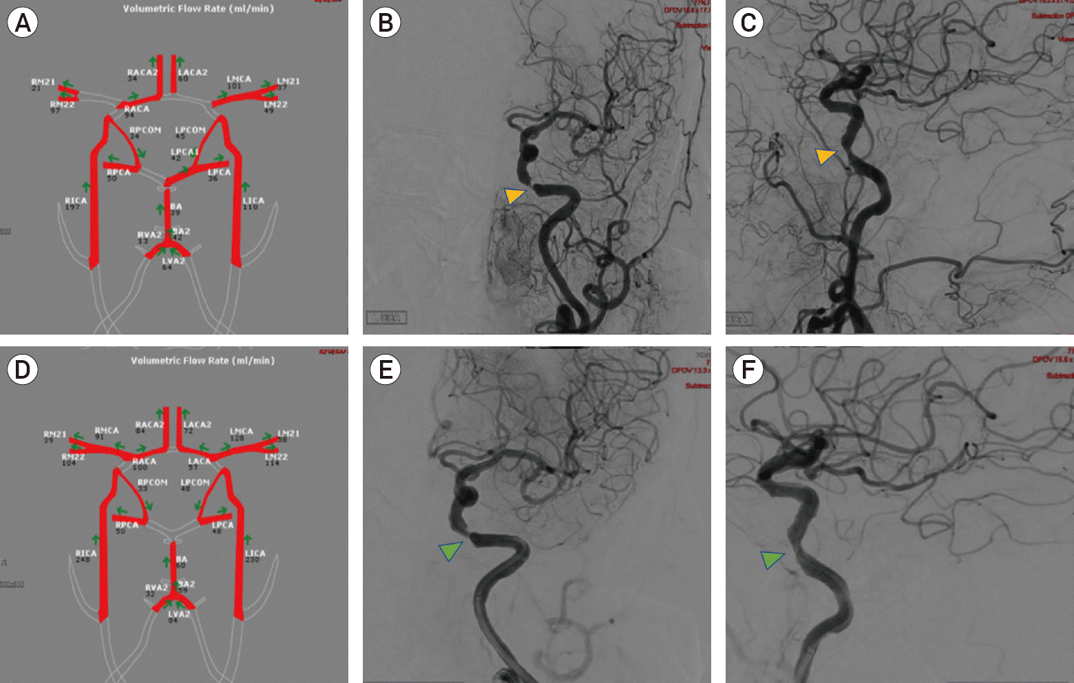 | Fig. 1.(A) MRI NOVA demonstrates decreased flow in left ICA. Frontal (B) and lateral (C) digital subtraction angiography images of the left ICA demonstrate a severe stenosis involving the petrous segment of the ICA (arrowheads). (D) Post procedure MRI NOVA demonstrates improved flow involving the left ICA. Post-balloon angioplasty and stenting frontal (E) and lateral (F) digital subtraction angiography images of the left ICA demonstrate improvement of the previously seen area of stenosis (arrowheads). MRI NOVA, magnetic resonance imaging Noninvasive Optimal Vessel Analysis; ICA, internal carotid artery.
|
Patient underwent diagnostic cerebral angiography, which showed severe stenosis (approximately 90%) of the petrous segment of the left ICA. Balloon angioplasty followed by Wingspan stent (Boston Scientific, Freemont, CA, USA) placement was performed. Repeat MRA NOVA following the procedure (1 week post-procedure) showed improved flow (230 mL/min) in the left ICA (Fig. 1). Hemodynamic augmentation was gradually tapered, and motor strength of right upper extremity remained stable. She was discharged to an acute rehabilitation facility. On 6-month follow-up, patient remained neurologically stable (mRS 2) and with no evidence of further strokes on MRI.
Case 2
A 52-year-old male with past medical history of HIV, asthma and frequent episodes of pneumonia was admitted in the emergency room of an outside hospital for confusion, dizziness and an episode of near syncope. On initial examination, patient was alert, oriented to person and place, and without any focal motor or sensory deficits. CT scan showed focal hypodensity involving the left basal ganglia. Serum glucose was noted to be elevated (666 mg/dL). Fluid hydration with normal saline and insulin was administered, and patient was transferred to our institution.
Upon arrival, patient had right-sided facial droop and right upper extremity paresis. National Institute of Health Stoke Scale (NIHSS) was 11. Patient was given tPA and admitted to the intensive care unit. Repeat CT showed interval mild increase in size of left basal ganglia infarction and acute left parietal infarctions. CT angiography showed focal high-grade stenosis of the proximal M1 segment of the left middle cerebral artery (MCA). Blood pressure was maintained at 140-180 mm Hg with vasopressors and patient was started on dual antiplatelet therapy. His examination improved the next day, with motor strength returning to 4/5 in the right upper extremity.
MRA NOVA was performed which showed reversal of flow in the left MCA M1 segment with low flow in the M2 branches of the left MCA (25 and 12 ml/min, compared to 127 and 52 mL/min in the M2 branches of the right MCA). Patient underwent cerebral angiogram which demonstrated a critical short segment left MCA stenosis. Balloon angioplasty followed by Wingspan stent insertion was performed, resulting in significant improvement of the stenosis. Post-procedure MRA NOVA performed 6 months later demonstrated improvement of flow in the left MCA M2 segments (30 and 61 mL/min) (Fig. 2).
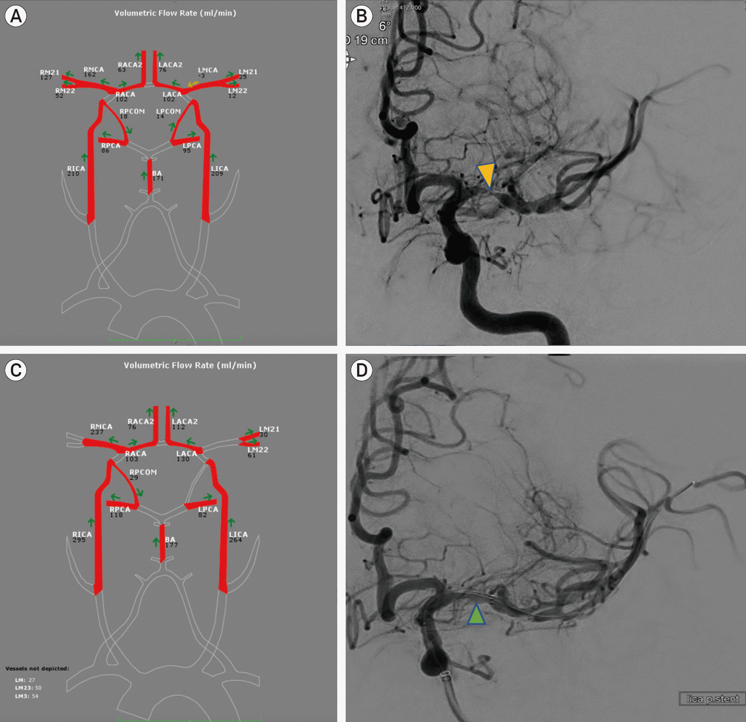 | Fig. 2.(A) MRI NOVA demonstrates decreased flow in the left MCA both M1 and M2 segments. (B) Frontal digital subtraction angiography image following left internal carotid artery injection demonstrates severe focal stenosis involving the proximal M1 segment of the left MCA (arrowhead). (C) MRI NOVA following angioplasty and stenting of the left MCA demonstrates improvement of flow through the left MCA. (D) Digital subtraction angiography immediately following angioplasty and stenting of the left MCA demonstrates improvement of the stenosis (arrowhead). MRI NOVA, magnetic resonance imaging Noninvasive Optimal Vessel Analysis; MCA, middle cerebral artery.
|
Patient remained stable and was discharged to an acute rehabilitation facility. At 3-month follow-up, patient demonstrated full strength of the right upper extremity, with mild dysarthria, and no facial droop (mRS 2).
Case 3
A 64-year-old male presented with aphasia and right-sided upper and lower extremity weakness while being evaluated for shortness of breath in the emergency department of an outside hospital. Past medical history was significant for multiple medical comorbidities including hypertension, diabetes, dyslipidemia, coronary artery disease and nephropathy. Initial CT scan did not show any bleed or acute transcortical infarction. However, CT angiogram showed significant stenosis of the left M1 segment of the MCA. Initial NIHSS was 17 and patient was given tPA and then transferred to our hospital.
Upon arrival, repeat CT showed a small volume of acute subarachnoid hemorrhage in the left frontal area. Prothrombin complex concentrate and cryoprecipitate were given to reverse tPA. Patient regained normal strength in the right upper and lower extremity, and partial improvement in aphasia was noted. He was then started on aspirin and ticagrelor and subsequent platelet reactivity assays were in therapeutic range.
MRA NOVA was performed, which demonstrated decreased flow in the left MCA (62 mL/min, compared to 101 ml/min in the right MCA) (Fig. 3). Patient underwent cerebral angiogram which showed severe focal left M1 segment stenosis. Balloon angioplasty and intracranial stenting with Wingspan stent was performed. Post-procedure MRA NOVA performed one day later showed increased flow within the left MCA (102 ml/min). The patient remained stable (mRS-3) and was discharged to an acute rehabilitation facility.
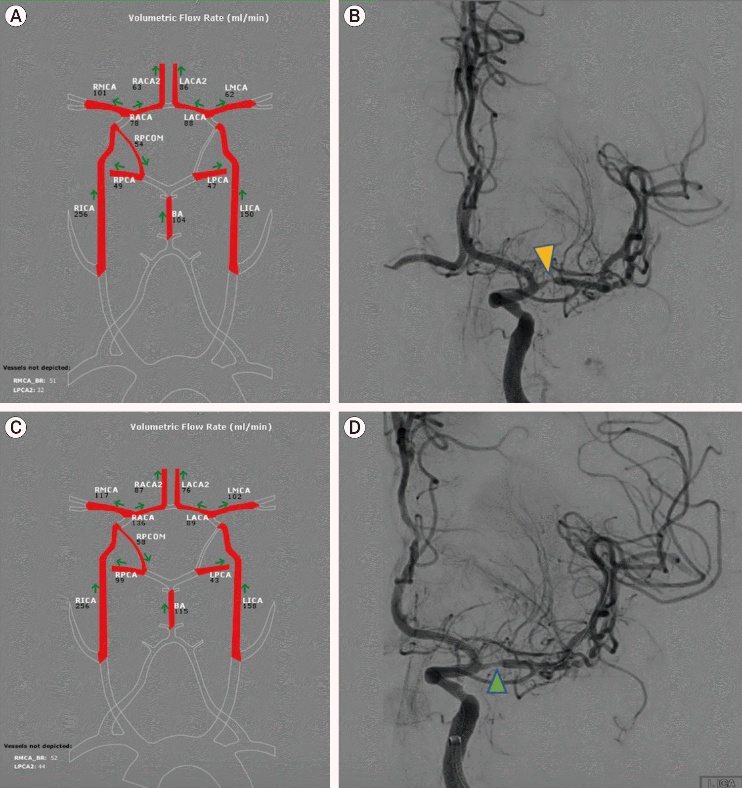 | Fig. 3.(A) MRI NOVA demonstrates decreased flow in the left MCA. (B) Digital subtraction angiography demonstrates severe focal occlusion of the proximal M1 segment of the left MCA (arrowhead). (C) MRI NOVA following balloon angioplasty and stenting of the left MCA demonstrates improvement in the flow of the left MCA. (D) Post balloon angioplasty and stenting angiography demonstrates improvement of the focal stenosis (arrowhead). MRI NOVA, magnetic resonance imaging Noninvasive Optimal Vessel Analysis; MCA, middle cerebral artery.
|
Go to :
DISCUSSION
ICAD can lead to ischemic stroke through several mechanisms which include hypoperfusion, artery-to-artery embolism, and plaque extension over small penetrating artery ostia [15]. Because of the heterogeneity of this condition, it is important to understand the mechanism of stroke in these patients, as treatment should be tailored to the specific mechanism of the disease. Traditionally, the underlying mechanism of stroke has been inferred by characteristics on imaging. For example, watershed distribution infarcts on MRI suggest hypoperfusion, whereas a distal wedge-shaped territorial infarct might indicate artery-to-artery embolism [12]. In this report, we have used Q-MRA to specifically evaluate the degree of hypoperfusion caused by intracranial stenosis, and propose that this quantitative imaging modality may help understand further the etiology of the stroke in these patients, and the potential impact of balloon and angioplasty and intracranial stenting.
Q-MRA is a technique that allows for non-invasive measurement of large vessel flow and velocities in the head and neck. The commercially available software, NOVA (Noninvasive Optimal Vessel Analysis, VasSol, Inc.), was utilized for calculating blood velocities within the vasculature of the head and neck for the patients in this report. This technique utilizes TOF imaging to provide 3D visualization of the vessels of interest, then the velocity and volume flow rates are measured using phase contrast MR technique [2,4,22,23]. The feasibility of blood flow measurements in individual cerebral vessels using phase contrast Q-MRA has been previously well demonstrated [19]. In addition, reference values for flow rates in all major cerebral arteries in healthy subjects across different ages using Q-MRA have been published in the past, and can serve as a guide for determining distal flow compromise in cases of intracranial arterial stenosis [1].
Different imaging techniques may allow for a qualitative measure of flow. Direct measure of the vessel lumen can give us information on the degree of stenosis, and may allow to qualitatively define the stenosis as mild, moderate or severe. However, there is no measure of collateral flow. Cerebral angiography may allow you to visualize some degree of collateral flow to the affected territory, however, this is subjective and to our knowledge not quantifiable. Direct, quantitative measurement of blood flow in the affected vascular territory with Q-MRA can provide information regarding collateral patters and hemodynamic effects of intracranial stenosis. Our premise is that patients without distal flow compromise, regardless of degree of stenosis, although at risk for stroke via local perforator ischemia, small vessel disease or emboli, are unlikely to benefit from interventions to augment flow [2].
Medical management of intracranial stenosis includes the use of antiplatelet medications and control of risk factors such as hypertension and dyslipidemia with antihypertensive and cholesterol-lowering medications. Aspirin is the most widely used antiplatelet agent. However, aspirin monotherapy has been associated with a relatively high rate of recurrent stroke in patients with symptomatic ICAD [11]. Dual antiplatelet therapy was used in the SAMMPRIS trial for patients with symptomatic ICAD, and demonstrated a lower rate of recurrent stroke in ICAD patients with 70-99% stenosis compared with historical controls [7]. However, despite maximized medical management, symptomatic ICAD has a high risk of stroke recurrence [11].
An early study of 45 patients with intracranial stenosis treated with angioplasty and Wingspan stent showed that the procedure was effective, reducing stenosis from 75% to 28%, and safe, with low periprocedural complications. The 30-day composite ipsilateral stroke or death rate was 4.5%, and 6-month composite ipsilateral stroke or death rate was 7% [21]. The encouraging results from prior studies prompted an expedited FDA approval of the device, and a large randomized controlled trial was conducted. SAMMPRIS Trial enrolled patients with a recent transient ischemic attack or stroke related to a 70-99% stenosis of a major intracranial artery, and randomized them to receive aggressive medical management therapy with or without percutaneous luminal angioplasty and stenting with the Wingspan stent system [8]. Recruitment was stopped early after 451 patients were enrolled (with intended enrolment of 764 patients) when interim analysis showed that the rate of stroke or death within 30 days was 14.7% in the stenting group compared to 5.8% in the medical management group. Furthermore, the probability of developing a stroke or death at 1 year was similarly higher in the stenting group compared to the medical management group (20% vs. 12.2%, respectively). The trial concluded that aggressive medical management is superior to stenting in high risk patients with severe intracranial stenosis.
Although the SAMMPRIS trial provides strong evidence to the superiority of medical management over stenting, the trial had some possible limitations. One critique is that patients were enrolled to the trial based on lesion severity, without considering the mechanism of stroke or the status of collateral supply and brain perfusion [21]. Sufficiently formed collateral supply has been demonstrated to play a crucial role in the avoidance of strokes in patients with high-degree of stenosis [18]. In addition, distal blood flow compromise has been associated with a higher risk of stroke [3]. Evaluating collateral supply and vascular reserve can perhaps select patients who are at higher risk of subsequent strokes, presumably a subset of patients that may benefit from stenting. Collateral supply can be investigated by measuring of oxygen extraction fraction on PET, vasodilator studies using breath-holding and transcranial doppler measurements of blood velocity, reduced blood flow and reactivity to acetazolamide, CT perfusion, as well as large-vessel quantitative MRA [14,24].
The risk of stroke from intracranial stenosis increases with the degree of luminal stenosis. The WASID (Comparison of Warfarin and Aspirin for Symptomatic Intracranial Arterial Stenosis) Trial showed that the probability of developing stroke at 1 year in patients with 70-79%, 80-89% and 90-99% stenosis and 100% occlusion was 17, 31, 5 and 33%, respectively. Patients with >70% stenosis of intracranial arteries and recent symptoms of ischemia have been identified to be at highest risk for subsequent strokes [13].
In the current report, analyzing blood flow with Q-MRA allowed us to determine the impact the stenosis had on blood flow and collateral supply. In fact, post-stenting Q-MRA demonstrated restoration of expected blood flow in the vascular territory affected. It is possible that angioplasty and stenting may be a suitable therapy for those ICAD patients where specifically blood flow is compromised, as opposed to those with ICAD where strokes occur secondary to other events such as artery-to-artery embolization. To this end, Q-MRA may be helpful in the workup of these patients.
Go to :
 XML Download