Journal List > J Cerebrovasc Endovasc Neurosurg > v.23(3) > 1147767

Oh, Kim, Kim, Lee, Sung, and Song: A case of an unruptured duplicated middle cerebral artery aneurysm–An unusual presentation of the distal internal carotid artery aneurysm

Abstract

The duplicated middle cerebral artery (DMCA) is an anatomic variation that arises from the distal internal carotid artery (ICA) and supplies blood to the middle cerebral artery (MCA) territory. Aneurysms of the DMCA have been reported in 36 cases in 2020. We also report a case of a 3.7 mm saccular aneurysm originating from the DMCA. A 52-year-old woman visited our hospital with worsening headache. She had no neurological abnormalities. Magnetic resonance imaging (MRI) and magnetic resonance angiography (MRA) revealed a right distal ICA aneurysm at the anterior choroidal artery. Cerebral angiography was performed to confirm the shape and the size of the aneurysm. Cerebral angiography revealed that the vessel that was originally identified as the anterior choroidal artery by the MRA was actually the duplicated MCA that was originating from the aneurysm neck and was supplying the MCA territory. The patient’s aneurysm was clipped using a transsylvian approach and she recovered without any neurological symptoms. DMCAs are rare and often associated with aneurysms and require preoperative evaluation to confirm the vascular status, aneurysm characteristics, and the shape of the parent artery.

INTRODUCTION

The duplicated middle cerebral artery (DMCA) arises from the distal internal carotid artery (ICA) and supplies blood to the middle cerebral artery (MCA) territory. This is a rare anatomical variation and is rarely associated with aneurysm [14]. The incidence of identifying the DMCA was 0.7-2.9% on autopsy and 0.24-1.5% on angiography [4]. In addition, the DMCA aneurysm is rare, and only 36 cases have been reported in the literature to this day [5,9]. DMCA was classified into two types: (1) Type A: an anomalous MCA at the ICA terminus and (2) Type B: an anomalous MCA arising between the origin of the anterior choroidal artery (AChA) and the ICA bifurcation [6]. Here, we report a case of a small saccular aneurysm originating from the type B DMCA, which was initially thought to be an anterior choroidal artery aneurysm in the MRA.

CASE REPORT

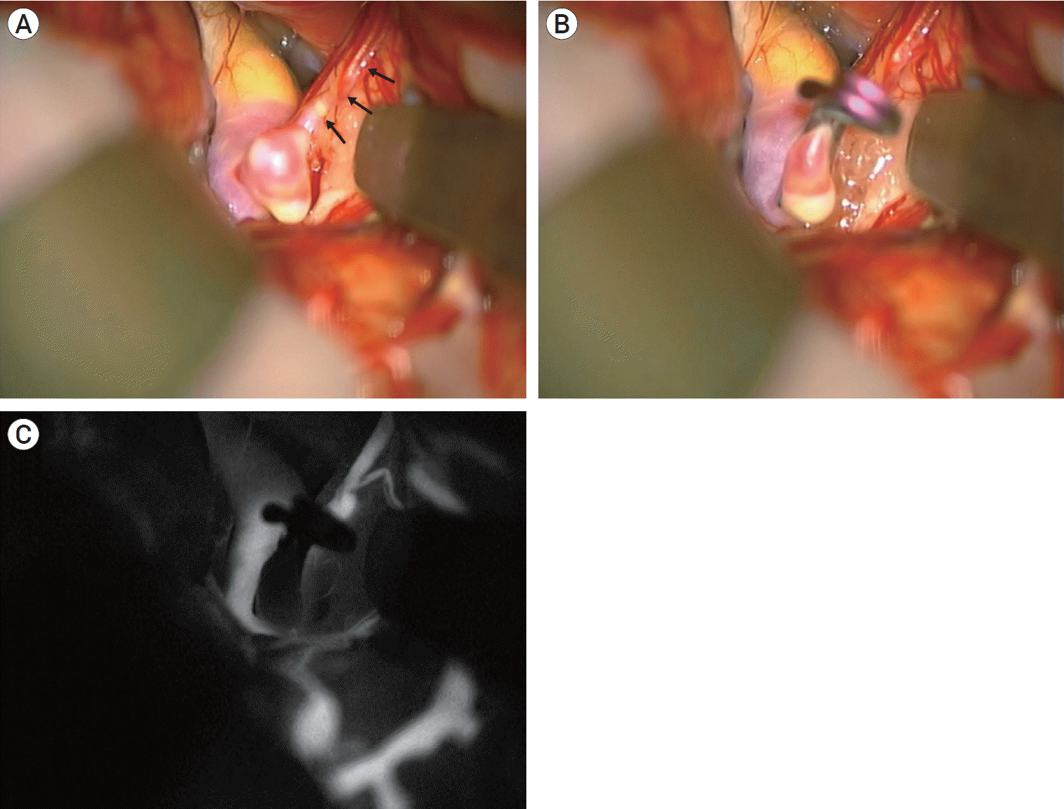
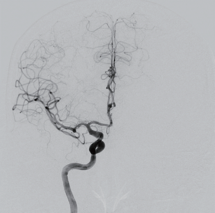
A 52-year-old woman visited our institute complaining of a headache. Magnetic resonance imaging (MRI) and magnetic resonance angiography (MRA) of the brain showed a saccular 3.2×3.8 mm aneurysm arising from the right distal ICA (Fig. 1). At first, it was diagnosed as an AChA aneurysm, which typically projects in the lateral direction, however, conventional cerebral angiography revealed an unusual artery arising from the distal ICA between its bifurcation and AChA, and an aneurysm arising from the origin of this artery (Fig. 2). Finally, we confirmed that the anomalous artery was not the AchA, but a type B DMCA. Due to the patient’s concerns, concerns about the possibility of rupture and the greater hemodynamic stress that is endured by the anomalous DMCA aneurysm, treatment, instead of a regular imaging follow-up, was recommended. Surgical clipping was performed because of the presence of stenosis at the DMCA origin. It was performed using the pterional approach, with opening of the Sylvian fissure. After dissection of the Sylvian fissure and the DMCA, the aneurysm, which was adherent to the temporal lobe cortex, was identified. The aneurysmal dome was detached from the cortex with careful dissection, and the temporal lobe was retracted. The aneurysm was successfully clipped after removing a small piece of the temporal lobe because the DMCA ran posteriorly and the origin of the DMCA was not confirmed (Fig. 3A, B). The DMCA passed into the Sylvian fissure along the anterior pole of the temporal lobe to supply the MCA territories. The flow of the DMCA was checked using Doppler and near-infrared indocyanine green video angiography (Fig. 3C). The patient recovered fully after the surgery; and was discharged without any neurological deficits. Postoperative cerebral angiography revealed a complete occlusion of the aneurysm (Fig. 4).

DISCUSSION

The presence of an anomalous artery originating between the anterior choroidal artery and the ICA bifurcation was described in 1959 [1] and Crompton et al. [3] named it an “accessory MCA.” [1,3] However, Teal et al. [13] reserved the use of the term “accessory MCA” only for the anomalous artery arising from the anterior cerebral artery, which passed laterally into the Sylvian fissure with the MCA [13]. They renamed the anomalous MCA, arising from the ICA, as the “duplication of the MCA” regardless of its size. There is little relevance of the occurrence of the aneurysms in the DMCA, because of their rarity [7].
Kai et al. [6] classified the DMCA into two types: type A and type B. According to Kai’s classification, type A DMCA arises at the top of the ICA, giving the impression of an early bifurcation [6]. Teal et al. [13] referred to this type as an early bifurcation of the MCA [13]. Type A DMCA has an outer diameter similar to that of the main MCA trunk; and also has perforating arteries, such as the lateral lenticulostriate arteries, which supply the basal ganglia [15]. In type B, the DMCA arises from the ICA between the AChA and the ICA bifurcation [6]. Teal et al. postulated that Type B DMCA is an anomalous early manifestation of an MCA branch. This type of DMCA has a smaller diameter than that of the main MCA trunk [8] and has no perforators [8]. In this case, we could confirm that type B DMCA was present in our case and it was supplying the anterior part of the temporal lobe.
Type A is the more common DMCA type; however, associated aneurysms are usually found in type B DMCA [6]. They postulated that type B DMCA is exposed to a higher hemodynamic stress because of its sharper separation angle from the ICA. Chang et al. [2] described a type B DMCA based on the separation angle [2]. Type A has a course parallel to the main MCA branch, and type B has a course that curves toward the temporal lobe with a sharper separation angle. They mentioned that only the DMCAs with a sharply curved course to the temporal lobe were associated with aneurysms originating from the origin of this vessel. The DMCA observed in the present case also demonstrated a sharp separation angle and was under a higher hemodynamic stress (Fig. 2A). Aneurysms associated with type A DMCA are rare; but when they are reported, they are usually associated with other pathological conditions such as dolichoectasia and stenosis of the main trunk [9,11]. Stojanović et al. has also mentioned the multiple cerebral aneurysms that are associated with the segmental DMCA also known as the fused DMCA; but these aneurysms have not been reported [12].
Identification of an anomalous DMCA before surgery is important for the surgical dissection of the aneurysm associated with type B DMCA because it may be similar to the AChA aneurysm, just like the present case. The endovascular technique for the repair of the type B DMCA aneurysms is similar in concept to the treatment of the small AChA aneurysms because of their similar location and projected direction [14]. However, care should be taken during the microscopic dissection, because the DMCA passes laterally through the Sylvian fissure and supplies the anterior temporal artery territories, unlike the AChA. Iwata et al. [5] also reported a case of a DMCA aneurysm, initially diagnosed as an unruptured aneurysm arising from the ICA, appearing to be an AChA aneurysm, on three-dimensional computed tomography. However, they found an unidentified vessel around the aneurysmal neck that could not be easily separated from the aneurysm during surgery; thus, coating of the aneurysm was completed without clipping. Postoperative cerebral angiography revealed a DMCA with an associated aneurysm, and coil embolization was performed.
Despite its anomalous origin, the DMCA contributes to the normal cerebral blood supply. DMCA usually branches into the temporopolar, the anterior and the middle temporal arteries and also has perforating arteries in some cases [10]. Regardless of whether a perforator is present or not, DMCA contributes to the normal cerebral blood supply, so that care should be taken not to damage it during surgery. We successfully clipped the aneurysm without damage to the DMCA, but bypass surgery should be considered when damage is expected [6].

CONCLUSIONS

DMCAs are rare and often related to aneurysms, especially type B DMCAs. Preoperative evaluation including cerebral angiography, should be considered. It is important to keep in mind the possibility of a DMCA when unexpected arteries are encountered around the distal ICA, particularly in cases of distal ICA aneurysm surgery based on the MRA or CTA findings.

Notes

Disclosure

The authors report no conflict of interest concerning the materials or methods used in this study or the findings specified in this paper.

REFERENCES

1. Alpers BJ, Berry RG, Paddison RM. Anatomical studies of the circle of Willis in normal brain. AMA Arch Neurol Psychiatry. 1959; Apr. 81(4):409–18.
crossref
2. Chang HY, Kim MS. Middle cerebral artery duplication: classification and clinical implications. J Korean Neurosurg Soc. 2011; Feb. 49(2):102–6.
3. Crompton MR. The pathology of ruptured middle-cerebral aneurysms with special reference to the differences between the sexes. Lancet. 1962; Sep. 2(7253):421–5.
crossref
4. Hori E, Kurosaki K, Matsumura N, Yamatani K, Kusunose M, Kuwayama N, et al. Multiple aneurysms arising from the origin of a duplication of the middle cerebral artery. J Clin Neurosci. 2005; Sep. 12(7):812–5.
crossref
5. Iwata M, Kawaguchi S, Manaka H. A case of unruptured cerebral aneurysm arising from duplicate origin of the middle cerebral artery. No Shinkei Geka. 2020; Jun. 48(6):515–20.
6. Kai Y, Hamada J, Morioka M, Yano S, Kudo M, Kuratsu J. Treatment of unruptured duplicated middle cerebral artery aneurysm: case report. Surg Neurol. 2006; Feb. 65(2):190–3. discussion 193.
crossref
7. Kim JS, Lee CH, Park H, Han JW. An unruptured cerebral aneurysm at the origin of the duplicated middle cerebral artery. J Cerebrovasc Endovasc Neurosurg. 2015; Sep. 17(3):223–6.
crossref
8. Komiyama M, Nakajima H, Nishikawa M, Yasui T. Middle cerebral artery variations: duplicated and accessory arteries. AJNR Am J Neuroradiol. 1998; Jan. 19(1):45–9.
9. Mori K, Tamase A, Seki S, Iida Y, Kawabata Y, Nakano T, et al. Duplicated middle cerebral artery associated with aneurysm at M1/M2 bifurcation: a case report. J Med Case Rep. 2018; Oct. 12(1):283.
crossref
10. Pico F, Labreuche J, Touboul PJ, Leys D, Amarenco P. Intracranial arterial dolichoectasia and small-vessel disease in stroke patients. Ann Neurol. 2005; Apr. 57(4):472–9.
crossref
11. Ren H, Ma L, Wei M, Li J, Yu M, Yin L. Duplicated middle cerebral artery origin with an aneurysm. Medicine (Baltimore). 2018; Mar. 97(9):e9947.
crossref
12. Stojanović NN, Kostić A, Mitić R, Berilažić L. Correlation between multiple cerebral aneurysms and a rare type of segmental duplication of the middle cerebral artery. BMC Neurol. 2020; Jan. 20(1):3.
crossref
13. Teal JS, Rumbaugh CL, Bergeron RT, Segall HD. Anomalies of the middle cerebral artery: accessory artery, duplication, and early bifurcation. Am J Roentgenol Radium Ther Nucl Med. 1973; Jul. 118(3):567–75.
crossref
14. Tsang COA, Smith L, Klostranec J, Orru E, Pereira VM. Ruptured duplicated middle cerebral artery aneurysm successfully treated by coil embolization with balloon remodeling. World Neurosurg. 2018; Dec. 120:509–10.
crossref
15. Umansky F, Dujovny M, Ausman JI, Diaz FG, Mirchandani HG. Anomalies and variations of the middle cerebral artery: a microanatomical study. Neurosurg. 1988; Jun. 22(6 Pt 1):1023–7.
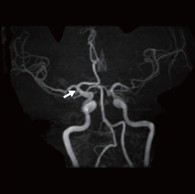
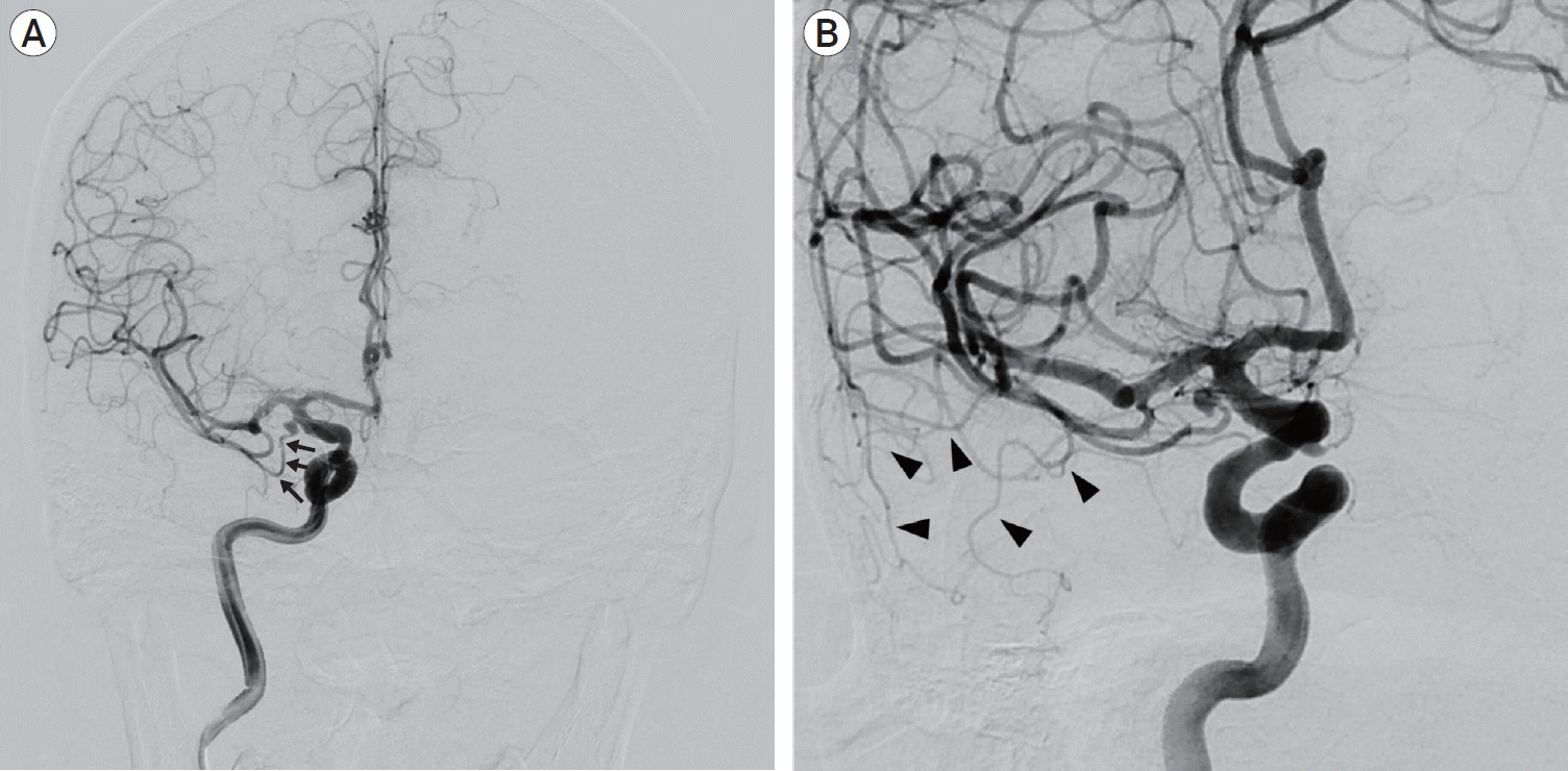
Fig. 1.
Magnetic resonance angiography showing a right distal ICA aneurysm (arrow) with lateral projection, suggestive of a typical AChA aneurysm. ICA, internal carotid artery; AChA, anterior choroidal artery.
jcen-2021-e2020-10-004f1.tif
Fig. 2.
Conventional cerebral angiogram of the right ICA (A) AP view of the right ICA angiogram demonstrating type B DMCA. Note the sharp separation angle from the ICA. (B) Oblique view of the right ICA angiogram showing the territories supplied by the DMCA (arrow heads). ICA, internal carotid artery; DMCA, duplicated middle cerebral artery.
jcen-2021-e2020-10-004f2.tif
Fig. 3.
Intraoperative findings (A) Intraoperative microscopic view showing the DMCA (arrows) and related aneurysm. Note the DMCA running laterally to supply the anterior pole of the temporal lobe. (B) Microscopic view showing well clipped aneurysm. (C) ICGVA demonstrating the patent DMCA flow. DMCA, duplicated middle cerebral artery; ICG-VA, indocyanine green video angiography.
jcen-2021-e2020-10-004f3.tif
Fig. 4.
Postoperative cerebral angiography revealing the complete occlusion of the aneurysm and the patent DMCA. DMCA, duplicated middle cerebral artery.
jcen-2021-e2020-10-004f4.tif
TOOLS
Similar articles