Abstract
Kirschner wire (K-wire) has been widely used for treatment of fracture for its cost-effectiveness and reliability. This case presents the K-wire breakage in distal interphalangeal joint (DIPJ) fixation. A 55-year-old male patient was injured by a knife and showed rupture of extensor tendon at 1/2 of middle phalanx. A 0.9-mm K-wire was implemented for DIPJ extension, and tenorrhaphy was done. After 6 weeks, we detected breakage of K-wire in the follow-up X-ray. The broken K-wire in the distal phalanx was removed. We removed the remaining K-wire through an incision on volar side of middle phalanx under C-arm after 2 weeks for the patient’s personal reasons. Breakage during postoperative K-wire maintenance is exceedingly rare. This patient is presumed to have ruptured because he continued using his finger. Therefore, while K-wire is present, continued use of finger without protection may cause breakage, so protective measures such as splint are required.
Go to : 
Kirschner wire (K-wire) has been widely adopted in treatment of fracture and dislocation of hand for its cost-effectiveness and proven reliability [1]. However, some studies have described the complication of K-wire fixation. Botte et al. [2] reviewed a series of 422 K-wire pins placed in the hand and wrist and reported an 18% complication rate, including pin loosing, pin bending, and pin tract infection. According to biomechanical study, K-wire offers a rigidity similar to that of interosseous wiring and other methods of fixation [3].
There were several case reports of K-wire breakage during removal after bone reduction [3–7]. However, this case study presents the first known case of K-wire breakage in internal fixation state.
Go to : 
A 55-year-old male patient was injured by a kitchen knife and showed total rupture of extensor tendon at 1/2 of middle phalanx.
Under brachial plexus block anesthesia, a tourniquet was applied to the upper arm at 250 mmHg. First, distal phalanx was extended in order to neutralize the subluxated joint and then was inserted with 0.9-mm K-wire from fingertip to middle phalanx shaft level. After opening the existing wound using 4-0 MERSILK (Ethicon Inc., Bridgewater, NJ, USA), ruptured extensor tendon was identified. We performed epitendinous suture using PDS 6-0, then Core suture was performed with the 4 strand method using 5-0 PDS (Ethicon Inc.) (Fig. 1). Splint was applied on the volar side after the skin suture. He was discharged 2 days after operation. He immediately started rehabilitation. He was given a new aluminum finger splint that protected his distal interphalangeal (DIP) joint. At the moment, there was no visible sign of pin site problem. X-ray follow-up at outpatients every 2 weeks confirmed that there was no problem with the fixation.
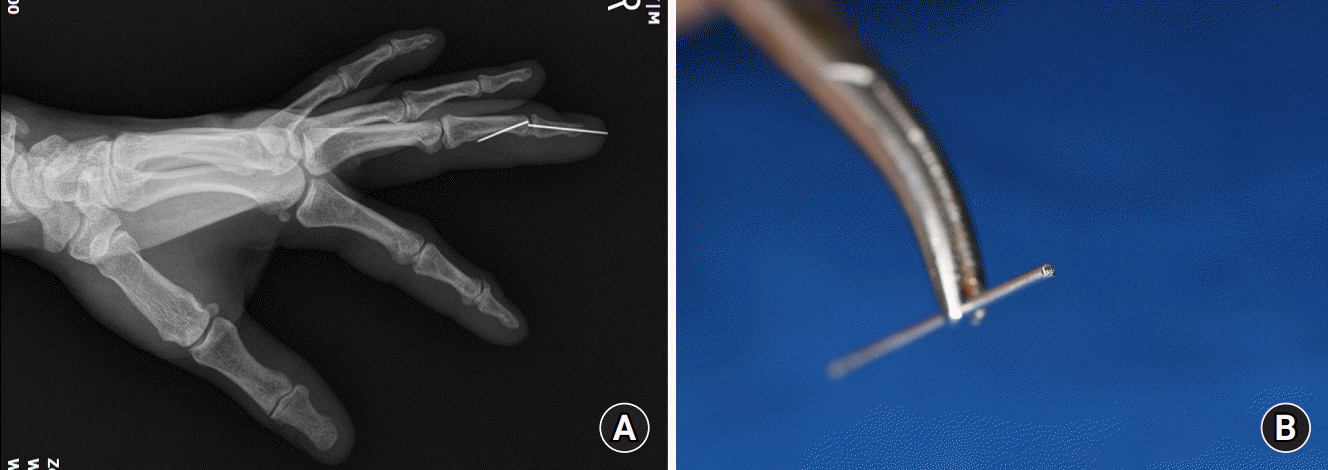
After postoperative 6 weeks, we detected breakage of K-wire in the follow-up X-ray image. At 5 weeks postoperatively, the patient did not wear finger splint and continued lifting heavy objects against the doctor’s instructions. Distal portion of the broken K-wire was immediately removed at outpatient clinic and antibiotics were used in order to prevent infection and aluminum splints were applied at neutral position to prevent further damage to DIP joint (Fig. 2).
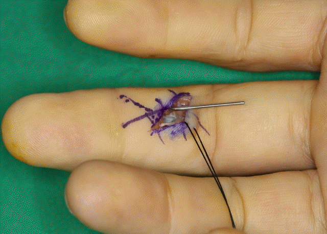
Surgery was recommended to the patient to remove the K-wire, but the removal was performed after 8 weeks postoperatively for personal reasons. The exact location of the K-wire was identified under C-arm and the periosteum was exposed while trying not to damage the arteries, nerves, and tendons after giving incision on the volar side. Distal segment of K-wire was partially exposed and removed with a wire holder. C-arm imaging confirmed that there was no remnant of K-wire (Fig. 3).
Written informed consent was obtained for publication of this case report and accompanying images.
Go to : 
K-wire is strong enough that only 14 cases of intraoperative breakage out of 11,856 surgeries were reported so far [4]. For this reason, K-wire is commonly and adaptably used for internal fixation in hand and wrist fractures and is also used for arthrodesis of digit joint [3].
According to Stahl et al. [5], minor complications caused by K-wire insertion occurred in only 15.2%. Pin loosing, pin tract infection, pin bending, or pin breakage were reported, but none of them caused permanent disability after the pin removal. Major complications such as osteomyelitis, tendon injury, and damage to nerve were noted in only 1.7%.
Pin loosing, the most common complication, occurs due to inability to accurately position the fixation level during reduction of fracture and can be prevented by accurately evaluating the level and location of reduction through intraoperative X-ray fluoroscopy and removing the incorrectly located pin immediately [5].
Pin tract infection is also common, but it can be treated with motion limitations and oral antibiotics, which can also be resolved by removing the pins. Osteomyelitis is the most serious complication. According to a study by Botte et al. [2], it occurs in only 1% of the entire pin tract infections. When signs of pin tract infection are seen, removing the internal fixator and giving proper treatment immediately can prevent infection from progressing into severe osteomyelitis.
There are studies on K-wire breakage used when fixing the toe joint. But, cases of broken K-wire after fixing the finger joint could not be found. There were 27 cases of K-wire breakage in 1,002 cases where the toes and metatarsophalangeal joints were fixed using 1.1-mm K-wires. As a method to reduce K-wire breakage, a K-wire with larger diameter was used; and considering that most of the K-wires were broken between 4 and 6 weeks, it was suggested that rapid K-wire removal could be a solution. However, when using a thick K-wire, the articular cartilage may be further damaged and it is difficult to place the K-wire in the medullary canal of the smaller bones. In addition, it was emphasized that the patient should wear splint and try to avoid direct trauma to the K-wire insertion site according to the doctor’s treatment instructions [6].
K-wire is widely used in bone fixation due to its rigidity, but breakage is rare. In order to avoid such complication, it is necessary to precisely perform closed reduction under fluoroscope and use K-wires of sufficient thickness [1,5].
In addition, the most important factor is the patient’s compliance [5,6]. Patients should keep their splints strictly and be careful not to be shocked according to the doctor’s exact treatment instruction, and they should check their progress on the given follow-up date in outpatient department.
Go to : 
REFERENCES
1. Black D, Mann RJ, Constine R, Daniels AU. Comparison of internal fixation techniques in metacarpal fractures. J Hand Surg Am. 1985; 10:466–72.

2. Botte MJ, Davis JL, Rose BA, et al. Complications of smooth pin fixation of fractures and dislocations in the hand and wrist. Clin Orthop Relat Res. 1992; (276):194–201.

3. Hong SJ, Lee HJ, Kim JY, Eo SR, Cho SH. K-wire breakage during metalware removal due to a defective k-wire shaft. Arch Plast Surg. 2013; 40:655–7.

4. Pichler W, Mazzurana P, Clement H, Grechenig S, Mauschitz R, Grechenig W. Frequency of instrument breakage during orthopaedic procedures and its effects on patients. J Bone Joint Surg Am. 2008; 90:2652–4.

5. Stahl S, Schwartz O. Complications of K-wire fixation of fractures and dislocations in the hand and wrist. Arch Orthop Trauma Surg. 2001; 121:527–30.

Go to : 


 PDF
PDF Citation
 Print
Print


 XML Download