INTRODUCTION
MATERIALS AND METHODS
1. Materials
2. Surgical technique
Scaphoid preparation
 | Fig. 1.(A) Anterior (Russe) scaphoid exposure shows the nonunion site. When the nonunion site was exposed, an oval cortical window was made using a 3-mm diameter high-speed burr on the volar aspect of the proximal and distal pole of the scaphoid. (B) Intraoperative photos of debridement of the nonunion site at the scaphoid and preparation to accept the vascularized medial femoral condyle graft. (C) The graft was further shaped to fit the nonunion site. The periosteal surface attached to the pedicle should lie volar. (D) Vascular repairs were end-to-side to the radial artery and end-to-end to a vena comitans. |
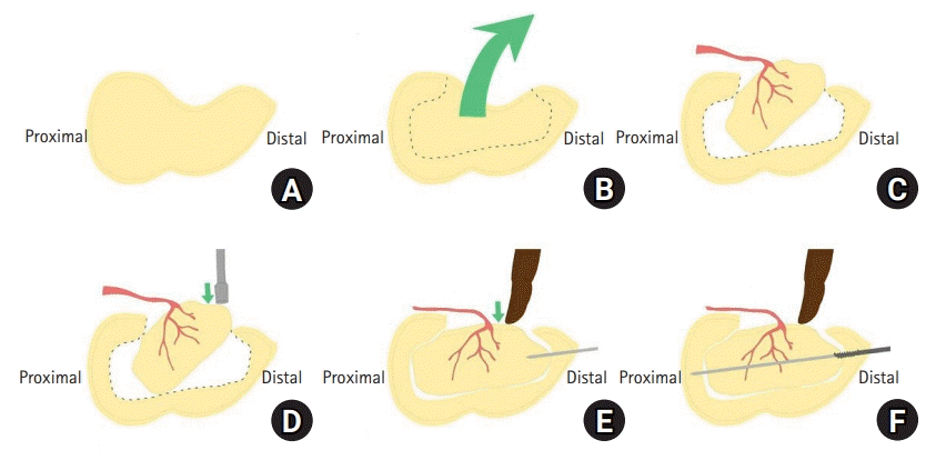
 | Fig. 2.(A) Lateral aspect of the scaphoid. (B) The surgeon excavated as much necrotic bone as possible if encountered while leaving the dorsal cortex intact. (C) The graft is inset obliquely into the scaphoid defect with the cortex facing palmarly. (D) Push the distal part of the graft with the impactor. (E) To fit graft properly without rotation or extrusion of graft and vessels, the surgeon holds the distal part of the graft with the index finger during temporary fixation with guide wire. (F) A 3.0-mm partially threaded headless compression screw was inserted along the guide wire and the guide wire was removed. |
Free vascularized medial femoral condyle bone graft harvest
 | Fig. 3.(A) Preparation and harvesting of the medial femoral condyle graft. The vastus medialis fascia was incised sharply at its posterior aspect, and the muscle retracted properly exposing the medial genicular artery system. (B) A rectangular shape of sufficient size to fill the gap in the scaphoid is marked out, the periosteum sharply incised, and the bone cut with a micro sagittal saw and osteotomes. (C) The corticocancellous graft was harvested with its vascular pedicle. |
Graft in setting, fixation and vascular anastomosis
3. Clinical and radiographic evaluation
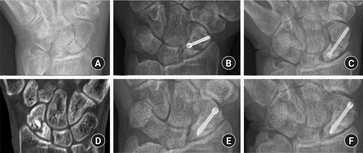
 | Fig. 4.(A) Scaphoid view of the wrist of a 51-year-old man, showing an scaphoid fracture. (B) Anteroposterior and (C) scaphoid view of wrist at 10 month after 1st operation in other hospital, showing an established scaphoid nonunion, sclerotic change around the fracture site. (D) Postoperative computed tomography at 10 month after 1st operation in other medical center, showing sclerotic change. (E) Anteroposterior and (F) scaphoid view of wrist 1-year after the last operation, showing achievement of union, correction, and maintenance of carpal height index. |


 PDF
PDF Citation
 Print
Print


 XML Download