Journal List > Korean Circ J > v.51(9) > 1147720

Xu, Huang, Liu, Bai, Ma, Cai, Zhang, and Zhang: Heart Failure Is Associated with Increased Risk of Long-Term Venous Thromboembolism

Author's summary

The risk of venous thromboembolism (VTE) in out-patients with heart failure (HF) in long-term period is still controversial, resulting in unclear recommendations for long-term treatment. In this analysis, we found that HF was an independent risk for VTE and pulmonary embolism but not deep vein thrombosis in long-term follow-up period. Patients with chronic HF were prone to have higher risk of VTE. This meta-analysis provided an evidence which is supportive for developing strategies for prevention of VTE in patients with HF.

Abstract

Background and Objectives

Venous thromboembolism (VTE), consisting of deep vein thrombosis (DVT) and pulmonary embolism (PE), is highly prevalent in in-hospital HF patients and contributes to worse prognoses. However, the risk of VTE in out-patients with HF in long-term period is controversial. This study aimed to evaluate the associations between HF and the risk of VTE in a long-term follow-up duration.

Methods

We searched for studies investigating the risk of VTE, PE, and DVT in patients with HF before April 15, 2020, in PubMed, MEDLINE, and Embase databases. Cohort studies and post hoc analysis of RCTs were eligible for inclusion if they reported relative risk of VTE, DVT or PE in patients with HF in more than 3-month follow-up period.

Results

We identified 31 studies that enrolled over 530,641 HF patients. Overall, patients with HF were associated with an increased risk of VTE (risk ratio [RR]=1.57, 95% confidence interval [CI]=1.34–1.84) and PE (RR=2.00, 95% CI=1.38–2.89). However, the risk of DVT was not significantly increased in HF patients (RR=1.33, 95% CI=0.67–2.63). Subgroup analysis showed that patients with chronic HF (RR=1.54, 95% CI=1.32–1.80) had a higher risk of VTE than those with acute HF (RR=0.95, 95% CI=0.68–1.32).

Conclusions

In conclusion, HF was an independent risk for VTE and PE but not DVT in a long-term follow-up period. Patients with chronic HF were prone to suffer from VTE than acute HF.

INTRODUCTION

Heart failure (HF) is a global public health problem that affects an estimated 26 million people worldwide,1) with an increasing prevalence and mortality rate.2)3) Due to immobility and bedridden status, the short-term risk of venous thromboembolism (VTE), including deep vein thrombosis (DVT), pulmonary embolism (PE), and thromboembolism in other veins, increased in patients with HF4)5) and may contribute to worse prognoses.6)7) A previous meta-analysis showed that the in-hospitalized incidence of VTE in patients with HF was 2.66% and the risk of VTE in in-hospitalized HF patients (<60 days) was almost 70% higher compared with non-HF patients.6) Clinical trials revealed that short-term thromboprophylaxis could be beneficial in HF patients.7) However, the risk of VTE in out-patients with HF in a long-term period is still controversial,8)9)10) which leads to unclear recommendations for long-term treatment in HF patients. Given these inconsistencies, we synthesized available data to quantify the risk of VTE in HF in long-term follow-up time.

METHODS

Search strategy

Our search was performed according to the recommendations of the Meta-Analysis of Observational Studies in Epidemiology Group.11) We searched for records published up to April 15, 2020, in PubMed, MEDLINE, and Embase databases, using keywords “venous thromboembolism” or “vein thromboembolism” or “vein thrombosis” or “venous thrombosis” or “deep venous thrombosis” or “deep vein thromboembolism” or “deep venous thromboembolism” or “deep vein thrombosis” or “pulmonary embolism” or “lung embolism” or “lung thromboembolism” or “pulmonary thromboembolism” and “heart failure” or “cardiac dysfunction” or “cardiac failure” (Supplementary Table 1). The search was restricted to human studies, but there were no language or publication form restrictions. The strategies for other databases were similar but adapted where necessary. Reference lists were also manually checked to identify other potential studies.

Selection criteria

Cohort studies or post hoc analyses of randomized controlled trials (RCTs) were included in this analysis. Patients who diagnosed HF (chronic HF: with the combination of symptoms [such as dyspnea] and cardiac dysfunction proven by an echocardiogram [left ventricular ejection fraction [LVEF] <50%] or defined by International Classification of Diseases, Ninth Revision [ICD-9] code, acute HF: defined by ICD-9 code) with an over 3-month followed-up time were eligible. We included datum if they presented original data on rates (number of events per follow-up period) or relative risks like risk ratios (RRs), odds ratios, and hazard ratios of all VTE (primary outcome), or DVT or PE alone (secondary outcomes) in HF patients, compared with patients without HF. Diagnoses of VTE, DVT or PE were based on ICD, or imaging examination such as compression venous ultrasound, ventilation-perfusion scan, gray-scale, and Doppler sonography or venography. Animal studies, cross-sectional studies, case-control studies, case reports, studies with follow-up periods <3 months, or data derived from the same studies were excluded. We did not exclude patients with cancer or patients undergoing surgery, who are already at high risk for VTE. Therefore, we were able to include all studies in our meta-analysis and comprehensively investigate the risk of VTE, PE or DVT, and HF in long-term follow-up duration.

Data extraction

Two investigators independently conducted literature searches, reviewed the potential articles, and abstracted data from eligible studies. Discrepancies were adjudicated by discussions with other investigators.
All available data were extracted from included studies, including country or region, study design, patient's population with HF, type of HF, follow-up time, use of anticoagulation therapy, number of participants with HF, ages of participants, adjusted relatives risks with 95% confidence intervals (CIs) and diagnostic criteria for VTE, PE, and DVT.

Quality assessment of studies

We assessed the risk of bias with an adapted and modified Newcastle-Ottawa Scale for observational studies, recommended by previous researchers.6) There were 6 items and 1 point was scored for each item. The judgments are diagnosis of VTE, PE, and DVT, HF diagnoses, study population (whether are restricted to patients with surgery or cancer), adjustment for age and sex, adjustment for recent major surgery and active malignancy and adjustment for other risk factors. Studies that received one point in all 6 items were judged as high quality (Supplementary Table 2).

Synthesis and analysis

Relative risks of VTE, PE, or DVT from each study and 95% CIs were logarithmically transformed and the corresponding standard errors (SEs) were calculated to stabilize the variance and normalize the distribution. We used the inverse variance method to combine the calculated log RRs and SEs. We investigated statistical heterogeneity across studies with the I2 statistic and did sensitivity analyses by omitting one cohort at a time. We assessed publication bias with Begg's test and inspecting funnel plots in which the natural log of RR was plotted against its SE.12) Meta-regression analysis was used to determine the impact of participants' age and follow-up duration upon the outcomes if data were reported in more than 10 studies according to Cochrane guidelines.13) We did subgroup analyses of primary outcomes based on study characteristics: type of heart failure (acute, chronic, and not reported), follow-up duration (3–6 months, 6 months–1 year, >1 year), anticoagulation therapy (no anticoagulation therapy, anticoagulation therapy in clinical practice, not reported), age (<60 years old, 60–70 years old, >70 years old), surgery (underwent, not underwent, not mentioned) and, cancer (suffered, not suffered, not mentioned). The p values were 2-tailed, and statistical significance was set at 0.05. All analyses were conducted using RevMan (version 5.3; The Cochrane Collaboration, Copenhagen, Denmark) and Stata software (version 15.0; Stata Corp LP, College Station, TX, USA).

RESULTS

Studies retrieved and characteristics

The search retrieved 4,242 manuscripts initially, and 621 duplicated articles were removed. After screening titles and abstracts, we took 105 records to be potentially eligible and gave an in-depth review of each full-text article. Finally, we included 31 (Supplementary Reference 1) studies, with over 530,641 patients with HF (Figure 1). The median quality of the included studies was 4.3 points. The sample size ranged from 34 to 270,535 and the mean age of patients of included studies was from 39.9 to 85.7 years. The follow-up period ranged from 3 months to 26 years, and the mean follow-up was 35.5 months (Table 1).
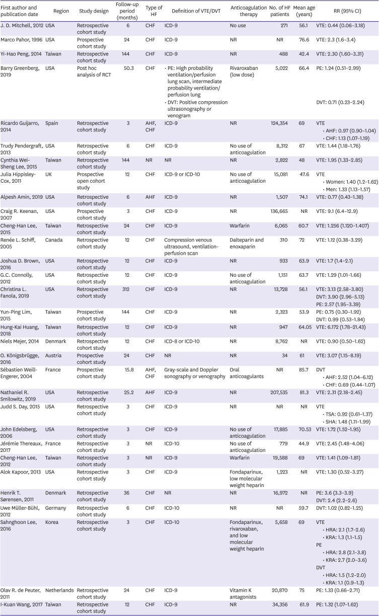
Figure 1

Flow of papers through review.

DVT = deep vein thrombosis; HF = heart failure; PE = pulmonary embolism; VTE = venous thromboembolism.
kcj-51-766-g001
Table 1

Characteristics of studies

kcj-51-766-i001
First author and publication date Region Study design Follow-up period (months) Type of HF Definition of VTE/DVT Anticoagulation therapy No. of HF patients Mean age (years) RR (95% CI)
J. D. Mitchell, 2012 USA Retrospective cohort study 6 CHF ICD-9 No use 271 56.1 VTE: 0.44 (0.06–3.18)
Marco Pahor, 1996 USA Prospective cohort study 24 CHF ICD-9 NR NR 76.6 VTE: 2.3 (1.6–3.4)
Yi-Hao Peng, 2014 Taiwan Retrospective cohort study 144 CHF ICD-9 NR 488 42.4 VTE: 2.30 (1.60–3.31)
Barry Greenberg, 2019 USA Post hoc analysis of RCT 50.3 CHF • PE: High probability ventilation/perfusion lung scan, intermediate probability ventilation/perfusion lung Rivaroxaban (low dose) 5,022 66.4 PE: 1.24 (0.51–2.99)
• DVT: Positive compression ultrasonography or venogram DVT: 0.71 (0.23–2.24)
Ricardo Guijarro, 2014 Spain Retrospective cohort study 3 AHF, CHF ICD-9 NR 124,354 69 VTE
• AHF: 0.97 (0.90–1.04)
• CHF: 1.13 (1.07–1.19)
Trudy Pendergraft, 2013 USA Retrospective cohort study 6 CHF ICD-9 No use of anticoagulation 8,312 67 VTE: 1.44 (1.18–1.76)
Cynthia Wei-Sheng Lee, 2015 Taiwan Retrospective cohort study 144 NR NR NR 2,822 48 VTE: 1.95 (1.33–2.85)
Julia Hippisley-Cox, 2011 UK Prospective open cohort study 12 CHF ICD-9 or ICD-10 No use of anticoagulation 15,081 47.6 VTE
• Women: 1.40 (1.2–1.62)
• Men: 1.33 (1.13–1.57)
Alpesh Amin, 2019 USA Retrospective cohort study 6 AHF ICD-9 NR 1,507 74.1 VTE: 0.77 (0.43–1.38)
Craig R. Keenan, 2007 USA Prospective cohort study 3 CHF ICD-9 NR 136,665 NR VTE: 9.1 (6.4–12.9)
Cheng-Han Lee, 2015 Taiwan Retrospective cohort study 24 CHF ICD-9 Warfarin 6,065 60.7 VTE: 1.256 (1.120–1.407)
Renée L. Schiff, 2005 Canada Retrospective cohort study 12 CHF Compression venous ultrasound, ventilation-perfusion scan Dalteparin and enoxaparin 310 72 VTE: 1.12 (0.38–3.29)
Joshua D. Brown, 2016 USA Retrospective cohort study 12 CHF ICD-9 NR 933 63.9 VTE: 1.7 (1.4–2.1)
G.C. Connolly, 2012 USA Retrospective cohort study 12 CHF ICD-9 No use of anticoagulation 1,151 63.7 VTE: 1.29 (1.01–1.66)
Christina L. Fanola, 2019 USA Retrospective cohort study 312 CHF ICD-9 NR 13,728 56.1 VTE: 3.13 (2.58–3.80)
DVT: 3.90 (2.96–5.13)
PE: 2.57 (1.95–3.39)
Yun-Ping Lim, 2015 Taiwan Prospective cohort study 144 CHF ICD-9 NR 2,323 53.9 PE: 0.75 (0.30–1.92)
DVT: 0.99 (0.53–1.84)
Hung-Kai Huang, 2018 Taiwan Retrospective cohort study 12 CHF ICD-9 NR 947 64.05 VTE: 6.172 (1.78–21.43)
Niels Mejer, 2014 Denmark Retrospective cohort study 12 CHF ICD-8 or ICD-10 NR 8,762 NR VTE: 0.90 (0.50–1.62)
O. Königsbrügge, 2016 Austria Prospective cohort study 24 CHF NR NR 34 61 VTE: 3.07 (1.15–8.19)
Sébastien Weill-Engerer, 2004 France Prospective cohort study 15.8 AHF, CHF Gray-scale and Doppler sonography or venography Oral anticoagulants NR 85.7 DVT
• AHF: 2.52 (1.04–6.12)
• CHF: 0.69 (0.44–1.07)
Nathaniel R. Smilowitz, 2019 USA Retrospective cohort study 25.2 AHF ICD-9 NR 207,535 81.3 VTE: 2.31 (2.18–2.45)
Judd S. Day, 2015 USA Retrospective cohort study 3 CHF ICD-9 NR NR NR VTE
• TSA: 0.92 (0.61–1.37)
• SHA: 1.48 (1.11–1.99)
John Edelsberg, 2006 USA Retrospective cohort study 3 CHF ICD-9 No use of anticoagulation 17,885 70.53 VTE: 1.72 (1.52–1.95)
Jérémie Thereaux, 2017 France Retrospective cohort study 3 NR ICD-10 No use of anticoagulation 779 44.9 VTE: 2.45 (1.48–4.06)
Cheng-Han Lee, 2012 Taiwan Retrospective cohort study 3 NR ICD-9 Warfarin 19,588 69 VTE: 1.41 (1.09–1.81)
Alok Kapoor, 2013 USA Retrospective cohort study 3 CHF ICD-9 Fondaparinux, low molecular weight heparin 1,223 NR VTE: 1.30 (0.52–3.27)
Henrik T. Sørensen, 2011 Denmark Retrospective cohort study 36 CHF NR NR 16,972 NR PE: 3.6 (3.3–3.9)
DVT: 2.4 (2.2–2.6)
Uwe Müller-Bühl, 2012 Germany Retrospective cohort study 6 CHF ICD-10 NR NR 59.7 DVT: 1.02 (0.82–1.25)
Sahnghoon Lee, 2016 Korea Retrospective cohort study 3 CHF ICD-10 Fondaparinux, rivaroxaban, and low molecular weight heparin 5,658 69 VTE
• HRA: 2.1 (1.7–2.6)
• KRA: 1.3 (1.1–1.5)
PE
• HRA: 2.8 (2.1–3.8)
• KRA: 2.7 (2.0–3.6)
DVT
• HRA: 1.5 (1.2–2.0)
• KRA: 1.1 (0.9–1.3)
Olav R. de Peuter, 2011 Netherlands Retrospective cohort study 24 CHF ICD-9 Vitamin K antagonists 20,870 75 PE: 1.33 (0.66–2.71)
I-Kuan Wang, 2017 Taiwan Retrospective cohort study 12 CHF ICD-9 NR 34,356 61.9 PE: 1.32 (1.07–1.62)
AHF = acute heart failure; CHF = chronic heart failure; CI = confidence interval; DVT = deep vein thrombosis; HF = heart failure; HRA = hip replacement arthroplasty; ICD = International Classification of Diseases; KRA = knee replacement arthroplasty; NR = no record; PE = pulmonary embolism; RR = risk ratio; SHA = shoulder hemiarthroplasty; TSA = total shoulder arthroplasty; VTE = venous thromboembolism.

Association of heart failure and risk of venous thromboembolism, pulmonary embolism, and deep vein thrombosis

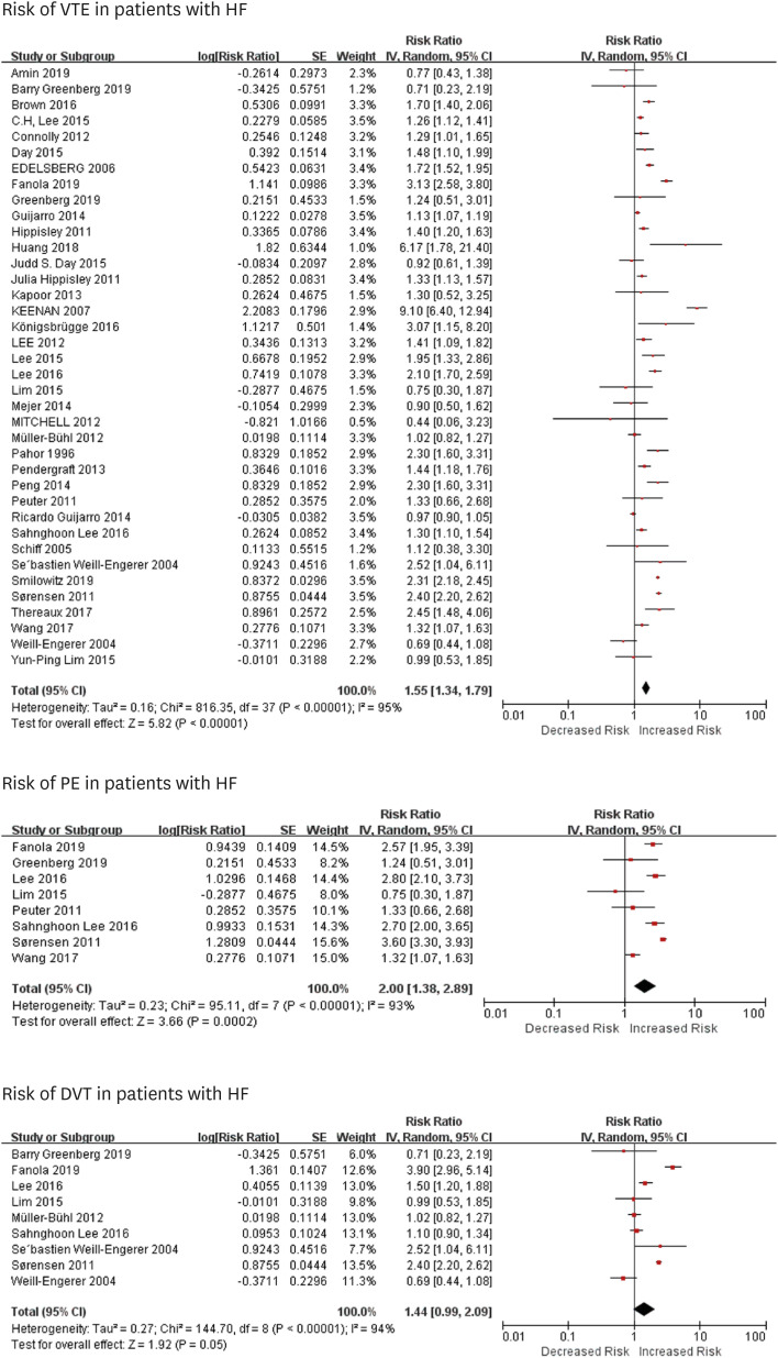
Among included studies, 24 (Supplementary Reference 2) articles reported rate or relative risks of VTE, 7 (Supplementary Reference 3) studies calculated about DVT, 7 (Supplementary Reference 4) manuscripts analyzed PE, and 5 (Supplementary Reference 5) recorded about three or 2 relative statistics of VTE, DVT or PE. The risk of VTE and PE were increased in patients with HF (VTE: RR=1.57, 95% CI=1.34–1.84, I2=95%; PE: RR=2.00, 95% CI=1.38–2.89, I2=93%) in the above 3 months follow-up period, but there was no a significant association between DVT and HF (RR=1.33, 95% CI=0.67–2.63, I2=94%) (Figure 2). No evidence of publication bias for all outcomes (VTE, PE, and DVT) was identified by Begg's test (all P>0.1) (Supplementary Figure 1). The sensitivity analyses confirmed that the association between HF and risk of endpoint events did not change with recalculation of the RRs by omitting one study at a time.
Figure 2

Risk of VTE, PE and DVT in patients with HF.

CI = confidence interval; DVT = deep vein thrombosis; HF = heart failure; PE = pulmonary embolism; RR = risk ratio; SE = standard error; VTE = venous thromboembolism.
kcj-51-766-g002

Subgroup analysis of venous thromboembolism in patients with heart failure and chronic heart failure

We did additional analyses on the associations between VTE in patients with HF, based on types of HF, follow-up duration, anticoagulation therapy, and age. We noted that patients with chronic HF (RR=1.54, 95% CI=1.32–1.80) had higher risk of VTE than those with acute HF (RR=0.95, 95% CI=0.68–1.32) (p for heterogeneity=0.009, I2=85.4%) (Figure 3). The risk of VTE in patients with HF did not show meaningful changes when analysis of follow-up period (p=0.09), with or without anticoagulation therapy (RR: 1.57 vs. 1.52, p=0.60), or ages (p=0.60). Also, surgery (R: 1.50 vs. 1.95, p=0.68) or cancer (R: 1.74 vs. 1.63, p=0.94) did not increase the risk of VTE in patients with HF. There were no significant differences when the subgroup analyzed in patients with chronic HF, neither did after 1-year follow-up (Tables 2 and 3, Supplementary Figures 2, 3, 4, 5, 6, 7, 8, 9, 10, 11, 12, 13, 14, 15, 16, 17, 18, 19, 20). Meta-regression indicated that the risk of VTE was not modified by age and follow-up duration (Supplementary Figure 21).
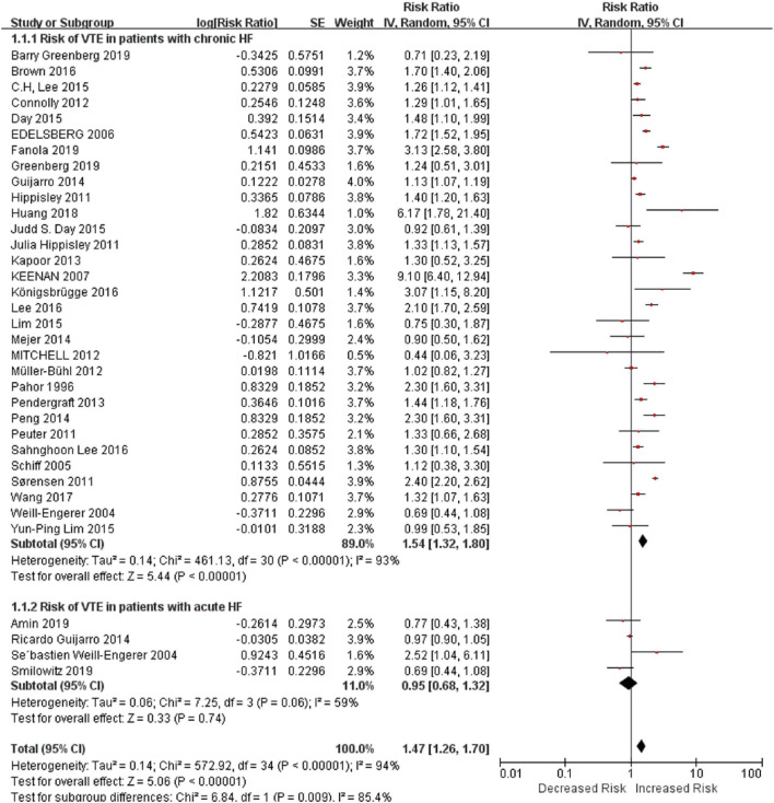
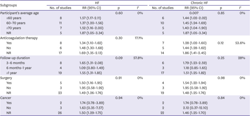
Figure 3

Risk of VTE in patients with acute HF and chronic HF.

CI = confidence interval; HF = heart failure; SE = standard error; VTE = venous thromboembolism.
kcj-51-766-g003
Table 2

Subgroup analyses of the association between HF and VTE

kcj-51-766-i002
Subgroups HF Chronic HF
No. of studies RR (95% CI) p I2 No. of studies RR (95% CI) p I2
Participant's average age 0.60 0% 0.007 0.85 0%
≤60 years 8 1.57 (1.17–2.11) 6 1.44 (1.02–2.02)
60–70 years 11 1.37 (1.20–1.56) 10 1.45 (1.24–1.69)
≥70 years 7 1.52 (1.16–2.00) 5 1.40 (1.04–1.90)
NR 5 1.87 (1.05–3.34) 5 1.87 (1.05–3.34)
Anticoagulation therapy 0.30 17.1%
Yes 8 1.34 (1.10–1.62) 7 1.28 (1.02–1.60) 0.12 53.6%
No 6 1.48 (1.30–1.68) 5 1.44 (1.28–1.62)
NR 17 1.69 (1.35–2.13) 14 1.86 (1.41–2.45)
Follow-up duration 0.09 57.8% 0.25 28%
3–6 months 8 1.65 (1.31–2.08) 6 1.79 (1.23–2.59)
6 months–1 year 4 1.09 (0.80–1.49) 3 1.18 (0.85–1.65)
≥1 year 19 1.55 (1.31–1.85) 17 1.51 (1.25–1.82)
Surgery 0.91 0% 0.98 0%
Yes 5 1.50 (1.16–1.95) 4 1.54 (1.22–1.94)
No 3 1.95 (0.58–1.92) 3 1.95 (0.58–1.92)
NR 23 1.49 (1.26–1.76) 19 1.48 (1.25–1.76)
Cancer 0.94 0% 0.84 0%
Yes 2 1.74 (0.78–3.89) 2 1.74 (0.78–3.89)
No 3 1.63 (0.35–7.57) 2 2.12 (0.37–12.10)
NR 26 1.50 (1.29–1.75) 22 1.46 (1.25–1.70)
CI = confidence interval; HF = heart failure; NR = no record; RR = risk ratio; VTE = venous thromboembolism.
Table 3

Subgroup analyses of the association between HF and VTE above 1 year follow-up period

kcj-51-766-i003
Subgroups HF Chronic HF
No. of studies RR (95% CI) p I2 No. of studies RR (95% CI) p I2
Participant's average age 0.87 0% 0.90 0%
≤60 years 7 1.65 (1.18–2.30) 6 1.60 (1.10–2.32)
60–70 years 8 1.42 (1.19–1.70) 8 1.42 (1.19–1.70)
≥70 years 6 1.59 (1.02–2.47) 4 1.27 (0.79–2.05)
NR 2 1.54 (0.59–4.00) 2 1.54 (0.59–4.00)
Anticoagulation therapy <0.05 88.4% <0.05 78.5%
Yes 7 1.14 (0.86–1.51) 6 1.17 (1.00–1.38)
No 3 1.35 (1.22–1.50) 3 1.35 (1.22–1.50)
NR 13 1.94 (1.65–2.27) 11 1.85 (1.45–2.34)
CI = confidence interval; HF = heart failure; NR = no record; RR = risk ratio; VTE = venous thromboembolism.

DISCUSSION

In this study, we noted that HF was an independent risk factor for VTE and PE in long-term follow-up duration, with an RR of about 1.57 and 2.00, respectively, while it was not significant for DVT. Patients with chronic HF had a higher risk of VTE. Subgroup analysis showed that after stratified by follow-up time, anticoagulation therapy, age or surgery, chronic HF was still a risk factor for VTE.
Links between HF and VTE are not well reported. Several mechanisms, such as stasis of blood because of dilatation of cardiac chambers, reduced myocardial contractility and increased intracardiac and central venous pressures, increased viscosity and coagulability of plasma, inflammation, neurohormonal activation, and endothelial dysfunction could contribute to the hypercoagulable state in patients with HF, raising the risk of VTE.14)
Our findings have several clinical implications. First, we found that HF could be a risk for VTE, and PE, but not for DVT in extended follow-up duration. It may be caused by several reasons. One is that much attention was paid to screen the lower extremity DVT (LEDVT), without realizing DVT in other location such as upper extremity, pelvis, and abdominal organs.15) Studies showed that upper extremity DVT (UEDVT) had a higher prevalence of HF when compared to patients with LEDVT (20% vs. 6.6%).16) Hospitalized HF patients often undergo central venous puncture and catheterization to receive therapy, most of which are through the upper extremity such as internal jugular vein, and above 10% of them would develop DVT.17) Also, some patients with undefined cardiomyopathy or who needed pulmonary artery pressure monitoring often undergo right heart catheterization, which may cause right ventricular thrombus18) and lead to severe PE.19) Moreover, patients who were treated with left ventricular assist devices or suffered from cardiomyopathy, like arrhythmogenic right ventricular cardiomyopathy, and dilated cardiomyopathy, were prone to develop right ventricular thrombus.20)21) Studies demonstrated that the risk of PE was at least twice in patients with HF and the risk increased as LV systolic function declined,22) especially in those severe decompensated HF patients.23) These results remind us that we should not neglect the risk of those patients with uncommon venous thrombus especially for those who are frequently rehospitalized, with long-term in-hospitalized stay or receiving catheter therapy. Another explanation is that DVT is hard for some patients to recognize and is often neglected by physicians because it could be asymptomatic. Nonetheless, symptoms of DVT are similar to symptoms of HF,24) so physicians may mistake them as worsening HF without realizing DVT.
Second, we noted that patients with chronic HF were more prone to have VTE than acute HF in the long-term period. Acute HF consists of new-onset HF, caused by acute primary cardiac dysfunctions (such as acute myocardial dysfunction and acute valve insufficiency), and decompensated chronic HF triggered by precipitant factors like infection.25) For those patients with new-onset HF, part of them can reverse with timely treatment,26) thus might not have the risk for VTE. Moreover, acute HF patients who should receive surgery such as myocardial infarction, are often prescribed thromboprophylaxis therapy to prevent thrombotic events,25)26) which may further reduce the occurrences of VTE. As for chronic HF, different factors are prone to result in different types of HF. An epidemiological study suggested that myocardial infarction more often causes HF with reduced ejection fraction (HFrEF), while atrial fibrillation is likely to cause HF with preserved ejection fraction (HFpEF).25) However, we still do not have studies on differential effect on VTE depending on HF type.
Third, we identified that the risk for VTE still existed in prolonged follow-up period (above 1 year). It reminds us that we should not ignore the risk of VTE in patients even after discharge from hospitals. Anticoagulation therapy is more prone to be prescribed in-hospitalization27) as most hospitalized HF patients in hospitalized are immobile and bedridden or are applied central venous puncture and catheterization17) and few patients will continue to receive anticoagulation therapy after discharge.28) This study implies that we should focus more on thromboprophylaxis in HF patients to prevent VTE out-of-hospitalization. However, we noticed that anticoagulation therapy could reduce the risk of VTE, but the effect was not significant. Another study showed that low-dose (2.5 mg rivaroxaban) did not reduce the incidence of VTE.9) It prompts us that a higher dosage and an extended duration might benefit patients with HF.28)
Our study had several limitations. First, we were unable to analyze the impact of severities and types of chronic HF on VTE to provide more insights into the risk of VTE in sub-group patients, as data (such as level of plasma N-terminal pro-brain natriuretic peptide, LVEF and etiology like atrial fibrillation) were not available in most of the included studies. Second, there were still few studies about the risk of DVT in patients with HF, so our analysis may underestimate the risk of DVT in HF patients. Third, our analysis only included 4 studies about acute HF and only 1 reported the risk of VTE in patients with new-onset HF. Thus, we lack enough data of acute HF patients, especially those new-onset HF patients, to distinguish whether acute HF or chronic HF would have a noticeable effect on the VTE risk. Forth, there was substantial heterogeneity among most analyses due to their different follow-up durations, types of diseases and complications, population, etc. Therefore, there may be large uncertainty of VTE rates overall and in subgroups. Fifth, we was not able to show the associations of activity level and the risks of VTE, PE, and DVT, since the data of included studies was limited. Finally, most of our studies were retrospective studies, and prospective studies with high quality such as RCTs are needed to testify our results.
In conclusion, HF was an independent risk for VTE and PE but not DVT in the long-term follow-up period. Patients with chronic HF were prone to have a higher risk of VTE. Further designated and prospective studies are needed for our better understandings of impacts of HF on the risk of VTE, PE and, DVT. RCTs about anticoagulation therapy in HF patients to prevent VTE, PE, and DVT are also needed for better management of HF patients.

Notes

Funding: This study was supported by the National Key Research and Development Program of China (2017YFC1308300).

Conflict of Interest: The authors have no financial conflicts of interest.

Data Sharing Statement: The data generated in this study is available from the corresponding authors upon reasonable request.

Author Contributions:

  • Conceptualization: Zhang Y, Zhang J.

  • Funding acquisition: Zhang J.

  • Investigation: Xu T, Huang Y.

  • Methodology: Huang Y, Liu Z, Bai Y, Ma Z.

  • Supervision: Cai X.

  • Visualization: Cai X.

  • Writing - original draft: Xu T.

  • Writing - review & editing: Zhang Y, Zhang J.

References

1. Ambrosy AP, Fonarow GC, Butler J, et al. The global health and economic burden of hospitalizations for heart failure: lessons learned from hospitalized heart failure registries. J Am Coll Cardiol. 2014; 63:1123–1133. PMID: 24491689.
2. Hao G, Wang X, Chen Z, et al. Prevalence of heart failure and left ventricular dysfunction in China: the China Hypertension Survey, 2012-2015. Eur J Heart Fail. 2019; 21:1329–1337. PMID: 31746111.
crossref
3. Khera R, Dharmarajan K, Krumholz HM. Rising mortality in patients with heart failure in the United States: facts versus fiction. JACC Heart Fail. 2018; 6:610–612. PMID: 29914774.
4. Alikhan R, Cohen AT, Combe S, et al. Prevention of venous thromboembolism in medical patients with enoxaparin: a subgroup analysis of the MEDENOX study. Blood Coagul Fibrinolysis. 2003; 14:341–346. PMID: 12945875.
5. Cohen AT, Davidson BL, Gallus AS, et al. Efficacy and safety of fondaparinux for the prevention of venous thromboembolism in older acute medical patients: randomised placebo controlled trial. BMJ. 2006; 332:325–329. PMID: 16439370.
crossref
6. Tang L, Wu YY, Lip GY, Yin P, Hu Y. Heart failure and risk of venous thromboembolism: a systematic review and meta-analysis. Lancet Haematol. 2016; 3:e30–44. PMID: 26765646.
crossref
7. Klein L, O'connell JB. Thromboembolic risk in the patient with heart failure. Curr Treat Options Cardiovasc Med. 2007; 9:310–317. PMID: 17761116.
crossref
8. Fanola CL, Norby FL, Shah AM, et al. Incident heart failure and long-term risk for venous thromboembolism. J Am Coll Cardiol. 2020; 75:148–158. PMID: 31948643.
9. Greenberg B, Neaton JD, Anker SD, et al. Association of rivaroxaban with thromboembolic events in patients with heart failure, coronary disease, and sinus rhythm: a post hoc analysis of the COMMANDER HF trial. JAMA Cardiol. 2019; 4:515–523. PMID: 31017637.
10. Lim YP, Lin CL, Hung DZ, Ma WC, Lin YN, Kao CH. Increased risk of deep vein thrombosis and pulmonary thromboembolism in patients with organophosphate intoxication: a nationwide prospective cohort study. Medicine (Baltimore). 2015; 94:e341. PMID: 25569651.
11. Stroup DF, Berlin JA, Morton SC, et al. Meta-analysis of observational studies in epidemiology: a proposal for reporting. Meta-analysis Of Observational Studies in Epidemiology (MOOSE) group. JAMA. 2000; 283:2008–2012. PMID: 10789670.
12. Huang Y, Cai X, Mai W, Li M, Hu Y. Association between prediabetes and risk of cardiovascular disease and all cause mortality: systematic review and meta-analysis. BMJ. 2016; 355:i5953. PMID: 27881363.
crossref
13. Deeks JJ, Higgins JP, Altman DG. Chapter 10. Analysing data and undertaking meta-analyses. Higgins JP, Thomas J, Chandler J, editors. Cochrane Handbook for Systematic Reviews of Interventions. London: Cochrane Collaboration;2019. p. 241–284. DOI: 10.1002/9781119536604.ch10.
14. Ng TM, Tsai F, Khatri N, Barakat MN, Elkayam U. Venous thromboembolism in hospitalized patients with heart failure: incidence, prognosis, and prevention. Circ Heart Fail. 2010; 3:165–173. PMID: 20086221.
15. Heit JA. Epidemiology of venous thromboembolism. Nat Rev Cardiol. 2015; 12:464–474. PMID: 26076949.
crossref
16. Rokosh RS, Ranganath N, Yau P, et al. High prevalence and mortality associated with upper extremity deep venous thrombosis in hospitalized patients at a tertiary care center. Ann Vasc Surg. 2020; 65:55–65. PMID: 31669473.
crossref
17. Citla Sridhar D, Abou-Ismail MY, Ahuja SP. Central venous catheter-related thrombosis in children and adults. Thromb Res. 2020; 187:103–112. PMID: 31981840.
crossref
18. Yang H, Chen F, Jiao H, et al. Management of tunneled-cuffed catheter-related right atrial thrombosis in hemodialysis patients. J Vasc Surg. 2018; 68:1491–1498. PMID: 29804743.
crossref
19. Suratkal V, Ahmed A. Right atrial thrombus and challenges in its management. J Assoc Physicians India. 2018; 66:65–68. PMID: 31313553.
20. Zghaib T, Bourfiss M, van der Heijden JF, et al. Atrial dysfunction in arrhythmogenic right ventricular cardiomyopathy. Circ Cardiovasc Imaging. 2018; 11:e007344. PMID: 30354672.
crossref
21. Long B, Robertson J, Koyfman A, Brady W. Left ventricular assist devices and their complications: a review for emergency clinicians. Am J Emerg Med. 2019; 37:1562–1570. PMID: 31072684.
crossref
22. Beemath A, Stein PD, Skaf E, Al Sibae MR, Alesh I. Risk of venous thromboembolism in patients hospitalized with heart failure. Am J Cardiol. 2006; 98:793–795. PMID: 16950187.
crossref
23. Darze ES, Latado AL, Guimarães AG, et al. Incidence and clinical predictors of pulmonary embolism in severe heart failure patients admitted to a coronary care unit. Chest. 2005; 128:2576–2580. PMID: 16236926.
crossref
24. Rogers C, Bush N. Heart failure: pathophysiology, diagnosis, medical treatment guidelines, and nursing management. Nurs Clin North Am. 2015; 50:787–799. PMID: 26596665.
25. Ponikowski P, Voors AA, Anker SD, et al. 2016 ESC Guidelines for the diagnosis and treatment of acute and chronic heart failure: the task force for the diagnosis and treatment of acute and chronic heart failure of the European Society of Cardiology (ESC) developed with the special contribution of the Heart Failure Association (HFA) of the ESC. Eur Heart J. 2016; 37:2129–2200. PMID: 27206819.
26. Harjola VP, Mullens W, Banaszewski M, et al. Organ dysfunction, injury and failure in acute heart failure: from pathophysiology to diagnosis and management. A review on behalf of the Acute Heart Failure Committee of the Heart Failure Association (HFA) of the European Society of Cardiology (ESC). Eur J Heart Fail. 2017; 19:821–836. PMID: 28560717.
crossref
27. Goldhaber SZ. Venous thromboembolism in heart failure patients: pathophysiology, predictability, prevention. J Am Coll Cardiol. 2020; 75:159–162. PMID: 31948644.
28. Imberti D, Pierfranceschi MG, Falciani M, Prisco D. Venous thromboembolism prevention in patients with heart failure: an often neglected issue. Pathophysiol Haemost Thromb. 2008; 36:69–74. PMID: 19127085.
crossref

SUPPLEMENTARY MATERIALS

Supplementary Reference 1

Studies included in the analysis
kcj-51-766-s001.doc

Supplementary Reference 2

Studies reported risk of venous thromboembolism in heart failure patients in the analysis
kcj-51-766-s002.doc

Supplementary Reference 3

Studies reported risk of deep vein thrombosis in heart failure patients in the analysis
kcj-51-766-s003.doc

Supplementary Reference 4

Studies reported risk of pulmonary embolism in heart failure patients in the analysis
kcj-51-766-s004.doc

Supplementary Reference 5

Studies reported two or three relative risk of venous thromboembolism, deep vein thrombosis or pulmonary embolism in heart failure patients in the analysis
kcj-51-766-s005.doc

Supplementary Table 1

Literature search strategy for PubMed
kcj-51-766-s006.xls

Supplementary Table 2

Quality assessment of article
kcj-51-766-s007.xls

Supplementary Figure 1

Publication biases of HF and VTE, PE and DVT.
kcj-51-766-s008.doc

Supplementary Figure 2

Subgroup analysis of risk of VTE in patients with HF according to type of HF.
kcj-51-766-s009.doc

Supplementary Figure 3

Subgroup analysis of risk of VTE in patients with HF according to follow-up time.
kcj-51-766-s010.doc

Supplementary Figure 4

Subgroup analysis of risk of VTE in patients with HF according anticoagulation therapy.
kcj-51-766-s011.doc

Supplementary Figure 5

Subgroup analysis of risk of VTE in patients with HF according anticoagulation therapy (with anticoagulation vs. without anticoagulation).
kcj-51-766-s012.doc

Supplementary Figure 6

Subgroup analysis of risk of VTE in patients with HF according age.
kcj-51-766-s013.doc

Supplementary Figure 7

Subgroup analysis of risk of VTE in patients with HF according to age above 1 year follow-up.
kcj-51-766-s014.doc

Supplementary Figure 8

Subgroup analysis of risk of VTE in patients with HF according to anticoagulation therapy above 1 year follow-up.
kcj-51-766-s015.doc

Supplementary Figure 9

Subgroup analysis of risk of VTE in patients with HF according to anticoagulation therapy above 1 year follow-up (with anticoagulation vs. without anticoagulation).
kcj-51-766-s016.doc

Supplementary Figure 10

Subgroup analysis of risk of VTE in patients with chronic HF according to follow-up time.
kcj-51-766-s017.doc

Supplementary Figure 11

Subgroup analysis of risk of VTE in patients with chronic HF according to age.
kcj-51-766-s018.doc

Supplementary Figure 12

Subgroup analysis of risk of VTE in patients with chronic HF according to anticoagulation therapy.
kcj-51-766-s019.doc

Supplementary Figure 13

Subgroup analysis of risk of VTE in patients with chronic HF according to anticoagulation therapy (with anticoagulation vs. without anticoagulation).
kcj-51-766-s020.doc

Supplementary Figure 14

Subgroup analysis of risk of VTE in patients with chronic HF according to anticoagulation therapy above 1 year follow-up.
kcj-51-766-s021.doc

Supplementary Figure 15

Subgroup analysis of risk of VTE in patients with chronic HF according to anticoagulation therapy above 1 year follow-up (with anticoagulation vs. without anticoagulation).
kcj-51-766-s022.doc

Supplementary Figure 16

Subgroup analysis of risk of VTE in patients with chronic HF according to age above 1 year follow-up.
kcj-51-766-s023.doc

Supplementary Figure 17

Subgroup analysis of risk of VTE in patients with HF according to surgery.
kcj-51-766-s024.doc

Supplementary Figure 18

Subgroup analysis of risk of VTE in patients with HF according to cancer.
kcj-51-766-s025.doc

Supplementary Figure 19

Subgroup analysis of risk of VTE in patients with chronic HF according to surgery.
kcj-51-766-s026.doc

Supplementary Figure 20

Subgroup analysis of risk of VTE in patients with chronic HF according to cancer.
kcj-51-766-s027.doc

Supplementary Figure 21

Meta regressions about age and follow-up time in risk of VTE in patients with HF.
kcj-51-766-s028.doc
TOOLS
Similar articles