

 PDF
PDF Citation
 Print
Print


I. Introduction
The surgeon’s dilemma regarding the management of condylar fractures has always revolved around the approach, timing, patient’s age, implant design selection, and surgeon’s experience. The criteria for open reduction of condylar fractures given by Zide and Kent1 marked one of the turning points in the decision-making and case selection process of condylar fractures. High condylar (intra-capsular/diacapitular) fractures pose a different surgical challenge, with the majority of surgeons preferring a closed reduction protocol for its management followed by physiotherapy2. The general consensus around the management of sub-condylar and low condylar fractures points towards open reduction and internal fixation. Even though the debate generally surrounds the incision, surgical approach, and implant design, we lack a detailed protocol-driven stepwise approach towards reduction and fixation of subcondylar fractures.
This article aims to devise a detailed and standardized protocol, common to any surgical approach or implant design, that once followed, would lead to predictable, reproducible, and repeatable results in every surgeon’s hands thereby eventually eliminating the uncertainty and difficulty in reduction and fixation of subcondylar fractures of the mandible.
Go to : 
II. Technical Note
A total of 34 condylar neck and condylar base (subcondylar) fractures, classified according to Loukota et al.3, were treated using our protocol under general anesthesia. In 21 cases, the proximal segments were antero-medially displaced and 13 cases had laterally displaced proximal segments. All the subcondylar fractures were approached using a retro-mandibular (antero-parotid transmassetric) approach.
As advocated by Orabona et al.4, fixation of concomitant fractures in the dentate part of the mandible were done first. Following which, our protocol was utilized for the reduction and fixation of the condylar neck and condylar base fractures.
1. Step 1
The mandible is antero-inferiorly distracted manually by placing a thumb over the ipsilateral molars intra-orally.
2. Step 2
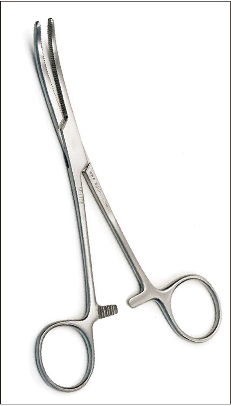
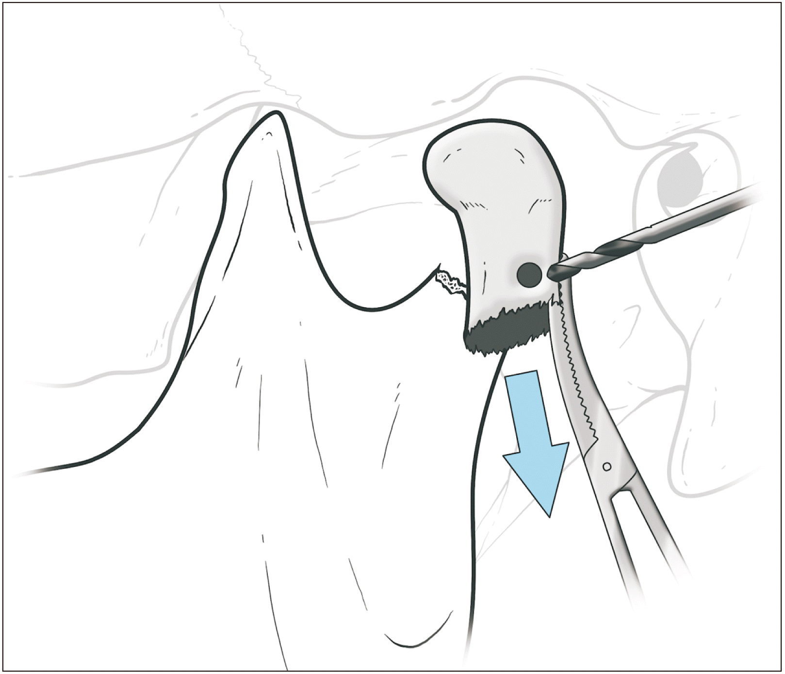
The posterior border of the proximal segment is identified and secured using a 16 cm Rochester-Pean curved hemostatic forceps (Fig. 1), with one beak over the cortical portion and the other beak placed into the medullary portion. Following which, the proximal segment is lateralized overriding the distal segment.(Fig. 2) This maneuver serves a dual purpose of giving stability to the proximal segment during drilling and fixation of the first screw, as well as aiding the stripping of some portion of the insertion of inferior head of the lateral pterygoid at the pterygoid fovea using the flat end of a periosteal elevator to minimize the pull exerted by the lateral pterygoid on the proximal segment.
3. Step 3
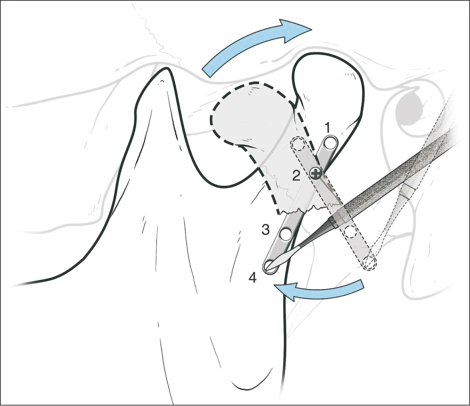
The antero-inferior force on the mandible is released to allow the mandible to passively distract postero-superiorly. For ease of understanding, the holes of the miniplate are numbered from 1 to 4 as illustrated in Fig. 3.
4. Step 4
A hole is drilled 3 mm from the fracture line on the postero-lateral border of the proximal segment with a 1.6 mm drill bit in a supero-medial direction.(Fig. 2) A 2 mm four-hole with gap titanium miniplate is secured to the proximal segment at hole No. 2 with a 2 mm×6 mm miniscrew. This makes the miniplate jut out laterally along the long axis of the proximal segment.(Fig. 3)
5. Step 5
The mandible is antero-inferiorly distracted manually and the miniplate is pulled inferiorly towards the distal segment with the sharp end of the periosteal elevator in hole No. 4 of the miniplate.(Fig. 3)
This maneuver orients the proximal segment of the condyle in the anatomic position and realigns the posterior border with a seamless contact of the proximal and distal segments thus ensuring the restoration of condyle and glenoid fossa relationship. Caution should be exercised to avoid any bend in the miniplate due to injudicious forces that may jeopardize the final alignment outcome of the fractured segments.
7. Step 7
Maintaining the downward pressure on the hole No. 4, hole No. 3 is drilled and a 2 mm×8 mm screw is placed in the infero-medial direction helping in mild compression at the fracture line.
8. Step 8
Periosteal elevator from hole No. 4 is released, the same hole is drilled, and a 2 mm×8 mm screw is placed in infero-medial direction.
9. Step 9
The last hole No. 1 is drilled in supero-medial direction and a 2 mm×6 mm screw is placed.
10. Step 10
The anterior border of the proximal segment is fixed to neutralize the anterior tension zone either with a 2 mm 2-hole with gap or 4-hole with gap miniplate.
Standard layered closure is done using 3-0 Vicryl followed by 6-0 prolene for the skin.
Go to :
III. Discussion
Reduction and fixation of medially displaced subcondylar fractures have always posed a challenge to a young surgeon. Peterson et al.5 described the use of a maxillo-mandibular fixation screw on the proximal segment to aid in its reduction, and also mentioned the use of temporary suture or wire around the condylar head as alternatives. However, there is no stepwise go-to guide mentioned in the literature to help surgeons in the decision making process after surgically identifying the proximal and distal segments. Open reduction and internal fixation of the subcondylar fracture helps in the restoration of ramal height, stable occlusion, and thus in early recovery to normal function6-10.
Two straight plates or three-dimensional plates are advocated for subcondylar fixation as single plate fixations have proven to cause fixation failure11. The distribution of compressive stress on the posterior border of the ramus, and tensile stress inferior and parallel to the sigmoid notch of the mandible were demonstrated by the photoelastic analysis put forth by Meyer et al.12. Two miniplates, fixed in a triangular geometry, one at the posterior ramal border and one following the curvature of the sigmoid notch, are ideal in fixation of subcondylar fractures13.
The authors employed the retromandibular transmasseteric anteroparotid approach as it is considered to be a surgeon-friendly approach6,14.
The protocol that the authors have described here does not necessitate the use of specialized instruments and its stepwise approach lends clarity and predictability to the entire procedure. The authors encountered no complications such as sialocele formation, facial nerve paralysis, infection, or post-operative restricted mouth opening in the 34 cases in which they employed this protocol. Hence, the authors believe this protocol would help all surgeons, young or experienced, to perform open reduction and internal fixation of the mandibular subcondylar region with ease and accuracy.
Go to :
 XML Download