

 PDF
PDF Citation
 Print
Print


I. Introduction
Mastoid lymph nodes (MLNs), or retroauricular lymph nodes1, are a small group of usually two nodes that belong to the superficial lymph node group of the head2. They are located in both retroauricular areas, in the mastoid region and are more numerous in children3. Lengelé et al.4 reported that, “draining the temporal region of the hairy scalp, the posterior surface of the auricle and the posterior wall of the external acoustic meatus, MLNs have efferent vessels which traverse the upper insertions of the sternocleidomastoid muscle to join the superior deep lateral cervical lymph nodes”.
Currently, only a limited number of publications has highlighted the pathological conditions that can involve the MLNs as a result of different etiologies: cat scratch disease lymphadenopathy5, post-microneedling mastoid lymphadenopathy1, and a pathologic process in a mastoid area can often mimic mastoiditis6-8.
Normal, metastatic9, reactive10, and suppurated lymph nodes show different ultrasound patterns, which help medical workers to establish correct diagnosis and management approach and to prevent complications. Suppurative lymphadenopathy in different parts of the human body demonstrates very specific ultrasound features, including anechoic regions, septations, peripheral vascularity, and posterior acoustic enhancement artifact11 that are related to necrosis within lymph nodes, also known as intranodal abscess formation or purulent fusion of the lymph nodes12.
To our knowledge, there are no publications that have highlighted the clinical, ultrasonographic, intra-, and postoperative data for suppurative mastoid lymphadenitis that presented as mastoiditis. The purpose of the report is to present an extremely rare case of the pathologic process that can be referred to both oral-maxillofacial and head neck surgeons for treatment.
Go to : 
II. Case Report
The patient was an 18-year-old Caucasian male who was referred to the Center of Maxillofacial Surgery from the Kyiv region with painful right retroauricular swelling and fever. The patient reported that the symptoms had started about one week prior; after experiencing hypothermia, swelling of the soft tissue of the right neck was noted. A physician diagnosed acute cervical lymphadenitis and prescribed treatment (oral Augmentin) after which the edema of the neck resolved, although edema in the right mastoid persisted and became painful and red.
Examination revealed painful swelling of the soft tissue above the right mastoid process (Fig. 1) with significant erythema. Simultaneously, the patient noted no ear pain or pressure, and purulent discharge from the ear and hearing loss were absent. Emergency gray scale (B-mode) and color Doppler ultrasound (US) (HD11 XE; Philips, Amsterdam, The Netherlands) showed soft tissue edema above the mastoid process, a 1.19×0.61-cm reactive upper posterior MLN with marked hilar vascularity (Fig. 2) and no ultrasound signs of suppuration. Color Doppler US in a slightly anterior and lower transducer position compared to the previous scan demonstrated a limited collection of pus (Fig. 3) at a place where the lower anterior MLN was presumably located. US of the right parotid area and neck also showed an increase in the size of one intraparotid and multiple cervical reactive lymph nodes with hilar vascularity. A diagnosis of right cervical and suppurative right mastoid lymphadenitis was made after the US investigation. The patient and his parents declined a surgical treatment approach; however, he was referred to our Center again after only two days. At his second visit, increase of mastoid swelling, local skin erythema (Fig. 4. A), and fever were noted. The local pain became unbearable for the patient. Fluctuation upon additional examination informed the diagnosis, which was changed to “abscess of the right mastoid area”. Abscess lancing (under local anesthesia [2.0 mL of 4% Ultracain D-S forte; Aventis Pharma Deutschland, Frankfurt, Germany]) revealed ~4.0 mL of pus (Fig. 4. B), and curettage (Fig. 4. C) showed only purulent content with no residual parts of the suppurated MLNs. The patient received intravenous ceftriaxone 1.0 g two times daily and wound bandages with Oflocain-Darnitsa ointment (components: lidocaine hydrochloride and ofloxacin; Darnitsa, Kyiv, Ukraine), which have hyperosmolar properties. The postoperative period was smooth, and the wound healed by secondary intention.(Fig. 5)
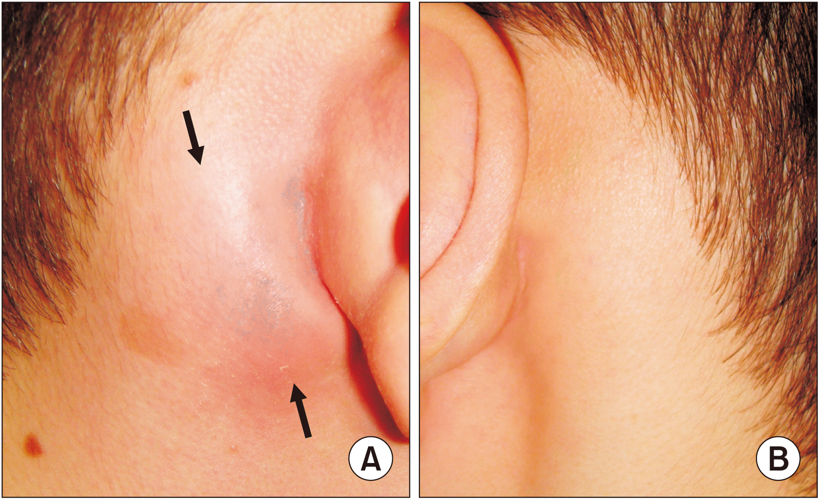 | Fig. 1Right lateral view (A) shows a right suppurative mastoid lymphadenitis (arrows) and left view (B) demonstrates the non-symptomatic side.
|
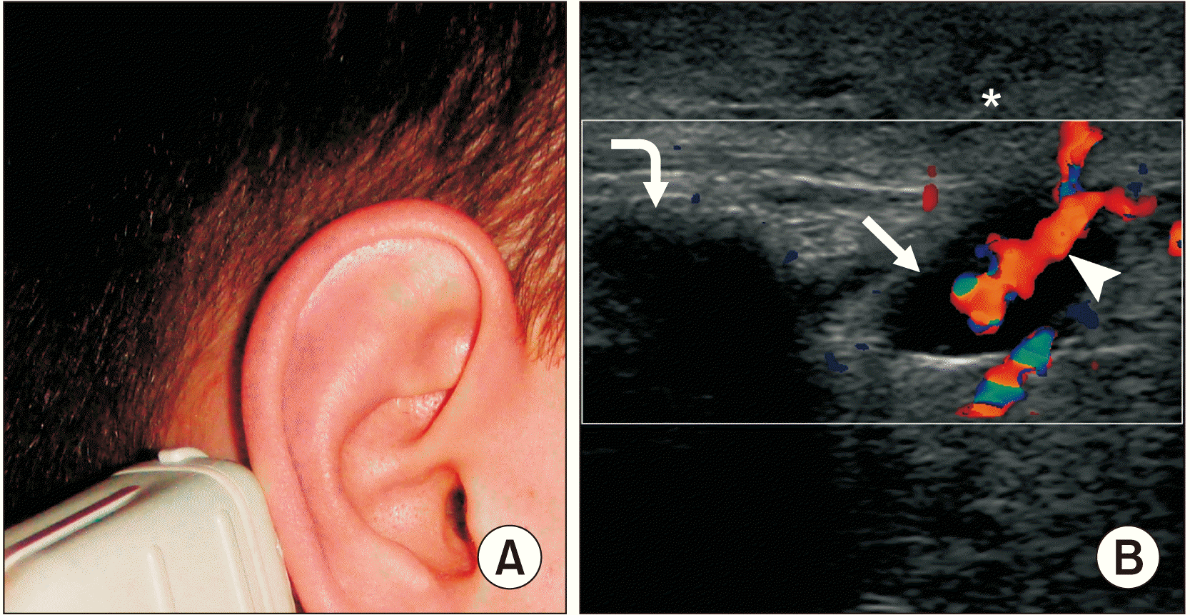 | Fig. 2The left image (A) shows the patient and symptomatic area from a longitudinal transducer position. Color Doppler sonogram (B) demonstrates soft tissue edema (asterisk) above the mastoid process (curved arrow), a 1.19×0.61-cm reactive upper posterior mastoid lymph node (arrowhead) with a marked hilar vascularity (arrow) and no ultrasound signs of suppuration. The oval shape of the node corresponds to reactive lymphadenitis. Depth of ultrasonography is 3.0 cm.
|
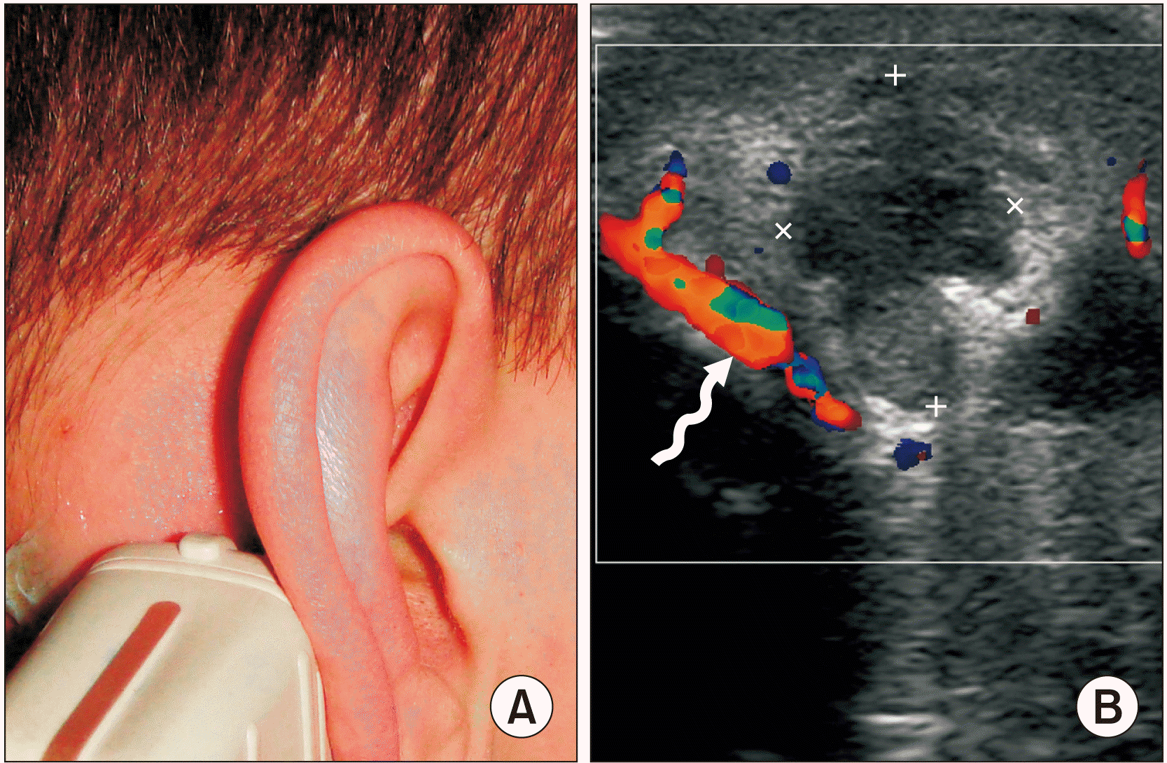 | Fig. 3Image (A) shows the symptomatic area in a longitudinal transducer position. Color Doppler ultrasound image (B) demonstrates a limited collection of pus (the borders are indicated by “+” and “×” calipers) at a place where the lower anterior mastoid lymph node was presumably located. Posterior auricular vessel is indicated by a waved arrow. Depth of ultrasonography is 3.0 cm.
|
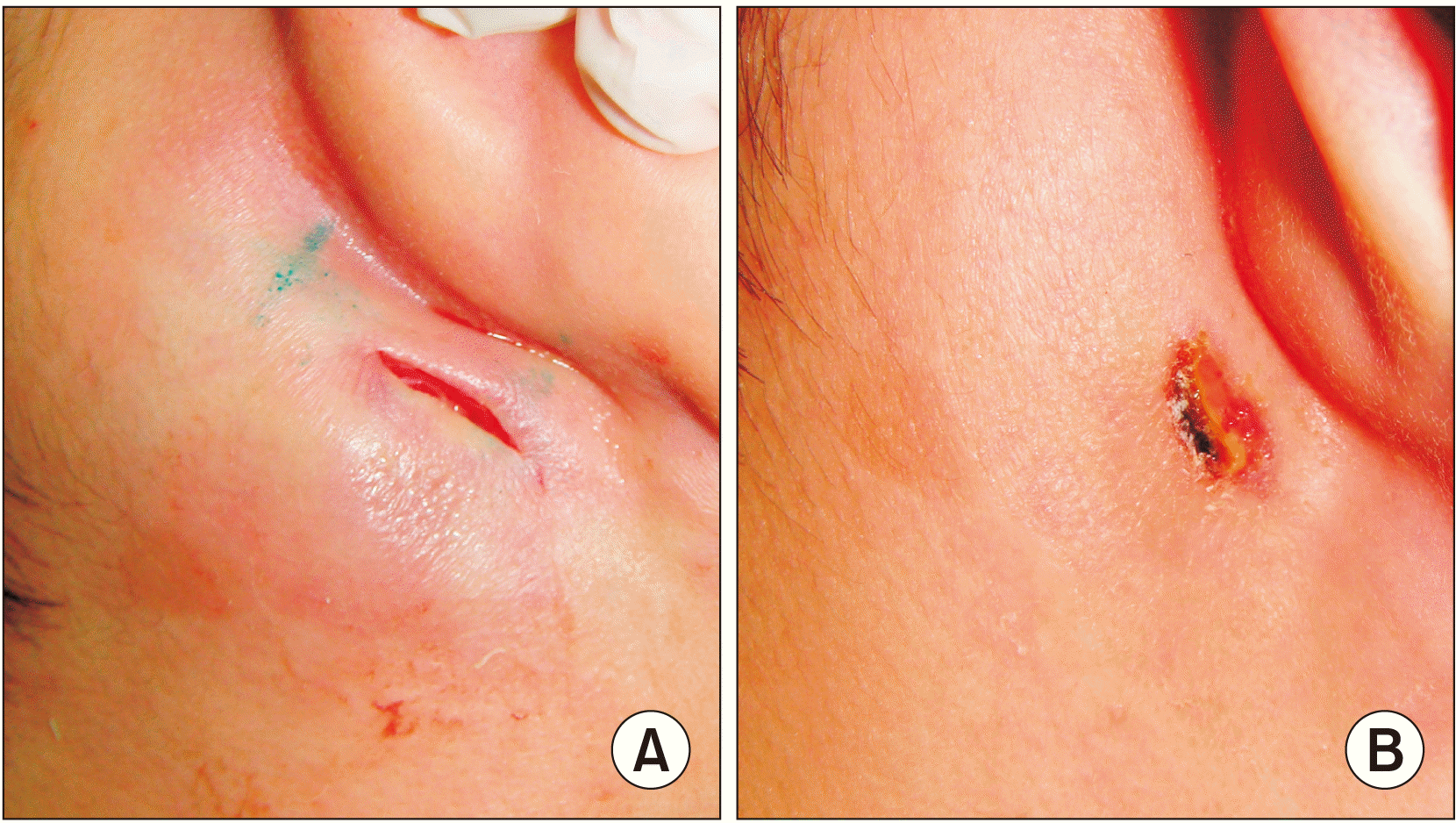
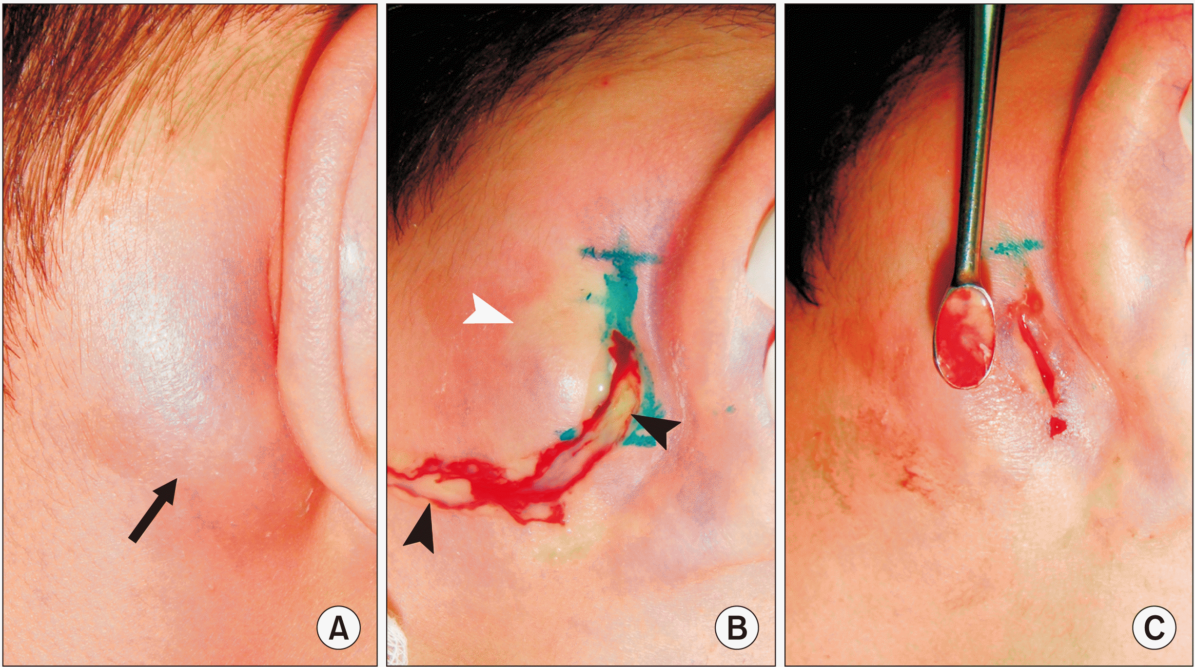 | Fig. 4Preoperatively (A) the lesion was noted with painful swelling, erythema, and fluctuation in the right mastoid area (arrow). A mastoid area abscess lancing (B) revealed ~4.0 mL of pus (black arrowheads) (white arrowhead indicates on skin ischemia as a result of local anesthesia) and curettage (C) showed only purulent content with no residual parts of the suppurated mastoid lymph nodes.
|
Go to :
III. Discussion
Infection of the MLNs or “lymph glands” occurs rarely, and cases that have reported an abscess following suppurative mastoid lymphadenitis are limited. To understand the pathologic condition, it is important to know the anatomy of MLNs and the ultrasound appearance of lymph nodes (normal, reactive, and lymph node metastases), which can easily differentiate mastoid lymphadenitis from mastoiditis, other types of abscesses, or acute otitis media.
Despite the data from Ahuja and Evans13 that MLNs in adults are usually involuted, our case clearly demonstrated the presence of two mastoid nodes, in a reactive and suppurated state, in an 18-year-old patient. Because parotid and submandibular nodes can be round in shape, this should not be used as the sole criterion for lymph node metastases14. Nodal size alone cannot be used as a criterion to differentiate between reactive and metastatic lymph nodes15. Calcification within normal and reactive nodes lymph nodes is uncommon15. Typically, on color and power Doppler, normal nodes show hilar vascularity or appear avascular, and reactive nodes predominantly show hilar vascularity16,17.
Hernandez et al.18 emphasized that acute lymphadenitis develops over days, whereas subacute or chronic lymphadenitis develops over weeks to months. McMillan еt al.19 reported that statistically one-third of infected nodes suppurate and become fluctuant. Further, for cases of suppurative lymphadenitis, a meticulous curettage approach is mandatory because it helps to remove residual parts of partially suppurated nodes. Abscess/phlegmon formation following a suppurative lymphadenitis in some East-European countries is referred to as “adenoabscess” and “adenophlegmon”20. “Abscess-forming lymphadenopathy” can be considered a synonym of “abscess following a lymphadenitis” and “adenoabscess”.
Dudkiewicz et al.21 described that acute mastoiditis becomes clinically significant when the infection spreads and induces periostitis, thus visually mimicking suppurative mastoid lymphadenitis. The term “Luc’s abscess” is applied only to a subperiosteal temporal abscess, or a limited collection of pus, as a complication of acute otitis media22. Another term, “Bezold’s abscess,” refers to an abscess that arises within the substance of the sternomastoid or digastric muscle following spread of pus through the tip of the mastoid process23,24.
Selecting the best diagnostic method is important for differential diagnosis between mastoid lymphadenopathy and mastoiditis; this process should be guided by the anamnesis morbi, a clinical picture of the pathologic process, and by the understanding that ultrasonography should have diagnostic priority over methods that use ionizing radiation, such as computed tomography (CT) or X-ray. Pattanayak et al.25 showed that both B-mode and color Doppler US can be used to detect reactive lymph nodes with almost perfect accuracy. According to their data, B-mode features for reactive lymph nodes had a sensitivity of 88%, a specificity of 97.3%, and color Doppler US had a sensitivity of 92% and a specificity of 97.3%25. CT is strongly recommended in cases when a soft tissue edema in the retroauricular area combines with otorrhea (ear discharge), otalgia and hearing loss in the affected side. Vazquez et al.26 reported that worldwide sensitivity of CT for assessing acute mastoiditis is between 87%-100%.
Mastoid suppurative lymphadenitis, similar to any purulent condition, should be treated quickly, with the intent to avoid secondary osteomyelitis of the mastoid process of the temporal bone (osteomyelitis of the mastoid) or mastoiditis. Osteomyelitis of the mastoid is very rare but commonly occurs secondary to otitis externa or suppurative otitis media. In some cases, acute mastoiditis can result in a retroauricular subcutaneous abscess, which presents with the same lateral characteristics as this case report, at the mastoid area with swelling and erythema.
In conclusion, suppurative lymphadenitis of the mastoid nodes is a rare entity. When encountered, pus draining with curettage of the parts of the lymph nodes which have not degraded should be considered as the first treatment modality. For this case of abscess that followed lymphadenitis, lancing resulted in a quick recovery.
Go to :
 XML Download