I. Introduction
In orthognathic surgery, sagittal split ramus osteotomy (SSRO) and intraoral vertical ramus osteotomy (IVRO) are representative surgical methods for treating mandibular prognathism, retrognathism, and facial asymmetry. Each surgical procedure has advantages and disadvantages, and the selection of the technique depends on the skill and preference of the operator and the preoperative condition of the patient’s jawbone and temporomandibular joint
1,2.
SSRO was first proposed by Trauner and Obwegerser
3. After various modifications, the method was developed into the current surgical design and rigid fixation using plates and screws or bicortical screws
3,4. The advantages of this procedure include the positioning of the distal segment of the mandible is facilitated, bone healing is promoted by allowing overlap of wide bony surfaces, and early function after surgery is enabled by using rigid fixation of the bone fragment
5,6. Caldwell and Letterman compared IVRO to SSRO
7 and found IVRO was relatively easier with a lower risk of damage to the inferior alveolar nerve and blood vessels
2. Because fixation is not required between the segments, condylar positioning is simplified. In previous studies, the mandibular condyle is reportedly repositioned to a physiologically equilibrated position during the postoperative physical therapy period, showing a therapeutic effect in most patients with temporomandibular joint disorder (TMD)
8,9.
Bilateral SSRO (BSSRO) is the most often-used surgical procedure due to its advantages and good postoperative results
1,2. However, during surgery for mandibular asymmetry, BSSRO can create bony interference between the proximal and distal segments on the deviated side when the mandibular midline shift occurs to the opposite side, leading to condylar displacement from the mandibular fossa that may inadvertently change the lateral shape of the face and cause TMD
1,10,11.(
Fig. 1) Therefore, surgeons have devised various surgical methods to eliminate bony interference
1. Among the various surgical methods, IVRO on the deviated side that generates condylar torque and SSRO on the opposite side should be considered an alternative surgical method to solve this problem
12.
 | Fig. 1Changes in the position of the deviated side condyle during osteotomy and fixation in bilateral sagittal split ramus osteotomy (BSSRO). A. Asymmetric mandible before osteotomy (left deviation). B. Mandible after osteotomy (reposition of the mandible on the midline of the face). C. Lateral displacement in the left condyle after fixation. During fixation of the left proximal segment to the distal segment, the condyle was displaced laterally due to bony interference. 
|
In the present study, the effects of SSRO combined with IVRO for the treatment of asymmetric mandible in class Ⅲ malocclusion patients were assessed and the postoperative stability of the mandibular condyle and TMD symptoms evaluated.
Go to :

II. Materials and Methods
1. Patient selection
A total of 82 patients who met both inclusion and exclusion criteria were selected among patients who underwent orthognathic surgery for facial asymmetry or mandibular asymmetry at Dong-A University Hospital between 2016 and 2021.
Inclusion criteria:
1) Patients were over 18 years of age and had achieved complete skeletal growth.
2) Patients with facial asymmetry with occlusal canting or chin deviation due to asymmetric growth of the mandibular ramus.
3) Chin deviation 3 mm or more based on the face midline.
4) Receiving three-dimensional (3D) computed tomography (CT) scan of face within 1 month before and after surgery.
5) Patients with preoperative and postoperative TMD symptoms and progress recorded in medical records.
Exclusion criteria:
1) Patients who underwent bilateral IVRO.
2) Patients who had a congenital anomaly of the mandibular condyle and the change in the position of the condyle cannot be properly detected.
3) Among the patients who underwent BSSRO, those who additionally underwent distal segment posterior bending osteotomy (DSPBO).
The study participants were classified as follows:
Group I: Patients who underwent IVRO on the deviated side and SSRO on the non-deviated side (n=8).
Group II: Patients who underwent BSSRO with the bony interference grinding method (n=10).
Only eight people were assigned to Group I and not randomly selected. In Group II, 10 of 74 patients were randomly selected and served as a control group to evaluate the effectiveness of SSRO combined with IVRO.
2. Surgical method selection
The most important criterion for deciding between BSSRO and SSRO with IVRO is the presence of facial asymmetry. In particular, in the case of facial asymmetry accompanied by mandibular deviation, canting is eliminated for improved results. Furthermore, when the mandible is positioned in the middle, condylar torque is generated in the proximal segment on the deviated side. Therefore, SSRO combined with IVRO is considered the surgical method of choice to prevent condylar torque in patients with severe mandibular deviation. However, if the distal segment of the mandible moves forward after surgery, BSSRO with grinding is performed because osseous nonunion may occur due to the loss of contact between the segments during IVRO. We performed 3D model surgery to determine the presence and amount of bony interferences during distal segment movement. Surgical treatment objective (STO) was identified and used as a criterion for deciding when to perform IVRO. During surgical decision-making, we also focused on the presence of TMD symptoms on the deviated side. In the presence of TMD symptoms on the deviated side, the possibility of IVRO was evaluated.
3. 3D CT image acquisition and superimposition
Preoperative and postoperative 3D CT data (digital imaging and communications in medicine [DICOM]) were obtained from the patients and the best image was selected to identify the front, rear, and lateral position changes, and condylar rotation. Adobe Photoshop CS3 was used to read the DICOM file and superimpose the preoperative and postoperative 3D images. Both sides of the mastoid process (temporal bone) bone shape pattern, the posterior midpoint of foramen magnum, and zygomatic arch were anatomical landmarks for CT image superimposition.(
Fig. 2)
 | Fig. 2Three-dimensional (3D) computed tomography (CT) image superimposition for measuring condyle position change. A. Preoperative 3D CT image. B. Postoperative 3D CT image. C. Preoperative and postoperative image superimposition. The images were superimposed using important anatomical structures as landmarks. 
|
4. Condylar position change evaluation
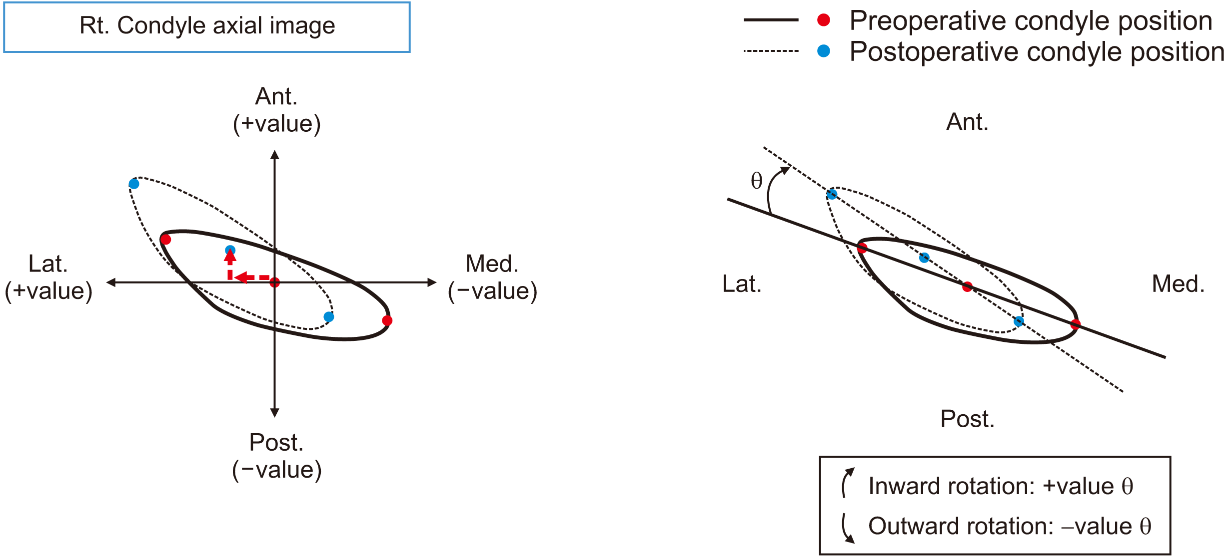
The x- and y-axes were formed on the superimposed CT image and changes in condylar position were observed by placing three points on the center, inner, and outer parts of the condyle. Mandibular rotation and bodily movement were measured.(
Fig. 3) For anteroposterior displacement, a positive value was defined as anterior displacement and a negative value as posterior displacement. For mediolateral displacement, a positive value was defined as lateral displacement and a negative value as medial displacement. For angle measurements, a positive angle was defined as inward rotation and a negative angle as outward rotation. Based on this, three values were measured in both the deviated and non-deviated side condyles on the superimposed images, and the relationship between the three values and the amount of chin movement during surgery was statistically analyzed.
 | Fig. 3Measurement of condyle position change. A. Mediolateral and anteroposterior displacement. B. Condyle rotation. (Rt.: right, Ant.: anterior, Lat.: lateral, Post.: posterior, Med.: medial) 
|
5. TMD symptom evaluation
Preoperative and postoperative medical records were used to evaluate the deterioration and incidence of preoperative and postoperative TMD symptoms. TMD symptoms were limited to joint disc displacement and masticatory muscle pain that did not significantly affect the results of orthognathic surgery. TMD, which may affect other surgical outcomes, was stabilized through treatment before orthognathic surgery.
One month before orthognathic surgery, the patient was examined for the presence of TMD symptoms. After surgery, the patient was re-evaluated for the decrease, disappearance, or development of new TMD symptoms based on the surgical method. In particular, the records were examined at more than 3 months after surgery to differentiate between temporary restricted mouth opening and masticatory muscle pain caused by surgical damage and intermaxillary fixation after surgery.
6. Statistical analysis
The data are presented as frequency and percentage for categorical variables and mean±standard deviation (SD)/median (interquartile range [IQR]) for numeric variables. Differences in study participant characteristics were compared across subgroups using the independent t-test or Mann–Whitney U test for continuous variables.
The differences in study participant characteristics were compared across subgroups using the ANOVA with Scheffé post-hoc test or the Kruskal–Wallis test with Dunn’s post-hoc test. To determine the normal distribution of data, Shapiro–Wilk test was used. For graphical visualization, error bar charts and scatterplots were presented.
All statistical analyses were performed using IBM SPSS Statistics (ver. 26.0; IBM, Armonk, NY, USA) and P<0.05 were considered statistically significant.
Go to :

III. Results
Changes in condylar position were measured in three aspects in Group I and Group II.
Table 1 shows the results of the statistical analysis of chin movement and condylar position change.
Table 1
Results of statistical analysis between chin movement and condylar position changes in the groups
|
Variable |
Group I |
Group II |
P-value |
|
Chin movement (mm) |
|
|
|
|
Mean±SD |
8.63±4.27 |
6.15±2.75 |
0.1551
|
|
Median (IQR) |
7.25 (5.25 to 13.63) |
5.75 (3.75 to 8.50) |
|
|
Mediolateral displacement |
|
|
|
|
Deviated side (mm) |
|
|
|
|
Mean±SD |
0.07±0.97 |
1.62±0.98 |
0.0041
|
|
Median (IQR) |
0.41 (−0.93 to 0.84) |
1.48 (0.89 to 2.26) |
|
|
Non-deviated side (mm) |
|
|
|
|
Mean±SD |
0.17±1.71 |
1.13±0.90 |
0.1441
|
|
Median (IQR) |
0.49 (−1.79 to 1.76) |
1.18 (0.65 to 1.90) |
|
|
Anteroposterior displacement |
|
|
|
|
Deviated side (mm) |
|
|
|
|
Mean±SD |
0.87±1.44 |
0.66±0.90 |
0.7001
|
|
Median (IQR) |
1.36 (−0.80 to 2.09) |
0.77 (0.09 to 1.18) |
|
|
Non-deviated side (mm) |
|
|
|
|
Mean±SD |
−0.30±0.97 |
0.27±0.82 |
0.1991
|
|
Median (IQR) |
−0.06 (−1.40 to 0.54) |
0.04 (−0.27 to 0.96) |
|
|
Condyle rotation |
|
|
|
|
Deviated side (°) |
|
|
|
|
Mean±SD |
−7.11±6.87 |
6.45±3.78 |
0.0001
|
|
Median (IQR) |
−6.00 (−13.55 to −2.00) |
5.80 (3.40 to 8.93) |
|
|
Non-deviated side (°) |
|
|
|
|
Mean±SD |
1.45±3.99 |
3.90±3.83 |
0.2041
|
|
Median (IQR) |
1.30 (−2.70 to 4.68) |
4.35 (0.78 to 7.13) |
|

1. Chin movement
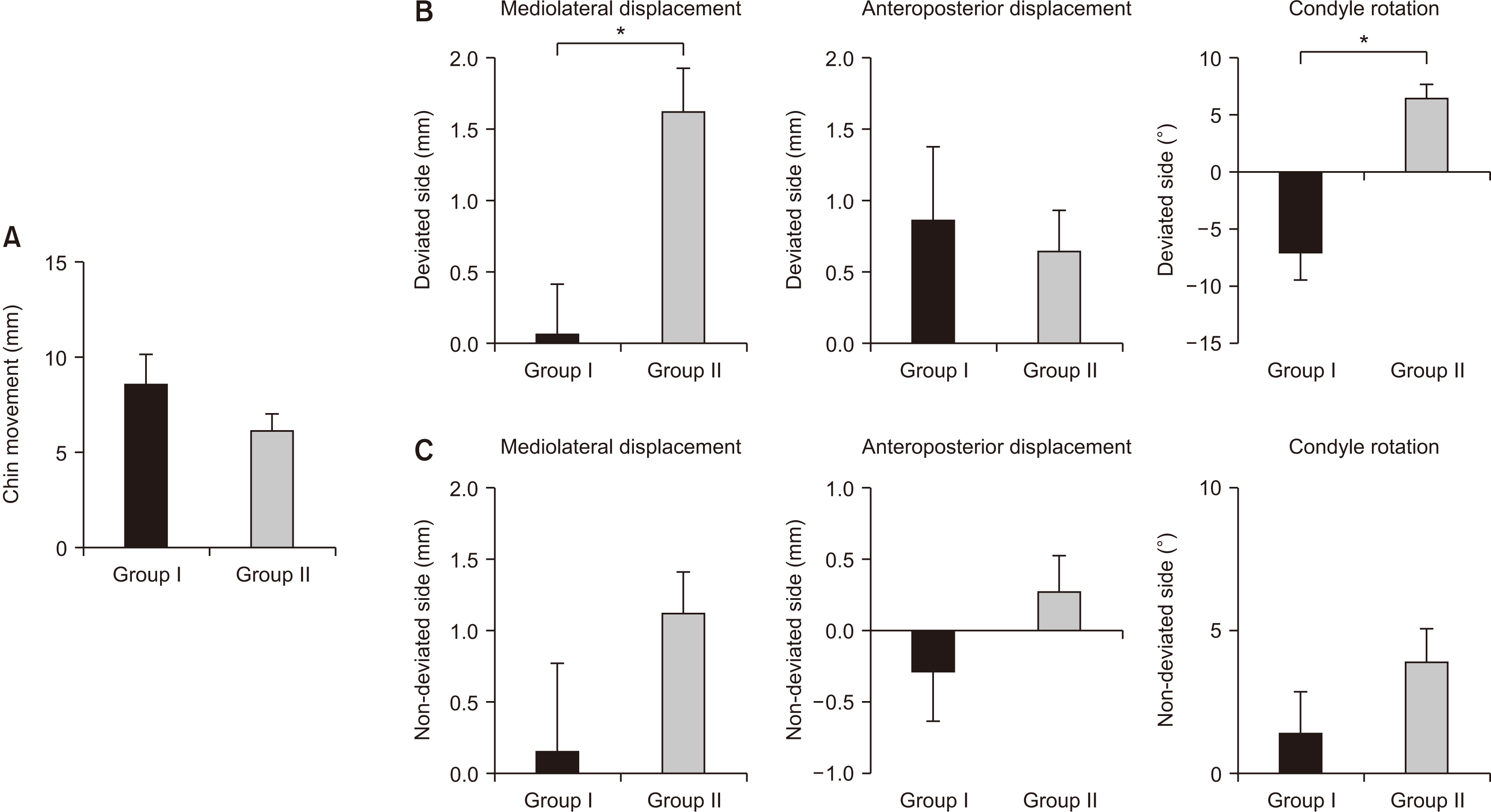
Chin movement was defined as the amount of movement of the mandibular midline (menton) in the coronal plane after surgery, and additional movement caused by genioplasty was excluded. Although statistical significance was not observed, postoperative chin movement tended to be greater in Group I as shown in
Table 1 and
Fig. 4. The mandibles moved an average of 8.63±4.27 mm and 6.15±2.75 mm in Groups I and II, respectively.
 | Fig. 4Result of condyle position change between the two groups. A. Chin movement after surgery. B. Amount of displacement and rotation of the condyles on deviated side. C. Amount of displacement and rotation of the condyles on non-deviated side. *P<0.05. (Group I: patients who underwent intraoral vertical ramus osteotomy [IVRO] on the deviated side and sagittal split ramus osteotomy [SSRO] on the non-deviated side, Group II: patients who underwent bilateral SSRO with the bony interference grinding method) 
|
2. Mediolateral displacement (+value: lateral movement/–value: medial movement)
The mediolateral displacement on the deviated side condyle was 0.07±0.97 mm in Group I and 1.62±0.98 mm in Group II and was statistically significantly correlated with chin movement (P=0.004).
The non-deviated side condyle measured 0.17±1.71 mm and 1.13±0.90 mm in Groups I and II, respectively, which were similar values to the deviated side but statistically non-significant (P=0.144).
3. Anteroposterior displacement (+value: anterior movement/–value: posterior movement)
The anteroposterior displacement of the deviated side condyle was 0.87±1.44 mm in Group I and 0.66±0.90 mm in Group II but was not statistically significantly correlated with chin movement (P=0.700).
The non-deviated side condyle displacement was −0.30±0.97 mm in Group I and 0.27±0.82 mm in Group II. The correlation with chin movement was statistically non-significant (P=0.199).
4. Condyle rotation (+value: inward rotation/–value: outward rotation)
The rotation of the deviated side condyle was −7.11°±6.87° in Group I and 6.45°±3.78° in Group II and was statistically significantly correlated with chin movement (P=0.000).
The rotation of the condyle on the non-deviated side was 1.45°±3.99° in Group I and 3.90°±3.83° in Group II but was not statistically significantly correlated with chin movement (P=0.204).
5. Preoperative and postoperative TMD symptoms
Table 2 shows the changes in TMD symptoms before and after orthognathic surgery. Most of the TMD symptoms in Group I patients who underwent SSRO with IVRO showed improvement. Group II patients, who underwent BSSRO, showed similar improvement in TMD symptoms. Newly developed TMD symptoms did not occur in any of the patients.
Table 2
Preoperative and postoperative temporomandibular joint disorder (TMD) symptoms
|
Group I (n=8) |
|
Group II (n=10) |
|
|
|
Preoperative |
Postoperative |
Preoperative |
Postoperative |
|
TMD symptom |
|
|
|
|
|
|
No symptoms |
1 (12.5) |
5 (62.5) |
|
3 (30.0) |
5 (50.0) |
|
TMD pain |
1 (12.5) |
0 (0) |
|
1 (10.0) |
0 (0) |
|
TMD noise |
1 (12.5) |
3 (37.5) |
|
4 (40.0) |
5 (50.0) |
|
LOM |
0 (0) |
0 (0) |
|
0 (0) |
0 (0) |
|
Myalgia |
1 (12.5) |
0 (0) |
|
0 (0) |
0 (0) |
|
TMD pain+noise |
3 (37.5) |
0 (0) |
|
2 (20.0) |
0 (0) |
|
TMD pain+LOM |
0 (0) |
0 (0) |
|
0 (0) |
0 (0) |
|
TMD pain+noise+LOM |
1 (12.5) |
0 (0) |
|
0 (0) |
0 (0) |
|
Result |
|
|
|
|
Improved |
7 (87.5) |
|
4 (40.0) |
|
Deteriorated |
0 (0) |
|
0 (0) |
|
No change |
1 (12.5) |
|
6 (60.0) |

Go to :

IV. Discussion
Among the complications that may appear after orthognathic surgery, TMD is reportedly the second most frequent occurrence after inferior alveolar nerve injury
13. Various symptoms such as resorption of condyle, development of arthralgia, and masticatory muscle pain can occur
13,14. Various opinions exist on postoperative TMD associated with orthognathic surgery. Some authors argue the incidence of TMD is high in skeletal symmetry and malocclusion before surgery, and the TMD symptoms can improve after orthognathic surgery
10,11,15,16. However, positional changes of the condyle may occur from the articular fossa during orthognathic surgery and excessive changes beyond the physiological limit may affect the masticatory muscles and the joint disc
17. Therefore, surgeons are attempting to minimize condylar torque and positional change due to the movement of the mandible
1 and several methods have been introduced to address this concern. The following four main methods are used: bony interference grinding, plate bending, BSSRO with DSPBO, and SSRO with IVRO
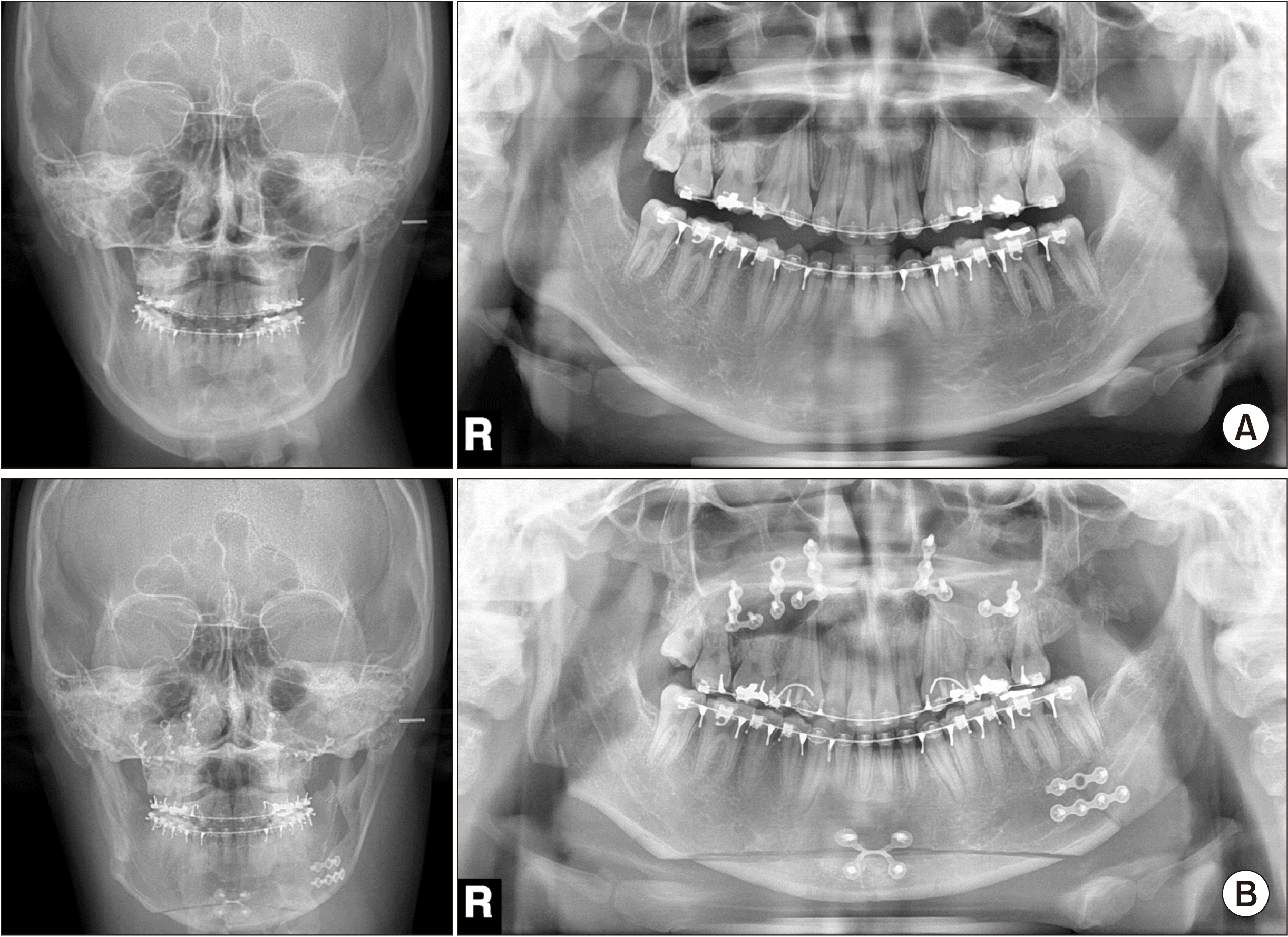
1,12.(
Fig. 5)
 | Fig. 5Digital panoramic and cephalometric radiograph of sagittal split ramus osteotomy (SSRO) with intraoral vertical ramus osteotomy (IVRO). A. Preoperative radiography. B. Postoperative radiography (right SSRO/left IVRO). 
|
Bony interferences are eliminated by grinding to achieve passive contact between the proximal and distal segments. If a large interference is present, it cannot be completely eliminated, and there is an increased risk of trauma to the inferior alveolar nerve, blood vessels, and surrounding soft tissues during grinding
1. Plate bending allows a gap at the end of the fracture section instead of rotation of the proximal segment to achieve contact between the proximal and distal segments and minimizes condylar torque during the fixation. However, malunion and nonunion (due to the lack of interface contact) and iatrogenic facial asymmetry in facial width are possible
1,18,19. BSSRO with DSPBO eliminates bony interference with additional osteotomy of the most posterior part of the distal segment on the side that causes condylar torque and displacement during fixation of the proximal segment. This method has disadvantages such as difficult access during secondary osteotomy, considerable risk of damage to the inferior alveolar nerve, and worsening of postoperative patient discomfort
1,20,21.
Because IVRO does not use rigid fixation, SSRO with IVRO causes less rotational displacement of the proximal segment on the deviated side and displaced or rotated condyles may return to their original physiologic position. Consequently, the additional effect of improved TMD symptoms can be obtained
2. Therefore, SSRO combined with IVRO is used as a surgical alternative to solve the spatial problem caused by BSSRO in cases of severe mandibular rotation and asymmetry
11.
Among the methods described above, grinding bony interferences to minimize condylar torque in BSSRO is usually considered the first-line option
22. However, bone grinding provides limited gains in the presence of large bony interferences or poor access due to the anatomical location
1.(
Fig. 6) Therefore, IVRO on the deviated side that generates condylar torque and SSRO on the opposite side as an alternative surgical method should be considered to solve this problem
11. In the present study, the practical value of SSRO combined with IVRO as a surgical technique was evaluated based on changes in condylar position and TMD symptoms after surgery in 18 patients who underwent orthognathic surgery.
 | Fig. 6Bilateral sagittal split ramus osteotomy (BSSRO) with bony interference grinding in a patient with asymmetric mandible (deviated side: left). A. Preoperative three-dimensional (3D) computed tomography (CT) image. B. Postoperative 3D CT image. Condylar displacement from the mandibular fossa is clearly observed on deviated side (left). (Rt.: right, Lt.: left) 
|
The amount of chin movement in Group I was higher than in Group II, indicating IVRO was more often used to minimize condylar torque on the deviated side condyle in patients with severe chin deviation. When determining the surgical approach, the decision to use IVRO with SSRO depends on the distance from menton to the midline of the face. Thus, when predicting the amount of bony interference on the deviated side during surgical preparation, grinding alone is insufficient to minimize torque generation in cases of severe menton deviation. Furthermore, for persistent TMD symptoms on the deviated side, IVRO is preferred. However, if IVRO is unsuitable for the facial contours, SSRO should be considered for the deviated side with additional DSPBO on the distal segment.
After measuring the change in condylar position between groups, the mediolateral displacement and rotation of the deviated side condyle were statistically significantly correlated with chin movement (
P=0.004 and
P=0.000, respectively). For mediolateral displacement of the deviated side condyle, Group I (0.07±0.97 mm) condyles treated with IVRO showed minimal movement, and Group II (1.62±0.98 mm) condyles showed significant lateral displacement.(
Fig. 4) IVRO prevented the lateral movement of the condyle. Conversely, in the case of SSRO with bone grinding alone on the deviated side, increased movement of the distal segment of the mandible prevented offset of condylar torque, indicating that additional DSPBO or plate bending is required. Notably, lateral displacement occurred on the deviated side in Group II indicating that lateral condylar displacement increases after SSRO.
For condyle rotation on the deviated side, Group I (−7.11°± 6.87°) treated with IVRO showed outward rotation, and Group II (6.45°±3.78°) treated with SSRO showed inward rotation. This finding can be attributed to the nature of IVRO; the proximal segment overlaps the buccal area of the distal segment and the condyle rotates outward in this process. However, due to the characteristics of SSRO, the operator applies a certain amount of force to align the proximal segment to the distal segment. The condyle shows inward rotation because it is fixed with a plate. This feature is observed when examining the rotational tendency of the non-deviated side condyle using SSRO.
The anteroposterior change did not show a significant difference between the two groups; Group I was 0.87±1.44 mm and Group II was 0.66±0.90 mm, indicating the deviated side condyle moved forward in both groups. Rotskoff et al.
9 showed the condyle moved anteriorly immediately after IVRO. Group II condyles that underwent SSRO moved slightly forward, which can be expected because the proximal segment was fixed while being moved slightly forward from its original position when the surgeon attached it to the distal segment.
As mentioned above, changes in the condylar position tended to differ depending on the surgical method. However, a distinct difference in the postoperative TMD symptoms between the groups was not observed, and the symptoms improved, as shown in
Table 2. Group II condyles that underwent BSSRO showed a greater amount of condylar position change than Group I, with improved TMD symptoms, probably because some bony interferences were eliminated by grinding to minimize condylar torque. Furthermore, physical therapy following surgery and the resolution of facial asymmetry resulted in improved masticatory muscle activity and resolution of several TMD symptoms. However, these results do not indicate the change in condylar location is independent of the occurrence and exacerbation of TMD symptoms. A total of 18 patients were analyzed in this study, limiting our ability to generalize the improvement in TMD symptoms. Further studies in which the 3D movement of the condyle, including the sagittal and coronal aspects, is investigated using a larger cohort are needed for a more accurate evaluation.
Go to :


 PDF
PDF Citation
 Print
Print


 XML Download