Abstract
Objectives
The purpose of this study was to evaluate the clinical and radiographic characteristics of idiopathic bone cavity (IBC) to determine the effect of surgical intervention on the process of healing.
Materials and Methods
All cases diagnosed with IBC during the period of 2011 to 2020 at our Department of Oral and Maxillofacial Surgery were searched. Ninety cases were retrieved. The features evaluated were sex, age, contour of the lesion, number of teeth involved, site, history of trauma, and postoperative healing pattern. The significance of differences was assessed by Mann–Whitney U test and chi-square test.
Results
The femalemale ratio showed no predilection toward either sex (0.90.8). The mean age of the collected sample was 22.05±14.38 years, and the age ranged from 10 to 58 years. All cases presented in the mandible and showed well-circumscribed radiolucency. Margins were either scalloped or round in shape, and the size varied from one tooth to six teeth involvement. Seventy cases involved three or fewer roots. Three cases showed bilateral lesion. Four cases had a history of trauma at the area of the lesion. Fifty-one cases were followed for six months after surgery, and all showed increased bone density at the lesion.
Conclusion
There is no definitive radiological or clinical feature of IBC. Considering the diversity of clinical and radiological features, such a diagnosis relies primarily on surgical findings of an empty bone cavity with no epithelial lining. Our data suggest that surgical intervention be the first choice of treatment as opposed to observation.
Idiopathic bone cavity (IBC), also known as simple or traumatic bone cyst, is defined by the World Health Organization as a non-neoplastic intraosseous pseudocyst devoid of epithelial lining1. With its lack of cystic lining, histopathological confirmation of IBC can be achieved in some cases where fibrous tissues are present, but is difficult since collecting tissue samples within an empty cavity often is not possible1,2. IBC is often asymptomatic, occurs in young patients in their second to third decade of life2, and is a solitary lesion3, in the posterior site of the mandible, and shows no association with sex4. Despite its clear description, the pathogenesis of idiopathic bone cavity remains unclear3,5. Thus, it is uncertain whether surgical intervention is necessary3 or if a surgically untreated lesion could yield spontaneous resolution. Few studies have reported cases of spontaneous resolution of surgically untreated IBC, but a longer follow-up period was needed to observe complete resolution5. Discacciati et al.6 suggest differentiation from a lesion with other maxillofacial cystic or neoplastic pathologies can be based on clinical and radiographical findings, but surgical exploration can serve as a definitive diagnostic tool. Its many names reflect the diverse clinical and radiological presentation of idiopathic bone cavity, and more extensive studies are needed to increase understanding of the lesion. The purpose of this study was to evaluate the clinical and radiographic characteristics of idiopathic bone cavity and the necessity of surgical intervention.
A search of all cases that were first diagnosed as idiopathic bone cavity among patients from 2011 to 2020, at the Department of Oral and Maxillofacial Surgery, Pusan National University Hospital (Yangsan, Korea) was conducted. Inclusion criteria for these cases were availability of diagnostic X-rays; clinical, radiographic, and surgical description of the lesion; and histopathology. The follow-up period differed by case so postoperative comparison of lesions was based on the cases that had a greater than six-month follow-up period and radiographic images.
Any case that was diagnosed as idiopathic bone cavity but did not undergo surgical treatment was not included in the study. Lesions that were misdiagnosed and cases where patients had systemic disease that might interfere with healing also were excluded.
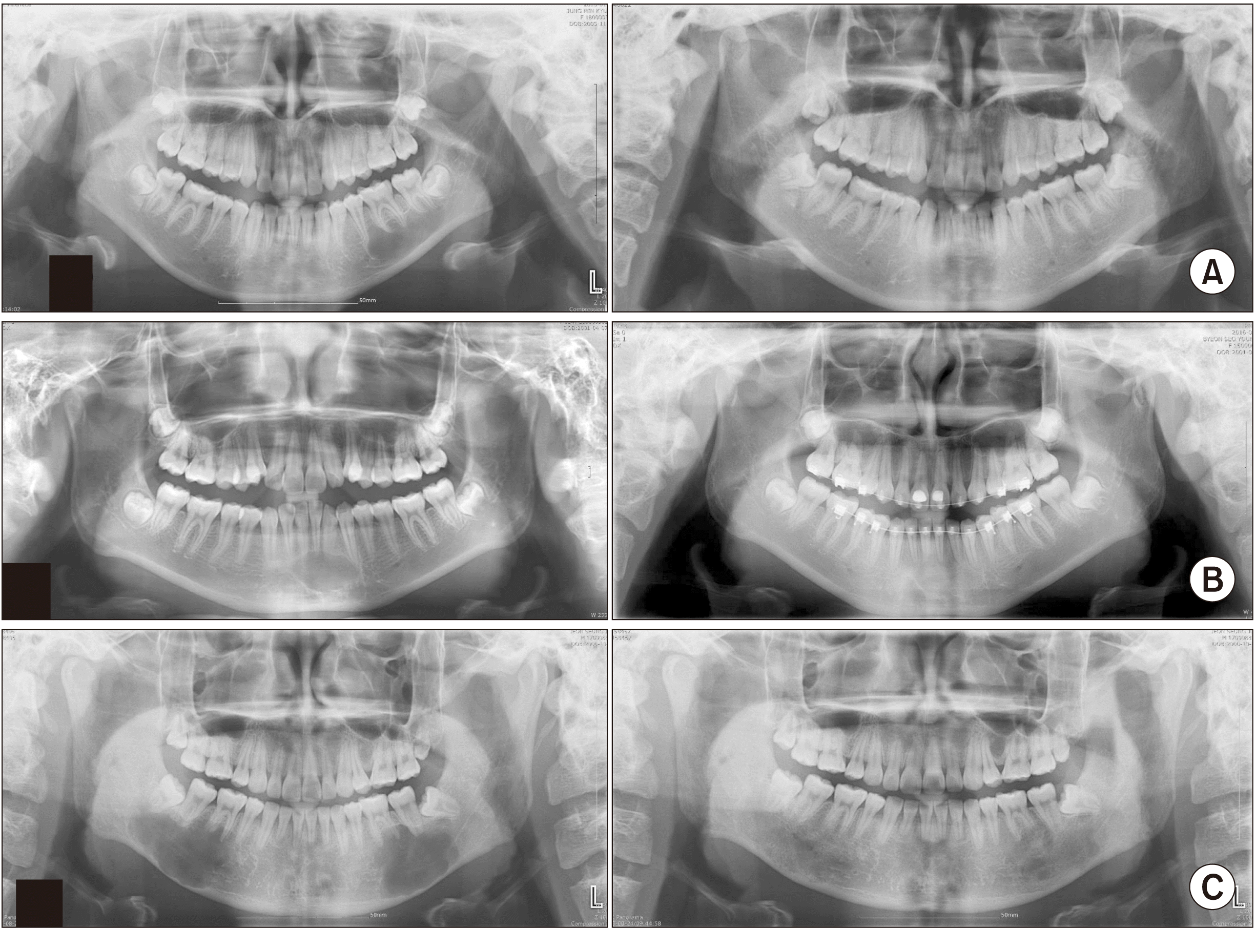
A total of 90 cases was included in this study.(Table 1) All cases had preoperative cone-beam computed tomography data and panoramic radiographs. The mean age at presentation of IBC was 22.05±14.38 years, which ranged from 10 to 58 years. There was no predilection between the two sexes. A single case was observed in the maxilla, with the remaining cases in the mandibular region. History of trauma associated with the lesion was acquired at the first visit and documented, but most cases showed no specific traumatic incidence. The legion size was measured in two categories with respect to the adjacent teeth involved, either of equal or smaller size than three teeth or greater than three teeth. The majority of cases (65 cases) involved three or fewer teeth. Patients younger than 20 years showed more lesions (21 cases) associated with a larger size than did the older (four cases) group (P<0.05).(Table 2) All asymptomatic IBCs were discovered incidentally during routine check-ups. However, 17 cases showed some clinical symptoms such as pain, swelling, idiopathic discomfort, or paresthesia. Involved teeth showed more frequent vitality than non-vitality. Cortical bone expansion was not found in 17 cases, and mild thinning of the cortex was seen in 51 cases (56.7%). Radiographic presentation of IBCs was evaluated as either scalloped or round in shape, and the proportions were equal (1:1). Radiographic examination over six months after surgical curettage or exploration showed reduction in size and evidence of increased bone density.(Fig. 3) Increased bone density of the lesion in 51 cases was observed during the six-month follow-up period, while 39 patients were lost postoperatively.
The pathogenesis of IBC remains theoretical despite its widely investigated clinical and radiological features. Among the major etiologic hypotheses, three have achieved some agreement and predominate: 1) abnormality of bone growth, 2) tumor degeneration, and 3) traumatism. The first two factors are based on clinical observation, and the third hypothesis considers the interaction between lesion pathogenesis and etiology. According to Harnet et al.7, traumatism is the most widely accepted theory, and it advocates vascular alterations caused by intramedullary hemorrhage after traumatic force on the lesion. Discacciati et al.6 also investigated these three factors, but they failed to observe significant results6.
The ambiguity of pathogenesis is partly due to various clinical and radiological features of IBCs. In many reported cases, presentation of IBCs varies by sex, size, preferential location, shape, recurrence, etc. Many studies have investigated clinical and radiological findings of IBCs in an effort to form differential diagnosis of the lesion from other benign intraosseous lesions, including odontogenic keratocyst, ameloblastoma, central giant cell lesions, lateral periodontal cysts, periapical cysts, and other odontogenic cysts4. Specifically, since radiological findings of IBCs mimic those of other cysts of the oral region, it is essential to investigate further to decide whether treatment intervention or follow-up observation is necessary. Some studies advocate conservative treatment as long as patients are free of symptoms since IBC is a benign lesion8. A sexual predilection exists in some reviews and case reports6,9-12, while others including Rushton13 reveal no such predilection14-16. Our study, however, is in accordance with the latter case (male to female ratio as 0.9:1.0). In general, it is agreed that IBCs often are found incidentally in the second to third decades of life, with a location predominantly in the mandible; this correlates with the data from our study10,12,17. You et al.10 reported the posterior mandible as the most common site, followed by the anterior region. Topographical distribution of all our cases, except one in the maxilla, was significantly higher in the posterior region of the mandible. In addition, 27 cases showed a lesion size greater than three teeth, and most of these patients were in their 20s, which confirmed a significant size difference between the younger and older groups. There was no association between degree of cortical bone expansion and existing symptoms, and a mild degree of expansion was most common. Although solitary and unilocular radiolucency is most common in IBCs18, multiple IBCs were found in about 11% of cases11. Most of these lesions were treated surgically, and recurrence was rare11,19. Three cases showed bilateral lesion in the mandible and were diagnosed originally as keratocystic odontogenic tumor, after which surgical exploration confirmed the diagnosis as IBC. A characteristic IBC trait is scalloping of the lesion projection into the interdental or interradicular spaces2. Scalloping borderlines were seen in about 40% of IBCs1; however, our study showed equal proportions of scalloped and rounded lesion contours.
Considering the atypical presentation of IBCs and the diversity of theories put forward in the literature, the preferred treatment option is clear. Especially when routine check-ups reveal enlargement of a lesion, a surgical approach is logical8. Since final diagnosis and treatment can be performed simultaneously in the same surgical procedure and as shown in our data, it is reasonable to consider surgical management of the lesion20. Surgical curettage or exploration can stimulate bony growth by generating new blood flow into the site2,3,9.
Multiple treatment modalities exist to stimulate the healing process of the emptied bone cavity. Treatment options include no surgical intervention with expectation of spontaneous healing and a surgical approach that can involve exploration, curettage, fenestration, autologous bone marrows injection, cavity packing, etc.1,14. The effectiveness of these treatment modalities is not known14, and there is no consensus on treatment protocol. Our data, however, suggest that simple curettage of the lesion was sufficient to yield bone regeneration that is detectable through X-rays. One surgical procedure was needed to confirm the tentative diagnosis and rule out other oral pathologies; treatment was spontaneous healing.
Like the many synonyms for idiopathic bone cavity, its etiology remains unclear. The diagnosis of idiopathic bone cavity relies primarily on radiographic interpretation or the surgical finding of an empty bone cavity with a lack of epithelial lining. Our data suggest that surgical intervention be the first choice of treatment for stimulating healing of idiopathic bone cavity.
Notes
Authors’ Contributions
J.R. participated in data collection and wrote the manuscript. I.N., Y.D.K., and S.H.S. participated in the study design and performed the statistical analysis. J.Y.L. participated in the study design and coordination and helped to draft the manuscript. All authors read and approved the final manuscript.
References
1. Zhang W, Chen M, Yang C, Han Z, Wei W, Chai Y. 2017; Does idiopathic bone cavity involving mandibular condyle need surgical intervention of bone cavity filling? J Craniofac Surg. 28:e539–43.
https://doi.org/10.1097/SCS.0000000000003851
. DOI: 10.1097/SCS.0000000000003851. PMID: 28796103.

2. Horne RP, Meara DJ, Granite EL. 2014; Idiopathic bone cavities of the mandible: an update on recurrence rates and case report. Oral Surg Oral Med Oral Pathol Oral Radiol. 117:e71–3.
https://doi.org/10.1016/j.oooo.2012.03.037
. DOI: 10.1016/j.oooo.2012.03.037. PMID: 22939327.

3. Chrcanovic BR, Gomez RS. 2019; Idiopathic bone cavity of the jaws: an updated analysis of the cases reported in the literature. Int J Oral Maxillofac Surg. 48:886–94.
https://doi.org/10.1016/j.ijom.2019.02.001
. DOI: 10.1016/j.ijom.2019.02.001. PMID: 30792085.

4. Perdigão PF, Silva EC, Sakurai E, Soares de Araújo N, Gomez RS. 2003; Idiopathic bone cavity: a clinical, radiographic, and histological study. Br J Oral Maxillofac Surg. 41:407–9.
https://doi.org/10.1016/s0266-4356(03)00145-1
. DOI: 10.1016/s0266-4356(03)00145-1. PMID: 14614871.

5. Battisti MPL, Soares MQS, Rubira CMF, Bullen IRFR, Lauris JRP, Damante JH. 2018; Assessment of spontaneous resolution of idiopathic bone cavity. J Appl Oral Sci. 26:e20170288.
https://doi.org/10.1590/1678-7757-2017-0288
. DOI: 10.1590/1678-7757-2017-0288. PMID: 29742261. PMCID: PMC5933833.

6. Discacciati ED, de Faria VM, Garcia NG, Sakai VT, Pereira AA, Hanemann JA. 2012; Idiopathic bone cavity: case series involving children and adolescents. J Investig Clin Dent. 3:103–8.
https://doi.org/10.1111/j.2041-1626.2011.0087.x
. DOI: 10.1111/j.2041-1626.2011.0087.x. PMID: 22522949.

7. Harnet JC, Lombardi T, Klewansky P, Rieger J, Tempe MH, Clavert JM. 2008; Solitary bone cyst of the jaws: a review of the etiopathogenic hypotheses. J Oral Maxillofac Surg. 66:2345–8.
https://doi.org/10.1016/j.joms.2007.08.035
. DOI: 10.1016/j.joms.2007.08.035. PMID: 18940504.

8. Assaf AT, Solaty M, Zrnc TA, Fuhrmann AW, Scheuer H, Heiland M, et al. 2014; Prevalence of Stafne's bone cavity--retrospective analysis of 14,005 panoramic views. In Vivo. 28:1159–64.
9. Velez I, Siegel MA, Mintz SM, Rolle R. 2010; The relationship between idiopathic bone cavity and orthodontic tooth movement: analysis of 44 cases. Dentomaxillofac Radiol. 39:162–6.
https://doi.org/10.1259/dmfr/12528297
. DOI: 10.1259/dmfr/12528297. PMID: 20203278. PMCID: PMC3520219.

10. You MS, Kim DY, Ahn KM. 2017; Surgical management of idiopathic bone cavity: case series of consecutive 27 patients. J Korean Assoc Oral Maxillofac Surg. 43:94–9.
https://doi.org/10.5125/jkaoms.2017.43.2.94
. DOI: 10.5125/jkaoms.2017.43.2.94. PMID: 28462193. PMCID: PMC5410434.

11. An SY, Lee JS, Benavides E, Aminlari A, McDonald NJ, Edwards PC, et al. 2014; Multiple simple bone cysts of the jaws: review of the literature and report of three cases. Oral Surg Oral Med Oral Pathol Oral Radiol. 117:e458–69.
https://doi.org/10.1016/j.oooo.2014.03.004
. DOI: 10.1016/j.oooo.2014.03.004. PMID: 24842451.

12. Cortell-Ballester I, Figueiredo R, Berini-Aytés L, Gay-Escoda C. 2009; Traumatic bone cyst: a retrospective study of 21 cases. Med Oral Patol Oral Cir Bucal. 14:E239–43. PMID: 19218905.
13. Rushton MA. 1946; Solitary bone cysts in the mandible. Br Dent J. 81:37–49. PMID: 20992458.
14. Tabrizi R, Karagah T, Shahidi S, Zare N. 2015; Does platelet-rich plasma enhance healing in the idiopathic bone cavity? A single-blind randomized clinical trial. Int J Oral Maxillofac Surg. 44:1175–80.
https://doi.org/10.1016/j.ijom.2015.05.011
. DOI: 10.1016/j.ijom.2015.05.011. PMID: 26074365.

15. Manganaro AM. 1997; Review of the idiopathic bone cavity of the jaws. Mil Med. 162:734–6. PMID: 9358719.

16. Roma LG, Lopes CB, Netto JNS, Miranda ÁMMA, Pires FR. 2021; Idiopathic bone cavity: clinicopathologic features from a case series with emphasis on differential diagnosis of chronic apical periodontitis. J Endod. 47:221–5.
https://doi.org/10.1016/j.joen.2020.11.004
. DOI: 10.1016/j.joen.2020.11.004. PMID: 33217470.

17. Copete MA, Kawamata A, Langlais RP. 1998; Solitary bone cyst of the jaws: radiographic review of 44 cases. Oral Surg Oral Med Oral Pathol Oral Radiol Endod. 85:221–5.
https://doi.org/10.1016/s1079-2104(98)90430-9
. DOI: 10.1016/s1079-2104(98)90430-9. PMID: 9503460.

18. Tong AC, Ng IO, Yan BS. 2003; Variations in clinical presentations of the simple bone cyst: report of cases. J Oral Maxillofac Surg. 61:1487–91.
https://doi.org/10.1016/j.joms.2003.05.003
. DOI: 10.1016/j.joms.2003.05.003. PMID: 14663817.

19. Suei Y, Taguchi A, Tanimoto K. 2007; Simple bone cyst of the jaws: evaluation of treatment outcome by review of 132 cases. J Oral Maxillofac Surg. 65:918–23.
https://doi.org/10.1016/j.joms.2006.06.297
. DOI: 10.1016/j.joms.2006.06.297. PMID: 17448841.

20. Resnick CM, Dentino KM, Garza R, Padwa BL. 2016; A management strategy for idiopathic bone cavities of the jaws. J Oral Maxillofac Surg. 74:1153–8.
https://doi.org/10.1016/j.joms.2015.12.014
. DOI: 10.1016/j.joms.2015.12.014. PMID: 26850870.

Fig. 1
Cone-beam computed tomography of different degrees of bone thinning pattern. A. Mild cortical bone thinning. B. Moderate cortical bone thinning. C. Severe cortical bone thinning.

Fig. 3
Radiographic changes six months postoperatively. All cases underwent surgical curettage of the lesion. A. Left: Initial lesion on the left posterior of the mandible. Right: Postoperative lesion. B. Left: Initial lesion on the anterior of the mandible. Right: Postoperative lesion. C. Left: Initial lesion on the right and left posterior mandible. Right: Postoperative lesion.

Table 1
Clinical variables of idiopathic bone cavity and statistical data (n=90)
| Variable | Value | P-value |
|---|---|---|
| Age (yr) | 22.05±14.38 (10-58) | - |
| Sex | 0.610 | |
| Male | 40 | |
| Female | 50 | |
| Jaw | ||
| Maxilla | 1 | <0.001* |
| Mandible | 89 | |
| Trauma | ||
| Yes | 4 | 0.803 |
| No | 9 | |
| Unknown | 77 | |
| Symptomatic | ||
| Yes | 17 | 0.656 |
| No | 72 | |
| Unknown | 1 | |
| Pulp vitality | ||
| Vital | 54 | 0.004* |
| Non-vital | 12 | |
| Vital+Non-vital | 4 | |
| Unknown | 20 | |
| Cortical bone thinning | ||
| None | 17 | |
| Mild | 51 | 0.405 |
| Moderate | 18 | |
| Severe | 4 | |
| Contour | ||
| Scalloped | 45 | 0.558 |
| Round | 45 | |
| Size | ||
| Equal to or less than three teeth involved | 70 | 0.735 |
| Greater than three teeth involved | 20 | |
| Location | ||
| Anterior | 33 | 0.922 |
| Posterior | 57 |
Table 2
Comparative analysis of distribution by lesion size and age (n=90)
| Size less or greater than three teeth involved | P-value | ||
|---|---|---|---|
|
|
|||
| Less (n=65) | Greater (n=25) | ||
| ≤20 yr (n=62) | 41 (45.6) | 21 (23.3) | 0.038* |
| >20 yr (n=28) | 24 (26.7) | 4 (0.4) | |


 PDF
PDF Citation
 Print
Print


 XML Download