I. Introduction
The mouth is the gateway to the body. Trauma to this region affects the airway, food intake, speech, facial aesthetics, and overall psychosocial well-being of patients. Mandibular fractures comprise 15.5%-59% of all maxillofacial fractures
1. The mandible is the second most often fractured adult facial bone because of its projecting and vulnerable position in the face
2.
Various countries across the globe have provided statistics for mandibular fractures, and the information provided is unique to the countries of origin and their residents. Several variables are related to the study of mandibular fractures due to differences in demographic characteristics reported in the literature
3. Socio-economic trends, geographic locations, and local behavior have a considerable impact on the etiology of the injury, which sequentially influences the distribution of fracture sites
4. No unanimity has been found for the common pattern of mandibular fractures because of disparate associated factors such as geographic area, population mass, socio-economic status, regional government, status of the population studied, and political era
5.
It is a paradox that, despite being adjacent to the information technology hub Gurugram and industrial hub Faridabad, Mewat (now renamed as Nuh) is the most underdeveloped district of not only Haryana state, but of all India. This region lies in the semi-arid zone and is inhabited primarily by Meo Muslims. According to the 2011 census, it has an estimated population of 10.89 lacs (1 lac=100,000) and an area of 1,507 km
2. A majority of the population (88%) resides in rural areas and is engaged in agriculture. Scarcity of water limits the industries in this region. This area lags significantly in various socio-economic parameters such as standard of living, education, health, sex, agriculture, livestock, and economy. The literacy rate is 69.94% among males and decreases to 37.6% in females. The available health data indicate a lack of good health care facilities
6. Before establishment of the medical college, there were limited health care facilities for management of facial trauma. SHKM Government Medical College, located in the Nalhar village of Nuh, was established in 2012, and is the only tertiary care health facility there. The institution caters to various blocks of Mewat and adjoining areas, namely, Firozpur Zirka, Taoru and Punhana, as well as adjoining districts of Haryana, Rajasthan, and Uttar Pradesh.
This retrospective study aimed to analyze mandibular fractures for incidence, age and sex distribution, etiology, anatomic distribution, occlusal status, and treatment provided and their correlation in patients who visited the Department of Dentistry, SHKM Government Medical College, Nalhar, Nuh, Haryana, from January 2013 to December 2019.
Go to :

IV. Discussion
The first description of mandibular fractures dates back to the 17th century B.C. in the “Edwin Smith Papyrus” bought by Smith in Luxor in 1862, and later translated by Breasted
7. In his manuscript, “Hippocratic Collection,” Hippocrates described manual reduction of mandibular fractures with monomaxillary dental fixation and bandaging
8,9. The importance of the concept of occlusion in management of mandibular fractures was found in a textbook written in Salerno, Italy
10. The concept of MMF was described by Guglielmo Salicetti in his book
Cyrurgia 11 in 1492. This concept was reintroduced by Gilmer
12 in the United States, in 1887. Until the 1900s, the concept of closed reduction was the mainstay of treatment for mandibular fractures. As the concepts in anesthesia and asepsis improved, a new era of open reduction and internal fixation began. From Luhr’s concept of compression plates to Spissel’s AO/ASIF (Association for Osteosynthesis/Association for Study of Internal Fixation) and the latest concept of non-compression plates introduced by Michlete and colleagues
13-16 and later refined by Champy et al.
17, the treatment of mandibular fractures has been in a constant state of evolution.
The basis of evolution in management strategies can be attributed to a better understanding of the biomechanics of the mandible, its behavior in response to traumatic forces, fracture patterns, etiology, epidemiology, mode of healing, and functional rehabilitation
18.
The mandible has varying strengths of bone in different regions, in correlation with stress distribution on function. It is a tubular V-shaped bone that articulates with the skull via paired temporomandibular joints
19.
Mandibular fractures constitute a significant percentage of maxillofacial fractures
1. In most studies, males are affected more commonly than females
2,5,20. In an extensive study by Ellis et al.
21, who analyzed over 3,400 mandibular fractures within 10 years between 1974-1983, there was a clear male preponderance.
This might be due to greater participation in outdoor activities and higher levels of physical activity in males. Furthermore, males are more likely to be involved in traffic accidents and altercations
22,23. Other reasons behind this sex pattern could be higher social activity, drug abuse, vehicular driving, industrial work, and sports issues among males
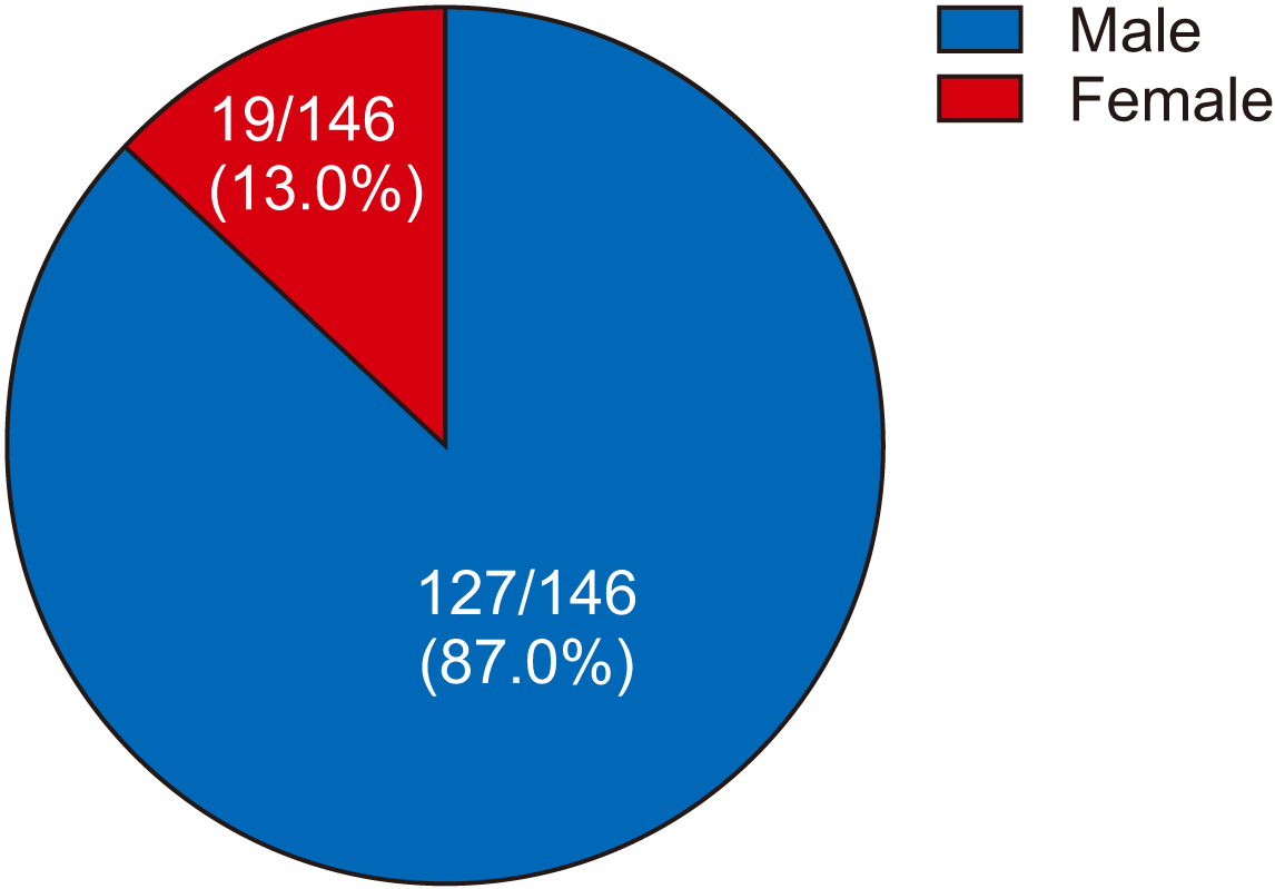
24,25. In the present study males also outnumbered females at a ratio of 7:1. The sex ratio observed in other studies was 3:1
18, 4:1
26,27, and 6.6:1
28. Mandibular fractures have been reported in all age groups (ranging from small children
22 to individuals of 95 years
29). In the present study, the range was from three to 70 years with an mean of 26 years; the mean age reported in other studies was 39.5 years
26, 31.5 years
18, and 32.1 years
27.
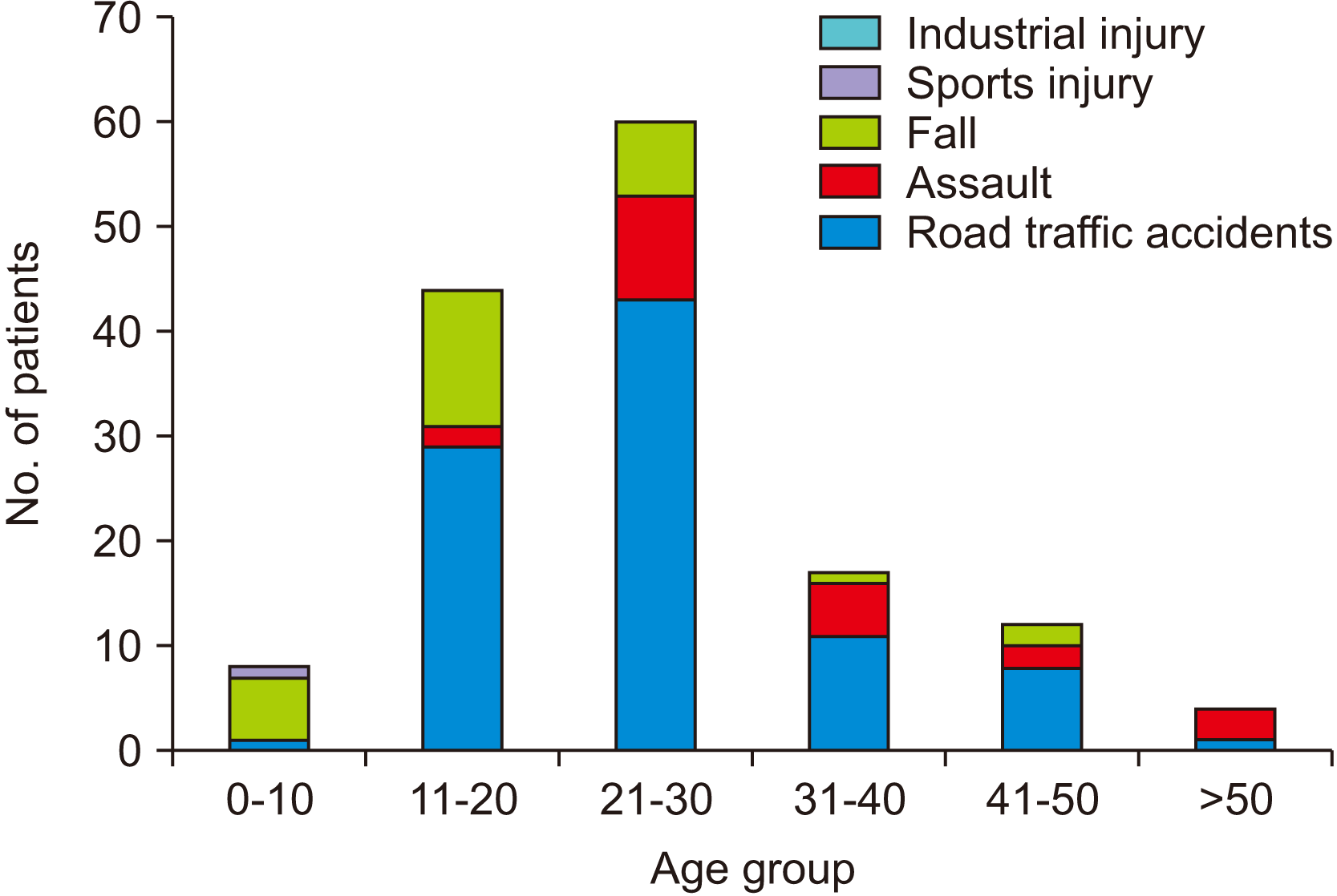
A majority of patients belonged to age group-III (41.1%) in the present study, similar to the 21- to 30-year-old group reported in other studies
21,26,28. The incidence of fractures increased from age group-I to -III and then decreased from age group-III to -VI. Thus, the incidence of mandibular fracture is lowest at both age extremes in the present study, which is similar to other studies
26. Among young persons, less frequent fracture likely is due to the percentage of time spent indoors and the elastic nature of young bone
2. Among individuals older than 60 years, comparatively less exposure to the outside world and a less active life can explain the lower incidence of mandibular fractures
20.
In a study by Shah et al.
26, the peak age group for mandibular fractures was 21-30 years in both males and females. In the present study, group-III was the peak age group in makes, while females age groups-III, IV, and V were affected equally.
There is a striking contrast in the etiology of mandibular fractures in developed and developing countries. The most common causative factor in developing countries is RTAs
3,22,23,30. This might be due to rash driving, speeding, subpar roads, unwillingness to follow road safety measures such as wearing of helmets or seatbelts, inadequate implementation of traffic rules, drunken driving, increased use of vehicles by minors, poor maintenance of vehicles, etc
3,20,22,23.
In the present study, the most common cause of mandibular fractures was RTAs (94/146, 64.4%), followed by fall (29/146, 19.9%), assault (22/146, 15.1%), and sports (1/146, 0.7%). This finding is consistent with previous studies
26,31-33. In India, the increasing incidence of alcohol dependence and drunken driving is the reason behind the increased incidence of fracture due to RTAs. Giri et al.
34 noted that the high prevalence of intoxication among the affected population points to the growing necessity of reliably documented and scientifically backed evidence of road traffic legislation, with compulsory use of seat belts and safety helmet regulations.
The only case of a sports injury was reported in age group-I, the first decade of life. Fall was the most common cause of mandibular fracture (75%) among the elderly (group-VI), likely because of medical conditions, poor muscular control and bodily response, and lack of support and care. Moreover, in geriatric patients, bones become more brittle and have a susceptibility to injury even after a minor fall
35.
In the current study, the most common fracture site was the parasymphysis (73/211, 34.6%). This finding is similar to previous studies. This could be explained by the horseshoe shape of the mandible, resulting in fractures of the parasymphysis under forces due to the large canine roots and convex structure
18,20,29,32,36. In this study, the second most common unilateral fracture site was angle (50/211, 23.7%), followed by condyle (43/211, 20.4%).
Various studies have different results for anatomical distribution of mandible fractures. Brasileiro and Passeri
37 reported the mandibular condyle as the most common site of fracture. Dongas and Hall
38 and Morris et al.
39 have shown mandibular angle to be the most common site of fracture, while Adi et al.
40 have shown the mandibular body as the most common site of fracture.
In most studies, the coronoid process of the mandible is the least affected site of mandibular fracture
20,26,41. This is true for the present study, with no reported case of coronoid fracture.
The most common combination fracture found in the study was angle with parasymphysis (26/146, 17.8%), which is similar to information reported in other studies
18,27,31,38,42. However, additional studies reported parasymphysis with condyle
18,20,24 and body with angle as the most common combinations
5,39.
Specific association between fracture locations is an important consideration when performing clinical assessment of a patient with a mandible fracture. Knowledge that one particular type of fracture is more likely with a fracture at another location can aid in diagnosis
39. In the present study, the second most common combination was parasymphysis with condyle (16/146, 11.0%).
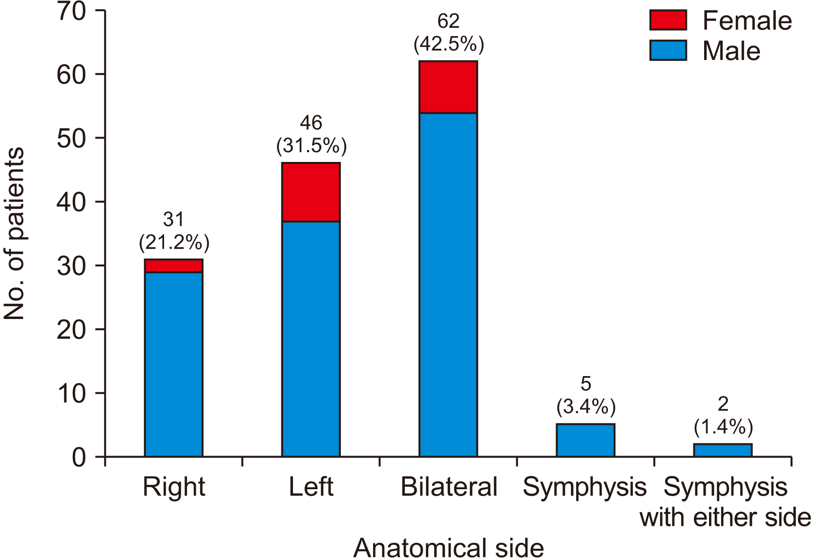
The left side was involved more commonly than the right, at a ratio of 1.4:1. This was true for males and females. Among assault patients, the left side of the mandible was affected more commonly, as most of the Indian population is right-handed
18. According to McManus
43, Professor of Psychology at University College London, approximately 90% of humans are right-handed, explaining why the left side of the face is the most common location of injury.
It is important to manage mandibular fractures to establish stable occlusion, preserve normal mandibular arch form, restore mandibular functions, retain the symmetry of the face, and avoid advancement of developmental disorders. Treatments generally vary according to fracture type, number and location, surgeon performance, and patient characteristics (e.g., age, dental profile, choice of treatment, financial status). The various treatment options available are intermaxillary fixation, open reduction and internal fixation, closed treatment with external fixation, and treatment with Kirschner wire
26.
In the present study, closed reduction was the predominant management method (139/146, 95.2%).
The Ivy eyelet method of intermaxillary fixation was used in patients with no or minimal displacement of fractured segments. In cases needing elastic traction to reduce displaced fracture segments, the Erich arch bar was used. Bridle wiring was performed in pediatric trauma patients. The mean period of intermaxillary fixation was four to six weeks. However, in delayed union, intermaxillary fixation was continued for up to eight weeks. The advantages of closed reduction include reduced morbidity as it does not traumatize the vascular envelope, lower expense, less invasiveness, need for less surgical experience, and can be carried out under local anesthesia with minimal equipment. The disadvantages include a significant period of immobilization and required prolonged closure of the oral cavity, to which the patient might not agree, and it requires intact dentition, maintenance of oral hygiene, and patient compliance. It cannot be utilized in certain situations such as underlying medical conditions and difficult occlusion. Moreover, once the intermaxillary fixation has been released, patients have difficulty in achieving full mouth opening for a few weeks, for which exercises and hot water fomentation are advised. Closed reduction was the preferred method in this study because of the location as a general dentistry department with limited faculty for advanced maxillofacial procedures and limited availability of general anesthesia. This indicates the need for a separate oral and maxillofacial surgical unit in each medical college. Moreover, most of the patients visiting the participating institute experienced financial limitations and preferred less expensive methods of treatment.
Go to :


 PDF
PDF Citation
 Print
Print


 XML Download