

 PDF
PDF Citation
 Print
Print


INTRODUCTION
Postoperative ileus (POI) is described as the decrease in gastrointestinal motility after intra-abdominal surgery. POI is accompanied by symptoms such as pain, nausea, vomiting, abdominal distension, absence of defecation, and intolerance to oral feeding [1,2]. Clinically, there is physiological POI and prolonged POI. Most patients after surgery recover within 2–3 days without any treatment, a process called physiological POI. However, approximately 10% of physiological POI persists for more than 3–7 days, called prolonged POI [3,4].
The pathophysiology of POI can be explained via pharmacological, neural, and immune-mediated mechanisms [5]. Immediately after abdominal surgery, gastrointestinal motility is decreased by anesthesia and opioids. Opioids affect the decrease in gastrointestinal motility via the μ-opioid receptors [6]. Neural and inflammatory mechanisms are the two main theories for the mechanism of current POIs. The neurogenic phase of POI is a reaction where the skin incision or the opened abdomen reduces gastrointestinal motility through adrenergic reflexes. When surgery of the small intestine proceeds, the motility of the gastrointestinal tract decreases due to nociceptive stimuli [7].
Gastrointestinal motility was impaired at 6–72 h after abdominal surgery by an immune-mediated cascade called the inflammatory phase of POI. Immediately after surgery, pro-inflammatory cytokines such as tumor necrosis factor α (TNF-α), interleukin (IL)-1α, and IL-6 are expressed; these cytokines are also expressed before the infiltration of neutrophils and monocytes. After infiltration of inflammatory cells, the levels of IL-1β and CCL2 increases [8,9]. Macrophages, neutrophils, and monocytes play an important role in POI [10,11]. When macrophages are activated, transcription factors, such as nuclear factor κB (NF-κB) and signal transducer and activator of transcription 3 (STAT3), are subsequently activated [12].
Neutrophils and monocytes mainly infiltrate the muscular layer of the intestine 3–4 h after abdominal surgery. Infiltrated neutrophils and monocytes cause inflammation of the intestine, which is the cause of the decrease in gastrointestinal motility [11,13]. Infiltrated neutrophils and monocytes induce the release of cyclooxygenase 2 (COX-2), which induces the production of prostaglandin E2 (PGE2). PGE2 promotes the production of inducible nitric oxide synthase (iNOS) [14]. Nitric oxide (NO) produced by iNOS causes impairment of smooth muscle of the intestine and a decrease in gastrointestinal motility [15].
Antioxidants, such as superoxide dismutase (SOD) and glutathione (GSH), protect the human body from oxidative damages by reducing the amount of free radicals [16]. SOD protects cells from oxidative stress by converting the superoxide anion radical (O2–) to H2O2 or H2O [17]. GSH prevents oxidative damage by reducing reactive oxygen species (ROS) such as free radicals, peroxides, and lipid peroxides [18]. Malondialdehyde (MDA), a product of lipid peroxidation of polyunsaturated fatty acid, is a highly reactive compound that is a biomarker of lipid peroxidation. MDA is formed by the degradation of polyunsaturated lipid by ROS [19]. MDA is a commonly used factor of lipid peroxidation owing to its high reactivity [20]. In this study, we sought to understand the effects of ilaprazole on oxidative stress in a POI model by measuring the levels of the enzymes and molecules related to oxidative stress.
The pharmacological treatment of POI is important for decreasing hospital cost and morbidity rates. Many pharmaceutical agents were investigated to elucidate their ability to relieve POI symptoms. 5-Hydroxytryptamine 4 (5-HT4) receptor agonist is one of these agents and is often used to treat gastrointestinal disorder, such as POI [21]. Itopride, mosapride, tegaserod, and metoclopramide are clinically used to treat gastroesophageal reflux disease, gastritis, and functional dyspepsia [22] by accelerating motor ability throughout the entire gastrointestinal tract in humans [23]. Mosapride, one of the selective 5-HT4R agonists, exhibits anti-inflammatory effect in the gastrointestinal motility disorder [24]. Clinically, mosapride exhibits pharmacological effects as a therapeutic agent for POI [25]. As a result, mosapride was used as the positive control in the present experiment.
Proton pump inhibitors (PPIs) are benzimidazole derivatives that combine with H+ to form the active product. These active forms are irreversibly bound to H+/K+ -ATPase as PPI receptors to inhibit gastric acid [26]. PPIs protect from acute systemic inflammation and induce long-term cross-tolerance by inhibiting TNF-α, IL-1β, and Toll-like receptor (TLR) [27]. In the present study, we sought to use the PPI, ilaprazole. Previously, in our laboratory, ilaprazole was demonstrated to exhibit anti-inflammatory effect. Therefore, this study aimed to confirm whether it suppresses the immune-mediated phase of POI.
Go to : 
METHODS
Materials
Ilaprazole and mosapride were purchased from Sigma-Aldrich Chemical Co. (St. Louis, MO, USA). The solvent of all reagents was 0.5% carboxy methylcellulose sodium (CMC-Na). CMC-Na was purchased from Sigma-Aldrich Chemical Co. In this experiment, phenol red solution in 0.5% CMC-Na solution was used to measure gastric emptying (GE) and gastrointestinal transit (GIT). Phenol red was purchased from Sigma-Aldrich Chemical Co.
Animals
A total of 36 healthy male Sprague-Dawley rats (7 weeks old, weighing 200–250 g) were purchased from Raon Bio (Yongin, Korea). Rats were housed in a temperature-controlled room with a 12-h light/dark cycle. Food and water were administered ad libitum. Before POI induction, rats were fasted for 24 h with access to tap water. After induction, rats were fasted for 24 h without water. All experimental procedures were in accordance with the guidelines established by the Institutional Animal Care and Use Committee of Chung-Ang University (IACUC-2019-00032).
Experimental design
Rats were randomly divided into six groups (n = 6 per each group): control (Normal rat + 0.5% CMC-Na), vehicle (POI rat + 0.5% CMC-Na), mosapride (POI rat + mosapride 2 mg/kg), ilaprazole 1 mg/kg (POI rat + ilaprazole 1 mg/kg), ilaprazole 3 mg/kg (POI rat + ilaprazole 3 mg/kg), and ilaprazole 10 mg/kg (POI rat + ilaprazole 10 mg/kg). In the control group and vehicle group, 4 ml/kg of 0.5% CMC-Na solution was administered. However, in the treatment groups, the reagents and 4 ml/kg of 0.5% CMC-Na were administered once after 30 min of surgical manipulation.
Surgical procedures
Rats were randomly divided into six groups and two rats were placed in each cage. The POI rat model was induced by abdominal surgery according to the method described below. Prior to the induction of POI, animals were fasted for 24 h with access to tap water. The operation was conducted as previously described method [28]. Briefly, animals were anesthetized by an intraperitoneal injection of tiletamine/zolazepam (30 mg/kg, Zoletil; Virbac, Carros, France) and the midline abdominal was incised. The small intestine to the cecum region was placed on a sterilized gauze through the opened abdominal cavity, and the entire region of the small intestine was massaged once or twice with a sterile cotton swab. At 15 min following the incision, the abdomen was sutured and at 30 min following surgical manipulation, the drug was orally administered. At 24 h after drug administration, a suspension of continuously stirred 1.5% methylcellulose and phenol red (50 mg/100 ml) was administered. Rats were sacrificed by CO2 inhalation 15 min after the administration of the phenol red solution.
Preparation of the gastrointestinal tract and terminal ileum tissue
At 24 h after drug administration, rats were sacrificed via CO2 inhalation. Thereafter, their stomach, small intestine, and terminal ileus were obtained and stored in a deep freezer (lower than –70°C) until use. The obtained stomach and small intestine were used for the GE and GIT studies. The obtained terminal ileus (3–4 cm from the cecum) was used for the histological and biochemical experiments. The end of the ileum (5 mm) was used for hematoxylin and eosin (H&E) staining [29] whereas the rest was used in the biochemical experiments.
Histological evaluation
The terminal ileus tissue was obtained and fixed in 10% buffered formalin and dehydrated with increasing concentrations of ethanol. Tissues were embedded in paraffin and cut into 5-μm thick sections. Structural damages in the terminal ileus were determined by staining with H&E. Images of the stained tissues were captured with a Leica DM IL LED (Wetzlar, Germany). Photomicrographs were captured at a magnification of ×200.
GE experiment
GE was measured using a previously described method [30,31]. Briefly, 15 min before sacrifice, 1 ml of phenol red solution was orally administered to rats. Thereafter, the stomach of rats was clamped from the pylorus to the cardia for removal. The stomach was homogenized in 5 ml of 0.1 N NaOH solution. This homogenate was precipitated with 0.5 ml of trichloroacetic acid (20% w/v) and centrifuged at 5,000 g for 10 min at 4°C. The supernatant was mixed with 4 ml of 0.5 N NaOH. The absorbance was measured at a wavelength of 590 nm with the Flexstation 3 devices (Molecular devices Co., Sunnyvale, CA, USA). The value of GE (%) was calculated with the following formula:
GIT experiment
GIT was measured using a previously described method [11]. At 15 min before sacrifice, 1 ml of phenol red solution was orally administered to rats. After sacrifice, the small intestine of rats was removed and divided into 10 segments. Each segment was homogenized in 0.7 ml of 0.1 N NaOH solution. This homogenate was precipitated with 0.2 ml of trichloroacetic acid (20% w/v) and centrifuged. The supernatant was mixed with 0.7 ml of 0.5 N NaOH. The absorbances at a wavelength of 590 nm were measured with the Flexstation 3 devices (Molecular devices Co.). Thereafter, mean geometric center (MGC) was calculated with the following formula:
Determination of the concentrations of TNF-α and IL-β in the terminal ileum tissue
The ileum tissues were homogenized and centrifuged at 10,000 g for 5 min at 4°C. Thereafter, the supernatants were transferred to EP tubes prior to the analysis. The protein of the supernatant was quantified by the BCA assay. The concentration of the cytokines, TNF-α and IL-1β, in the supernatants of the terminal ileum tissues was determined with an enzyme-linked immunosorbent assay (ELISA) kit for rats (TNF-α; SEA113Ra, IL-1β; SEA563Ra Cloud-Clone Corp., Fernhurst, TX, USA), according to the manufacturer’s instructions.
Determination of the concentrations of oxidative stress indexes in the ileum tissue
After the end of ileum tissue was homogenized and incubated in lysis buffer for 30 min, the sample was centrifuged at 10,000 g for 5 min at 4°C to obtain the supernatant. The protein in the supernatant was quantified with the BCA assay. The levels of SOD, GSH, and MDA in the supernatants of the ileum tissues were determined with ELISA kit for rats (SOD, SEB960Ra; Cloud-Clone Corp.) (GSH, E1101Ra; MDA, E0156Ra; Bioassay Technology Laboratory, Shanghai, China), according to the manufacturer’s instructions.
Statistical analysis
Statistical differences between groups were analyzed by one-way ANOVA. Dunnett’s test was performed post-hoc for the ANOVA. Data are expressed as mean ± standard error of the mean (SEM). Differences relative to the control group were considered significant at *p < 0.05, **p < 0.01, ***p < 0.001 while differences relative to the vehicle group were considered significant at #p < 0.05, ##p < 0.01, ###p < 0.001.
Go to :
RESULTS
Changes in GE in the stomach by ilaprazole
In this experiment, GE was used to measure motility in the stomach. As shown in Fig. 1, GE in the vehicle group was significantly decreased relative to that in the control group. However, in the mosapride group, GE was significantly increased relative to that in the vehicle group. Compared to the vehicle group, the ilaprazole-treated POI groups showed an increase in GE in the stomach. Moreover, the GE values of the 1 mg/kg, 3 mg/kg, and 10 mg/kg ilaprazole administered groups were found to be dose-dependently increased.
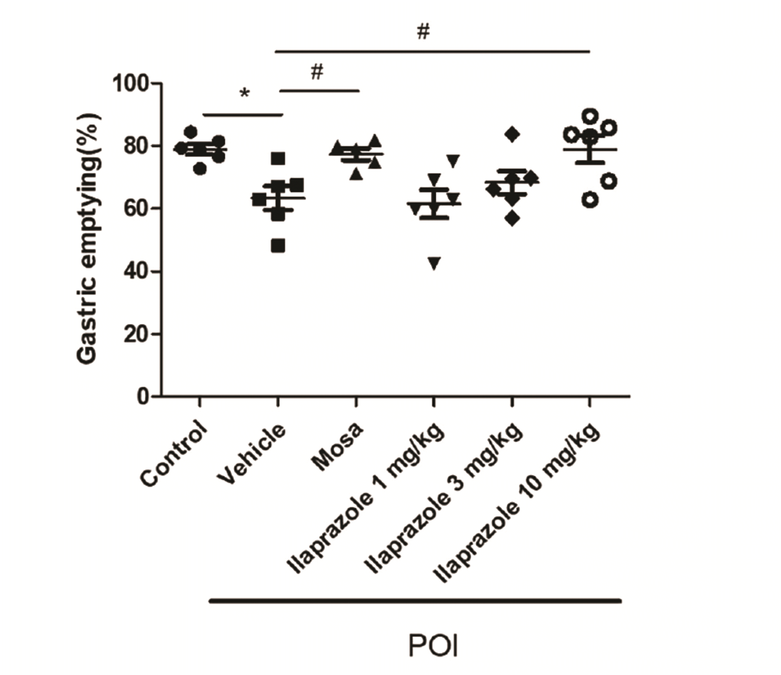 | Fig. 1Effect of ilaprazole on gastric emptying (GE) in stomach of each group.The recovery effect of the ilaprazole on GE in postoperative ileus (POI) model of rats. The GE value was measured from the rat stomach and the absorbance at 590 nm was measured (n = 5–6). Control: not open the abdomen treated with 4 ml/kg 0.5 % CMC-Na; vehicle: POI rats treated with 4 ml/kg 0.5 % CMC-Na; ilaprazole 1 mg/kg: POI rats treated with ilaprazole 1 mg/kg; ilaprazole 3 mg/kg: POI rats treated with ilaprazole 3 mg/kg; ilaprazole 10 mg/kg: POI rats treated with ilaprazole 10 mg/kg; Data were expressed as means ± SEM. Dunnett’s test was performed post-hoc for the ANOVA. *p < 0.05 vs. control group, #p < 0.05 vs. vehicle group.
|
Changes in GIT in the small intestine by ilaprazole
Gastrointestinal motility was estimated by measuring GIT in the small intestine (Fig. 2). GIT of the POI groups had a significantly lower transit ability than that of the control group. Compared to the vehicle group, gastrointestinal motility was increased in the ilaprazole-treated groups (1 mg/kg, 3 mg/kg, and 10 mg/kg groups), with the ilaprazole 10 mg/kg-treated POI group achieving a recovery similar to that of the mosapride 2 mg/kg treated POI group.
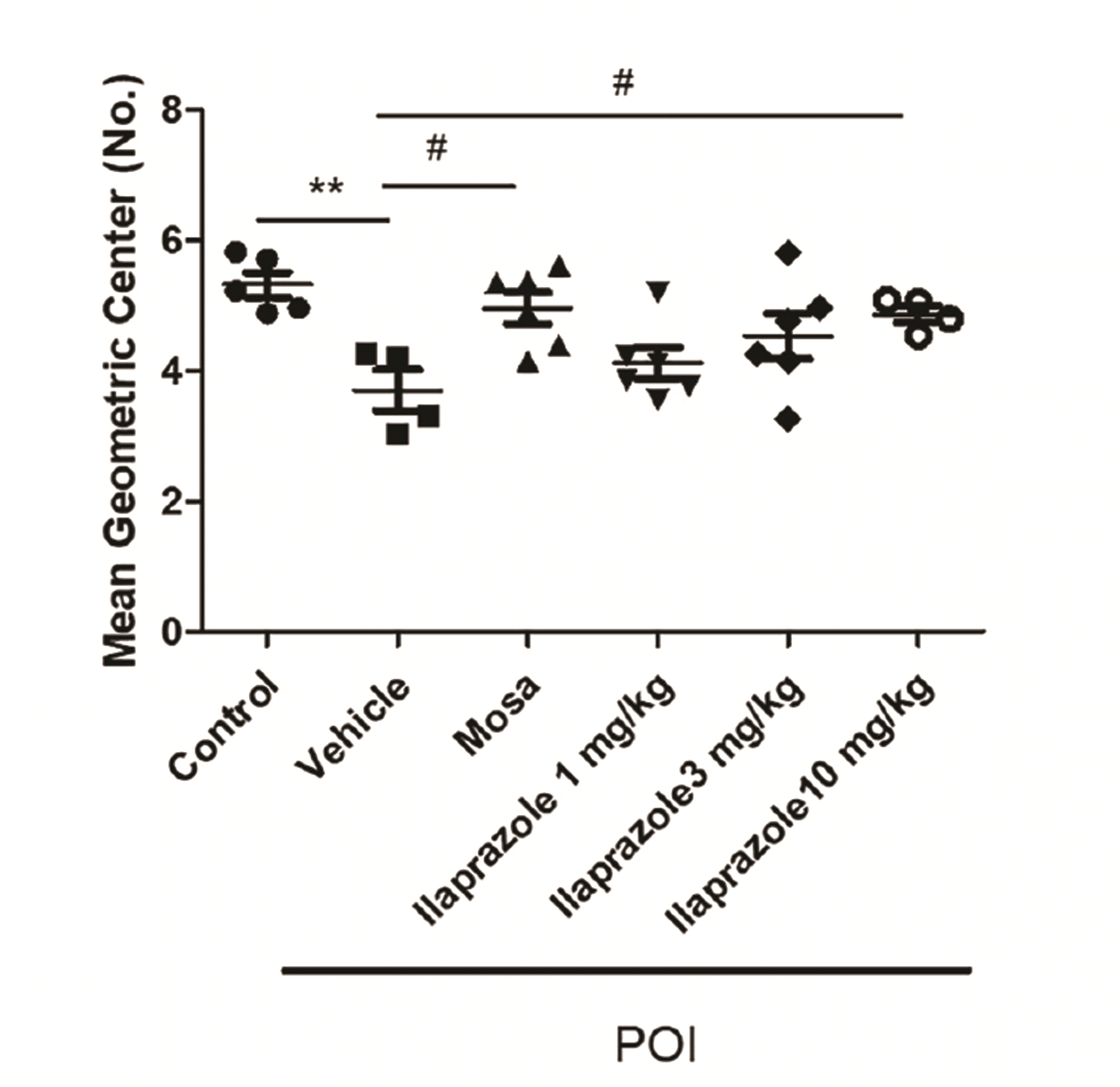 | Fig. 2Effect of ilaprazole on GIT in small intestine of each group.The MGC value was measured from the rat stomach and the absorbance at 590 nm was measured (n = 4–6). Control: not open the abdomen treated with 4 ml/kg 0.5 % CMC-Na; vehicle: POI rats treated with 4 ml/kg 0.5 % CMC-Na; ilaprazole 1 mg/kg: POI rats treated with ilaprazole 1 mg/kg; ilaprazole 3 mg/kg: POI rats treated with ilaprazole 3 mg/kg; ilaprazole 10 mg/kg: POI rats treated with ilaprazole 10 mg/kg; The data were expressed as means ± SEM. Dunnett’s test was performed post-hoc for the ANOVA. GIT, gastrointestinal transit; MGC, mean geometric center; POI, postoperative ileus. **p < 0.01 vs. control group, #p < 0.05 vs. vehicle group.
|
TNF-α expression in the terminal ileus tissue
TNF-α is an inflammatory cytokine that represents the acute phase of inflammation. The level of TNF-α in the terminal ileum was measured with an ELISA kit. As shown in Fig. 3, the level of TNF-α was decreased in the POI only group. In the group treated with mosapride, the increase in TNF-α was significantly alleviated relative to that in the POI only group. The level of TNF-α was decreased in the ilaprazole-treated group, with a significant decrease identified in the group administered 10 mg/kg ilaprazole.
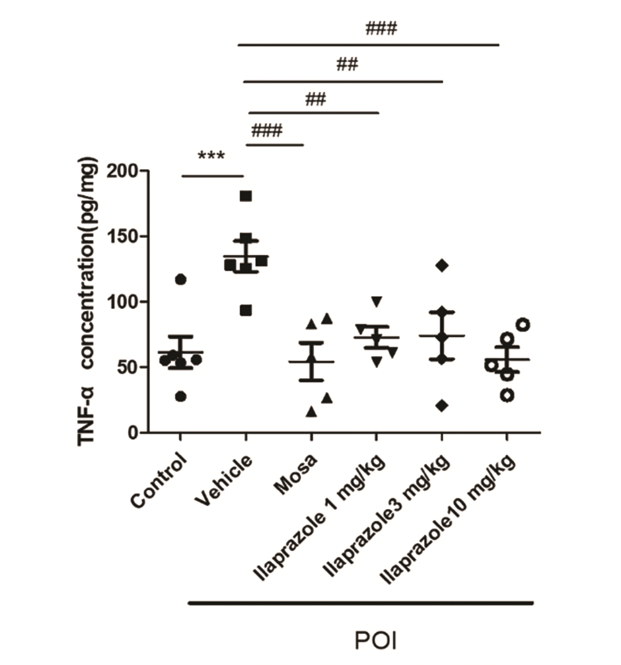 | Fig. 3Effect of ilaprazole on tumor necrosis factor (TNF)-α in terminal ileus of each group.Anti-inflammation effect of the ilaprazole at the terminal ileus of the postoperative ileus (POI)-induced rats. TNF-α concentration was detected absorbance in 450 nm (n = 5–6). Control: not open the abdomen treated with 4 ml/kg 0.5% CMC-Na; vehicle: POI rats treated with 4 ml/kg 0.5 % CMC-Na; ilaprazole 1 mg/kg: POI rats treated with ilaprazole 1 mg/kg; ilaprazole 3 mg/kg: POI rats treated with ilaprazole 3 mg/kg; ilaprazole 10 mg/kg: POI rats treated with ilaprazole 10 mg/kg; The data was expressed as means ± SEM. Dunnett’s test was performed post-hoc for the ANOVA. ***p < 0.001 vs. control group, ##p < 0.01 and ###p < 0.001 vs. vehicle group.
|
IL-1β expression in the terminal ileus tissue
IL-1β is an indicator of inflammation and a lymphocyte activating factor. As shown in Fig. 4, the level of IL-1β in the POI + vehicle group was significantly increased relative to that in the control group. However, in the mosapride group, its level was significantly decreased relative to that in the POI + vehicle group. Ilaprazole significantly attenuated IL-1β in all dosing groups (1 mg/kg, 3 mg/kg, 10 mg/kg). Furthermore, in the ilaprazole 3 mg/kg and 10 mg/kg treated POI groups, the level of IL-1β was similar to that found in the positive control (mosapride) group.
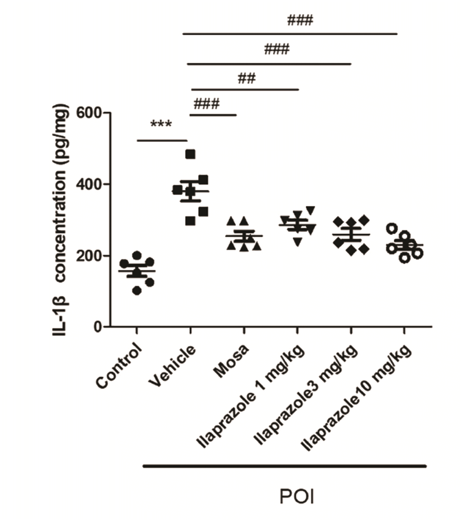 | Fig. 4Effect of ilaprazole on interleukin (IL)-1β in terminal ileus of each group.Anti-inflammation effect of the ilaprazole at the terminal ileus of the postoperative ileus (POI)-induced rats. IL-1β concentration was detected absorbance in 450 nm (n = 6). Control: not open the abdomen treated with 4 ml/kg 0.5 % CMC-Na; vehicle: POI rats treated with 4 ml/kg 0.5 % CMC-Na; ilaprazole 1 mg/kg: POI rats treated with ilaprazole 1 mg/kg; ilaprazole 3 mg/kg: POI rats treated with ilaprazole 3 mg/kg; ilaprazole 10 mg/kg: POI rats treated with ilaprazole 10 mg/kg; The data was expressed as means ± SEM. Dunnett’s test was performed post-hoc for the ANOVA. ***p < 0.001 vs. control group, ##p < 0.01, ###p < 0.001 vs. vehicle group.
|
Histological change in the terminal ileus by ilaprazole
Images of the terminal ileus in each group were captured with a microscope (Fig. 5). Structural damages were determined by H&E staining. As a result, cell swelling and necrosis were identified in the vehicle group (Fig. 5B) while in the mosapride group (Fig. 5C), structural damages were recovered relative to that in the vehicle group. In the ilaprazole 10 mg/kg group (Fig. 5F), a protective effect against structural damages to the terminal ileus tissue was revealed.
 | Fig. 5Effect of ilaprazole administration on structural damage of small intestine in each experimental group.Histology of terminal ileus of each group. The structural damage was identified histologically in terminal ileus of each group (black arrow: swelling, red arrow: structural damages). (A) Control group; (B) vehicle group; (C) mosa group; (D) ilaprazole 1 mg/kg group; (E) ilaprazole 3 mg/kg group; (F) ilaprazole 10 mg/kg group. Photomicrographs were taken at a magnification of ×200, H&E.
|
Effect of ilaprazole on oxidative stress
SOD is an enzyme that catalyzes the conversion of the superoxide radical to O2 and H2O2 and is used as an indicator of the degree of cellular oxidation; the lower the level of SOD, the greater the level of oxidation. As shown in Fig. 6A, the level of SOD decreased in the vehicle group compared to that in the control group and increased in the Ilaprazole + POI groups (1 mg/kg, 3 mg/kg, 10 mg/kg). In the ilaprazole 10 mg/kg group, SOD level was significantly increased, thereby restoring its level to that of the control group.
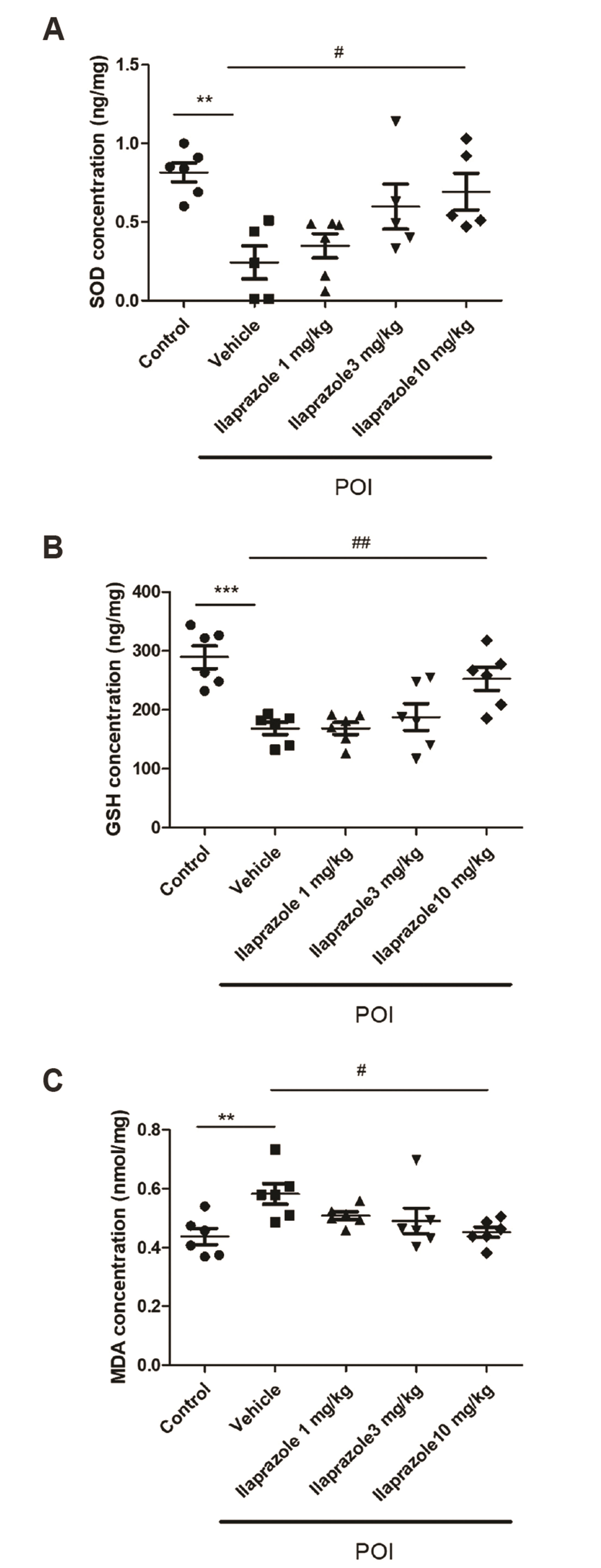 | Fig. 6Effect of ilaprazole on oxidative stress in terminal ileus of each group.Antioxidative effect of the ilaprazole at the terminal ileus of the POI-induced rats. (A) SOD, (B) GSH, and (C) MDA concentrations were detected absorbance in 450 nm (n = 5–6). Control: not open the abdomen treated with 4 ml/kg 0.5% CMC-Na; vehicle: POI rats treated with 4 ml/kg 0.5 % CMC-Na; ilaprazole 1 mg/kg: POI rats treated with ilaprazole 1 mg/kg; ilaprazole 3 mg/kg: POI rats treated with ilaprazole 3 mg/kg; ilaprazole 10 mg/kg: POI rats treated with ilaprazole 10 mg/kg; The data was expressed as means ± SEM. Dunnett’s test was performed post-hoc for the ANOVA. POI, postoperative ileus; SOD, superoxide dismutase; GSH, glutathione; MDA, malondialdehyde. **p < 0.01 and ***p < 0.001 vs. control group, #p < 0.05 and ##p < 0.01 vs. vehicle group.
|
GSH prevents cellular damage by reducing peroxides, lipid peroxides, and free radicals, which are products of ROS. In this study, GSH was used as an index of oxidative stress. As shown in Fig. 6B, the level of GSH was decreased in the vehicle group compared to that in the control group and significantly increased in the ilaprazole 10 mg/kg group compared to that in the vehicle group.
As MDA is an index of lipid peroxidation, its level was measured to determine oxidative stress. The level of MDA in the terminal ileus is shown in Fig. 6C. MDA level was increased in the vehicle group compared to that in the control group and decreased in the ilaprazole-treated POI rat model with an increase in ilaprazole concentration. In the ilaprazole 10 mg/kg group, a significant decrease in MDA level was found relative to that in the vehicle group.
Go to :
DISCUSSION
POI is an iatrogenic disorder that is characterized by bowel dysfunction symptoms post-op. POI increases the morbidity and length of hospital stays and the expenses accrued by patients [32,33]. Further, its complex pathophysiology involves pharmacological, neural, and immune-mediated mechanisms [4]. The immune-mediated mechanism, the main cause of POI, starts 3–6 h after abdominal surgery and last several days [8,11,34]. In this study, we focused on the immune-mediated mechanism of POI.
Ilaprazole is one of the clinically used PPIs for gastric ulcers, esophagitis, and Helicobacter pylori infection [35]. In the current study, the PPI, ilaprazole, displayed a protective effect against acute systemic inflammation [27]. Based on this effect, we sought to confirm the potential use of ilaprazole as a treatment agent for POI.
POI induces a decrease in motility in the stomach and small intestine after abdominal surgery [36-38]. In this experiment, GE and GIT were examined to verify gastrointestinal motility. First, GE was examined to elucidate the motility in the stomach. Thereafter, GIT was examined to elucidate motility in the small intestine. Based on our findings, GE was decreased in the vehicle group and increased in the ilaprazole-treated group. Additionally, GIT decreased in vehicle group and increased dose-dependently in the ilaprazole-treated group. Collectively, these findings indicate that ilaprazole improves gastrointestinal motility, which is the main symptom of POI.
Although the mechanism of POI is yet to be fully understood, recent studies have reported the changes identified in inflammatory factors in the mucosa of the POI rat model. Resident macrophages around the myenteric plexus are activated upon abdominal surgery, thereby activating transcription factors such as NF-κB, STAT3, and inflammatory factors. As a result, neutrophils are infiltrated and the activities of NO synthase and COX-2 promote the inflammatory response [39]. Although the inflammatory response of POI is yet to be established, many studies on POI have measured TNF-α and IL-1β levels as indexes of inflammation. In this study, the levels of TNF-α and IL-1β were measured in the terminal ileus to identify the inflammatory changes. TNF-α and IL-1β were increased in the vehicle group and decreased in the ilaprazole group. Moreover, the reduction in inflammation was most significant in the Ilaprazole 10 mg/kg group. Based on the findings, we confirmed that ilaprazole exerts a protective effect on inflammation caused by abdominal surgery.
Structural damages are induced by abdominal surgery [11,29]. The inflammatory phase of POI is initiated by mediators released from damaged tissue or luminal bacterial products crossing an impaired gastrointestinal epithelium [40]. To determine the structural damage depicted by cell necrosis and swelling of the villi in the present study, the terminal ileum tissues of each group were stained with H&E. As a result, we confirmed the swelling of cells and necrosis in the vehicle group, and the protective effect against structural damage in the ilaprazole 10 mg/kg group.
Herein, we measured the oxidative stress index, and SOD, GSH, and MDA levels. SOD protects cells from oxidative stress by converting the superoxide anion radical (O2–) radicals to O2 and H2O2 [17,41,42]. In a recent study, SOD level was reported to increase after abdominal surgery [43]. GSH prevents oxidative damage by reducing ROS such as free radicals, peroxides, and lipid peroxides [18]. GSH protects cells from exposure to numerous endogenous and exogenous factors [44]. In previous studies, GSH was decreased in a model of gastrointestinal disorder [45-48]. To evaluate the degree of oxidative stress, an indicator of lipid peroxidation, MDA, was measured at the end of the ileum tissue. MDA is an essential component of the body's natural defense system and protects the body from various harmful factors [49]. Owing to surgical manipulation, the level of MDA increases in the POI model [48,50].
In the present study, SOD level was decreased in the vehicle group and restored in the ilaprazole 10 mg/kg group. Similarly, the level of GSH was decreased in the vehicle group and increased in the ilaprazole 10 mg/kg group. However, MDA level was decreased in the ilaprazole 10 mg/kg group. Such findings indicate that ilaprazole decreases oxidative stress induced by surgical manipulation in the POI rat model.
In this study, all doses of ilaprzole (1 mg/kg, 3 mg/kg, and 10 mg/kg) had beneficial effects on inflammatory cytokines (TNF-α and IL-1β) releases. However, oxidative stress and physiological dysfunction were improved only in 10 mg/kg of ilaprazole. In previous study, it was demonstrated that postoperative ileus was associated with inflammation and oxidative stress [51]. These results mean that postoperative gastrointestinal dysfunction only can improve when both inflammation and oxidative stress are reduced.
In the present study, we demonstrated that ilaprazole exhibits a protective effect in POI by: 1) reducing both swelling and necrosis of the cells in the terminal ileus, 2) recovering gastrointestinal motility impaired by surgical manipulation, 3) exhibiting anti-inflammatory effects to decrease TNF-α and IL-1β expression, and 4) exerting antioxidative effects as demonstrated by the increases in SOD and GSH and decreases in MDA. However, further studies are still required as clinical data on ilaprazole remain lacking and the inflammatory mechanism that causes POI is yet to be clearly identified.
Go to :
 XML Download