

 PDF
PDF Citation
 Print
Print


INTRODUCTION
Tuberculosis of the cystic duct lymph node is very rare. To the best of the author’s knowledge, only four cases have been reported since 2004.1-4 All cases had right upper quadrant pain and were suspected of having cholecystitis preoperatively. The exact diagnosis of the cystic duct or pericholedochal tuberculous lymphadenitis is important for proper medical treatment and for avoiding unnecessary surgery. This paper presents a case of a patient with cystic duct tuberculous lymphadenitis who was diagnosed with cystic duct stones and gallbladder (GB) polyp preoperatively.
Go to : 
CASE REPORT
A 35-year-old male presented with intermittent abdominal pain, nausea, and vomiting for a 3-month duration. He was diagnosed twice with pulmonary tuberculosis 10 and 4 years earlier and had received anti-tubercular therapy for four and 9 months, respectively. His vital signs, including temperature, were normal at presentation; there were no signs of jaundice. He complained of tenderness in the right upper quadrant, and the Murphy’s sign was positive. The laboratory tests showed a hemoglobin level of 13.1 g/dL, white blood cell count of 7,650/μL, aspartate transaminase of 15 IU/L, alanine transaminase of 13 IU/L, and total bilirubin of 0.4 mg/dL. The posteroanterior chest radiograph revealed minimal fibrotic changes at the left apex that appeared to be post-tuberculosis sequelae. Abdominal CT (Fig. 1) revealed a minute GB polyp, a few small cystic duct stones with a partially collapsed GB, and few mottled calcifications at the periphery of the pancreatic uncinate process. Abdominal ultrasonography (US) detected a small cholesterol polyp in the GB and short segmental tubular thickening of the distal cystic duct (Fig. 2). The impression on US was that of a cholesterol polyp of the GB, and cystic duct cancer or segmental mural edematous thickening of the cystic duct with or without minute stones. Cholecystectomy was planned because the postprandial right upper quadrant pain persisted, and the diagnosis of cystic duct thickening was uncertain. Laparoscopy was performed initially, and the cystic duct was identified and dissected. The duct was grossly normal, but a lymph node measuring 2 cm in size was identified at the posterior junction of the cystic and common bile ducts. Although there was no cystic duct stone or thickening, the cystic duct lymph node had been misdiagnosed as a cystic duct stone or thickening on preoperative CT and US. The procedure was then converted to open cholecystectomy, and the lymph node was excised. The findings on a frozen section were suspicious of chronic granulomatous and tuberculous lymphadenitis. Chronic cholecystitis and a cholesterol polyp were finally diagnosed after histopathological analysis of the GB. The lymph node had chronic granulomatous lymphadenitis and focal extensive central caseous necrosis with acid-fast bacilli on Ziehl-Neelsen staining (Fig. 3). The postoperative course was uneventful. He began to eat a soft diet 2 days after surgery and did not perceive postprandial abdomen pain; he was discharged 11 days after surgery. He received anti-tubercular medication at an outpatient clinic with isoniazid, rifampicin, ethambutol, and pyrazinamide for 2 months, followed by isoniazid, rifampicin, and ethambutol for 7 months. CT did not reveal any newly developing lymphadenopathy for 5 years after surgery.
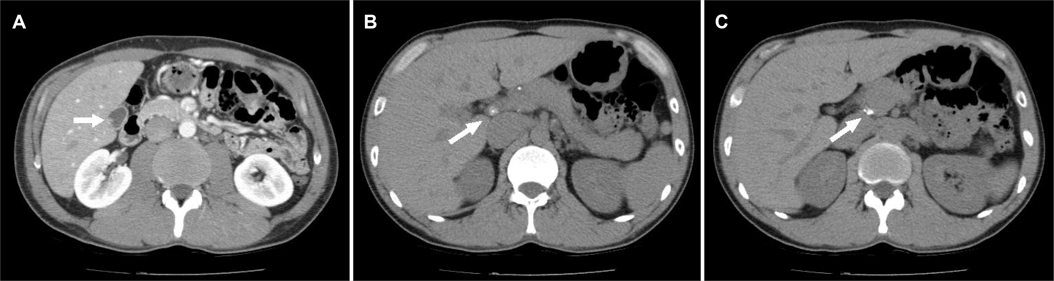 | Fig. 1Abdominal computed tomography shows (A) collapsed gallbladder with a minute cholesterol polyp (arrow), (B) a tuberculous cystic duct node preoperatively suspected of being a cystic duct stone (arrow), (C) mottled calcification at the periphery of the pancreatic uncinate process (arrow).
|
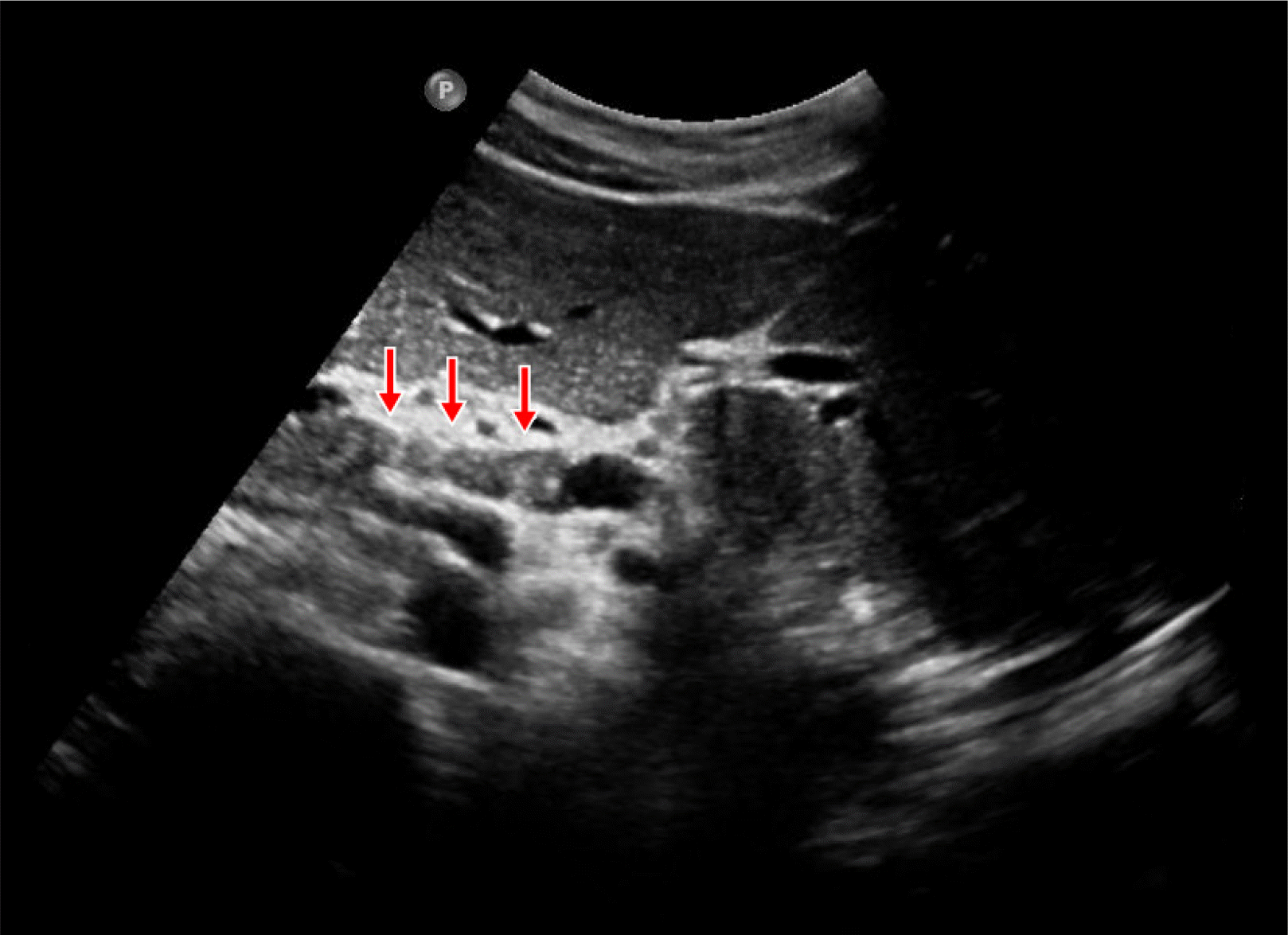 | Fig. 2Abdominal ultrasonography shows a cystic duct node, which was misdiagnosed preoperatively as short segmental tubular thickening of the distal cystic duct (arrows).
|
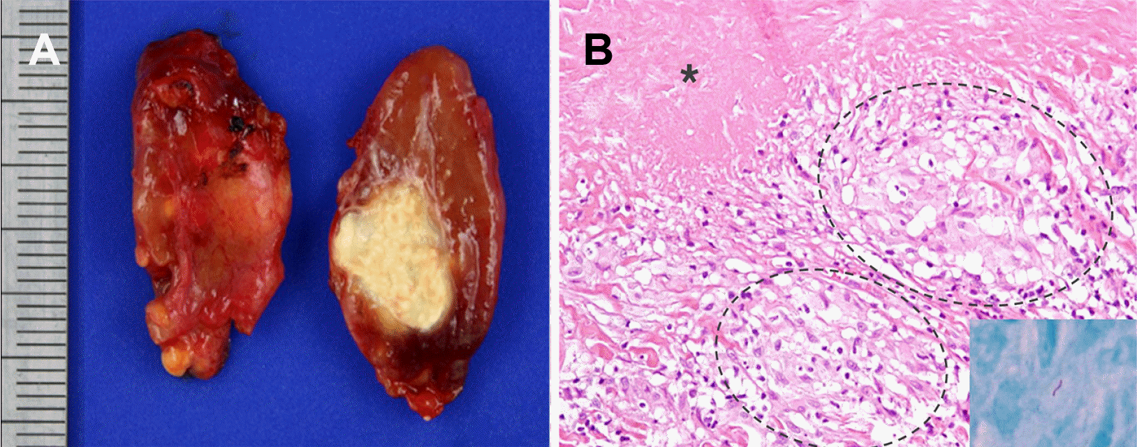 | Fig. 3(A) Cut surface of the enlarged peri-cystic duct lymph node showed a central area of pale yellowish caseous necrosis. (B) Microscopically, several epithelioid granulomas (dotted circles), and amorphous, granular, eosinophilic caseous necrosis (asterisk) were observed (hematoxylin and eosin, ×200). Ziehl-Neelsen staining revealed acid-fast bacilli (inlet, ×600).
|
Go to :
DISCUSSION
The present case demonstrated a tubercular cystic duct node that was diagnosed erroneously as a cystic duct stone or thickening on preoperative CT and US; the final diagnosis was only evident after a histopathological evaluation of the GB. As mentioned previously, a literature review revealed only four cases of tuberculous cystic duct lymph nodes.1-4 Table 1 summarizes the findings of these cases and the present case; there were two male and three female patients. Tuberculous lymphadenitis is more common in females than in males.5,6 On the other hand, pulmonary tuberculosis is more common in males; this can be explained by the immunological and hormonal differences between males and females.5 Three patients had concomitant GB stones, but the 4th and the present case only had minimal sludge in the GB. In particular, almost all cases of primary GB tuberculosis were reported to have coexisting gall stones.7 All patients had right upper quadrant pain, suggesting cholecystitis, except for the first case; histopathology of the GB in the other cases showed chronic cholecystitis. The patient had a history of pulmonary tuberculosis. Therefore, the route of infection was assumed to be by swallowing infected sputum, with consequent infection of the duodenum and lymphatic spread to the cystic duct node. On the other hand, the hematogenous route cannot be excluded. The four other patients had no history of tuberculosis or concomitant tuberculosis at other sites. Pericholedochal tuberculous lymphadenitis is relatively common compared to tubercular cystic duct nodes. Sixteen cases have been reported in Korea, and these cases had the characteristics of obstructive jaundice.8 The present case had a few calcified nodes at the periphery of the pancreatic uncinate process that were regarded as healed tuberculous nodes. In another case with a tuberculous cystic duct node, the periportal node was detected on postoperative CT.1 A tuberculous cystic duct node may be considered a type of pericholedochal tuberculous lymph node. Pericholedochal tuberculous lymph nodes have important clinical implications. They are associated with fatal complications, such as jaundice, portal hypertension,9 and biliary fistulas;10 a malignancy may also be suspected in some cases.8,11 Adenopathy showing peripheral rim-enhancement with relatively low attenuation centers and calcified lymph nodes may suggest a diagnosis of tuberculosis. However, these findings are also seen in metastatic lymph nodes.12 Unlike benign nodes, metastatic nodes have characteristic US and CT findings indicating a clustered distribution, large size, round shape, heterogeneous texture, irregular margins, extracapsular spread, ill-defined borders, necrosis, and ring enhancement. On the other hand, these features are not highly reliable.13
Table 1
Summary of the Reported Cases of Tuberculous Cystic Duct Lymph Nodes
![]()
In conclusion, tuberculous cystic duct nodes are quite rare. Accurate preoperative diagnosis is difficult but is essential for proper management. Excisional biopsy is recommended for the cystic duct or pericholedochal nodes detected during surgery, particularly in patients with a history of tuberculosis.
Go to :
 XML Download