

 PDF
PDF Citation
 Print
Print


INTRODUCTION
Gastrointestinal stromal tumors (GISTs) are mainly found as submucosal tumors resulting from mutations in the KIT or platelet-derived growth factor receptor protooncogenes in the interstitial cells of Cajal.1 GISTs are the most common mesenchymal tumors of the gastrointestinal tract. A surgical resection with a negative margin is the standard treatment for patients with primary GISTs. The treatment of rectal GISTs should consider the sphincter-sparing approach.2,3 On the other hand, there is no consensus regarding the amount and frequency of imatinib administration before surgery that allows the preservation of the anal sphincter. Preoperative imatinib may prevent an accurate assessment of the recurrent risk. Endoscopic resection is a novel treatment for rectal GIST that allows complete resection of the anal sphincter, but it is rarely reported. This paper reports the successful endoscopic resection of a huge rectal GIST without the administration of preoperative imatinib.
CASE REPORT
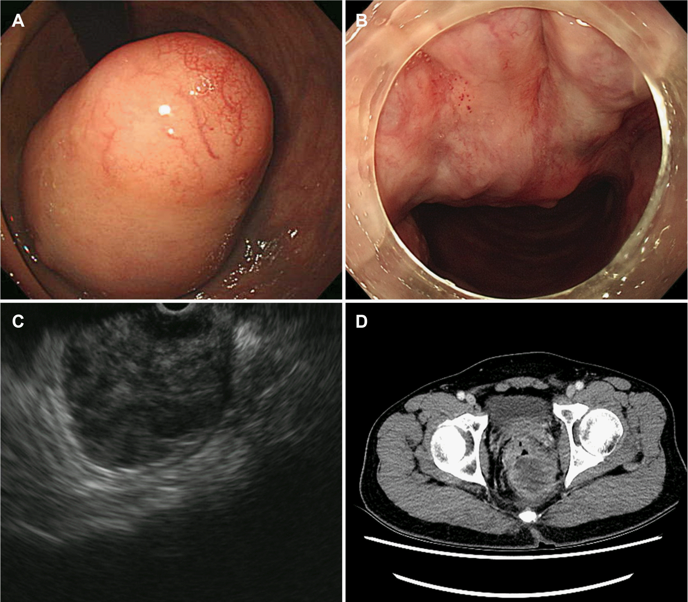
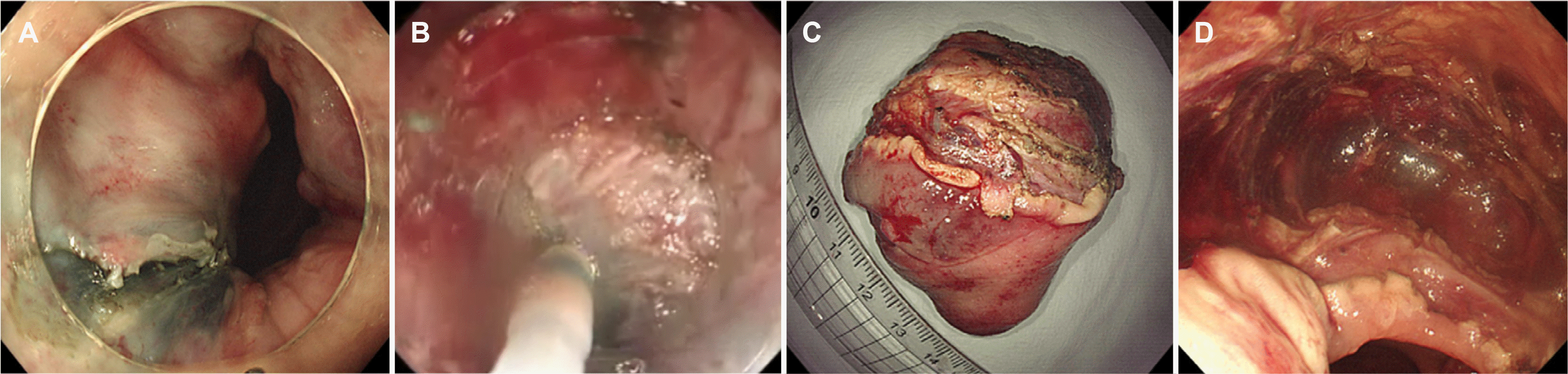
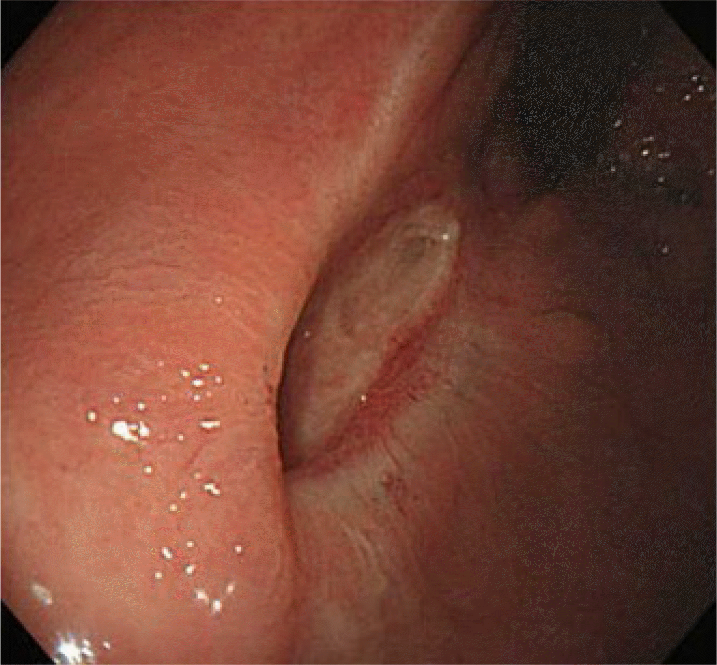
A 71-year-old man presented with recurrent constipation. He had no previous illnesses or family history. A digital rectal examination revealed a hard mass in the distal rectum. Colonoscopy revealed a huge submucosal tumor, approximately 5 cm in diameter, in the distal rectum involving the anal canal. Endoscopic ultrasonography revealed a 5.5×4.0 cm heterogeneous hypoechoic mass originating from the muscularis propria. Chest and abdominal computed tomography showed a 5.5×4.0 cm sized round mass of soft tissue density in the distal rectum without local or distant metastases (Fig. 1). The patient was presented with two treatment plans: imatinib treatment after surgical removal or preoperative imatinib treatment followed by surgery. The patient preferred removal first but was concerned about the possibility of anal sphincter injury. An endoscopic resection was performed to preserve the anal sphincter function using a single-channel waterjet upper gastrointestinal endoscope (GIF H290, Olympus, Tokyo, Japan) with a short-type ST hood (DH-28GR, Fujifilm, Tokyo, Japan) under deep sedation (midazolam 3 mg+propofol 20 mg, 20 mg added after consciousness evaluation per 20-30 sec). After injection with a mixture of glycerol, epinephrine, indigo carmine, and normal saline, Dual knife (KD-650Q, Olympus) and IT-knife nano (KD-612L/U, Olympus) were used to incise the mucosa and dissect the submucosal and proper muscle layer in the proximal direction, starting from the anal side (Fig. 2). The following procedures were performed: Endocut Q mode (effect 3, duration 2, interval 3) for incision, swift coagulation current (effect 3, 40 W) for dissection, and soft coagulation current (effect 3, 60 W) for hemostasis. The base of the tumor was located in the proper muscle layer and was excised completely by dissection to the inner circular muscle layer. The duration of the procedure was 110 min. The histopathology examination revealed a 5.5×4.0 cm sized, well-defined, encapsulated mass consisting of spindle and focally epithelioid cells arranged diffuse pattern from the mucosa to the circular layer of the proper muscle. The tumor cells were positive for KIT (CD117), CD34, and DOG1. More than 20 mitoses per 50 high-power fields were noted, corresponding to a high risk of malignant GIST recurrence (Fig. 3). The patient was prescribed oral imatinib (400 mg/day). Three months after surgery, a colonoscopy revealed a small ulcer and healed scar without recurrence (Fig. 4). To date, the patient is receiving imatinib treatment without tumor recurrence or complications.
DISCUSSION
Rectal GIST requires a multidisciplinary approach to determine the best treatment for complete tumor resection and preservation of the anal sphincter. Preoperative imatinib should be considered if a patient with rectal GIST undergoes an abdominoperineal resection.2,3 Although preoperative imatinib treatment can reduce the tumor size and increase the complete resection rate, it has some limitations. First, it reduces the tumor size and mitotic rate, making an accurate assessment of the risk of recurrence difficult. Therefore, preoperative imatinib should be considered only if the tumor can be downstaged. Second, there is controversy regarding the actual improvement in survival rates.4,5 Third, testing for mutations before imatinib administration is recommended to identify the genotypes likely to respond to treatment.6 Tumors without a genotype likely to respond to treatment cannot receive preoperative imatinib. Fourth, the maximum duration of the treatment response is not known. The guidelines recommend 6 months or longer, but the exact duration of treatment cannot be predicted in some patients.2,3
The standard treatment for GIST without distant metastases is complete surgical resection with a microscopically negative margin. Generally, minimally invasive, local resection without lymphadenectomy is performed because of the low incidence of lymph node metastasis. Similarly, transanal endoscopic microsurgery (TEM) is commonly used in rectal GISTs to preserve the anal sphincter function.7,8 On the other hand, TEM must be performed under either general or spinal anesthesia and requires high-cost equipment and a long hospital stay. A colorectal endoscopic resection is a novel procedure that enables the en bloc resection of colorectal tumors. An endoscopic resection can be performed under conscious or deep sedation, without anesthesia.
In a meta-analysis of rectal tumor treatment, endoscopic submucosal dissection had similar resection rates, side effects, and recurrence rates to TEM, but shorter treatment and hospitalization periods.9 Similar results were confirmed when comparing only rectal epithelial tumors in our previous retrospective study.10 There is a major limitation in reflecting the overall subepithelial tumor because the registered sample size of the study was small, and most of the registered patients were small neuroendocrine tumors.10 Therefore, future studies on the endoscopic treatment of rectal subepithelial tumors are needed.
While an endoscopic resection of gastric GISTs is relatively common, colorectal GISTs have rarely been reported.11 The reason for this is the relatively thin colon wall, which increases the risk of perforation. On the other hand, as the rectum is in the pelvic cavity, complications from perforation are exceedingly rare. Therefore, a more aggressive endoscopic deep resection, including the muscle layer, is possible. To date, there is no consensus regarding the maximum size of rectal GISTs that can be removed with an endoscopic resection. A huge rectal GIST (>5 cm) was removed by endoscopic resection without neoadjuvant imatinib.
Despite some limitations in securing the visual field and complications, such as bleeding or perforation, advances in endoscopic techniques and equipment are expected to increase the endoscopic treatment of rectal GISTs. Currently, there is no precise recommendation for the endoscopic resection of rectal GISTs. On the other hand, if further efforts to perform endoscopic treatment continue, endoscopic resection may be a suitable treatment option for rectal GISTs. In conclusion, an endoscopic resection should be considered for the treatment of rectal GIST as an anal sphincter preserving approach.

 XML Download