Journal List > Korean J Transplant > v.35(3) > 1147591

Kim, Hong, Lee, Eum, Kim, Jin, Han, Yang, Park, and Chung: Impact of delayed graft function on clinical outcomes in highly sensitized patients after deceased-donor kidney transplantation

Abstract

Background

We investigated whether the development of delayed graft function (DGF) in pre-sensitized patients affects the clinical outcomes after deceased-donor kidney transplantation (DDKT).

Methods

The study included 709 kidney transplant recipients (KTRs) from three transplant centers. We divided KTRs into four subgroups (highly sensitized DGF, highly sensitized non-DGF, low-sensitized DGF, and low-sensitized non-DGF) according to panel reactive antibody level of 50%, or DGF development. We compared post-transplant clinical outcomes among the four subgroups.

Results

Incidence of biopsy-proven acute rejection (BPAR) was higher in two highly sensitized subgroups than in low-sensitized subgroups. It tended to be higher in highly sensitized DGF subgroups than in the highly sensitized non-DGF subgroups. In addition, the highly sensitized DGF subgroup showed the highest risk for BPAR (hazard ratio, 3.051; P=0.005) and independently predicted BPAR. Allograft function was lower in the two DGF subgroups than in the non-DGF subgroup until one month after transplantation, but thereafter it was similar. Death-censored graft loss rates and patient mortality tended to be low when DGF developed, but it did not reach statistical significance.

Conclusions

DGF development in highly sensitized patients increases the risk for BPAR in DDKT compared with patients without DGF, suggesting the need for strict monitoring and management of such cases.

HIGHLIGHTS
  • The combination of high sensitization and delayed graft function (DGF) could increase the risk of biopsy-proven acute rejection, as the most important factor in allograft outcome, comparing with high sensitization or DGF alone.

  • In highly sensitized patients before kidney transplant (KT), enhanced desensitization and strict monitoring after KT may be necessary to prevent DGF.

INTRODUCTION

The term “delayed graft function” (DGF) is defined as the need for dialysis immediately after deceased-donor kidney transplantation (DDKT) mainly due to ischemic-reperfusion injury [1]. In previous studies, the development of DGF in DDKT was reported to be approximately 10%–50% [2,3] and entailed ischemic damage occurring during brain death, organ harvest and transplantation as is a major mechanism [3,4]. DGF is clinically important as it may worsen the clinical course after transplantation [5,6]. Various mechanisms have been proposed to explain the role of DGF in deteriorating clinical prognosis after DDKT, resulting in progressive chronic tissue damage of allograft kidney [7,8]. Further, the activation of innate immune response by ischemic damage in DGF eventually leads to the activation of adaptive immune system directly related to allograft rejection, resulting in the deterioration of allograft function [3,4].
Sensitization refers to the presence of alloantibody targeting human leukocyte antigen (HLA). Risk factors for HLA sensitization include prior transplantation, transfusion, and pregnancy [9,10]. In a previous study, pre-transplant sensitization in kidney transplantation (KT) was reported in up to 30% [9,11]. Pre-transplant sensitization to HLA is a key risk factor for the development acute allograft rejection and also for adverse long-term allograft outcomes [9,12]. In living-donor KT (LDKT), desensitization therapy has been shown to improve short-term and long-term prognosis after KT in highly sensitized patients because LDKT ensures sufficient time for desensitization [13,14]. However, in DDKT, procedures that require a lot of time and preparation, such as plasmapheresis, are difficult to perform before transplantation, and only rituximab (RTX), a B-cell depleting agent can be used for desensitization in DDKT [15,16]. Therefore, it is possible that many cases of DDKT may progress without enough removal of anti-HLA antibody.
In this regard, it is possible that the likelihood of activating an allogeneic immune response increases under DGF after DDKT in highly sensitized patients, and leads to a higher incidence of allograft rejection and worsening allograft survival. However, this additive effect of DGF on high sensitization in allograft outcomes has yet to be fully investigated. Therefore, the purpose of this study was to investigate whether the development of DGF in highly sensitized patients may synergistically affect the short- and long-term allograft outcomes in DDKT.

METHODS

This study was approved by the Institutional Review Boards of Seoul St. Mary’s Hospital (IRB. No. XC15RIMI0061K), Keimyung University School of Medicine, Dongsan Medical Center (IRB. No.2017–08-019), and Uijeongbu St. Mary’s Hospital (IRB. No. XC15RIMI0061U). The three transplant centers were exempted from informed consent, with the approval of institutional review boards, because the study was explained to all KTRs before KT. Personal data related to the patient's clinical course post-KT was used and personally identifiable information was protected. This study was a retrospective medical record study, and this manuscript did not contain personally identifiable information. In addition, all study methods were performed in compliance with relevant guidelines and regulations.

Study Population

In this study, we included 709 cases of DDKT who received kidneys from 614 deceased donors (DDs) at three transplant centers (Seoul St. Mary’s Hospital, Keimyung University Dongsan Medical Center, and Uijeongbu St. Mary’s Hospital) between October 2005 and December 2018. In all kidney transplant recipients (KTRs), the panel reactive antibody (PRA) level was measured via enzyme-linked immunosorbent assay (ELISA, LAT-M; One-Lambda Inc., Los Angeles, CA, USA) or the Luminex method (LIFECODES Life Screen Deluxe; Gen-Probe Inc., San Diego, CA, USA) as described previously before KT [15].
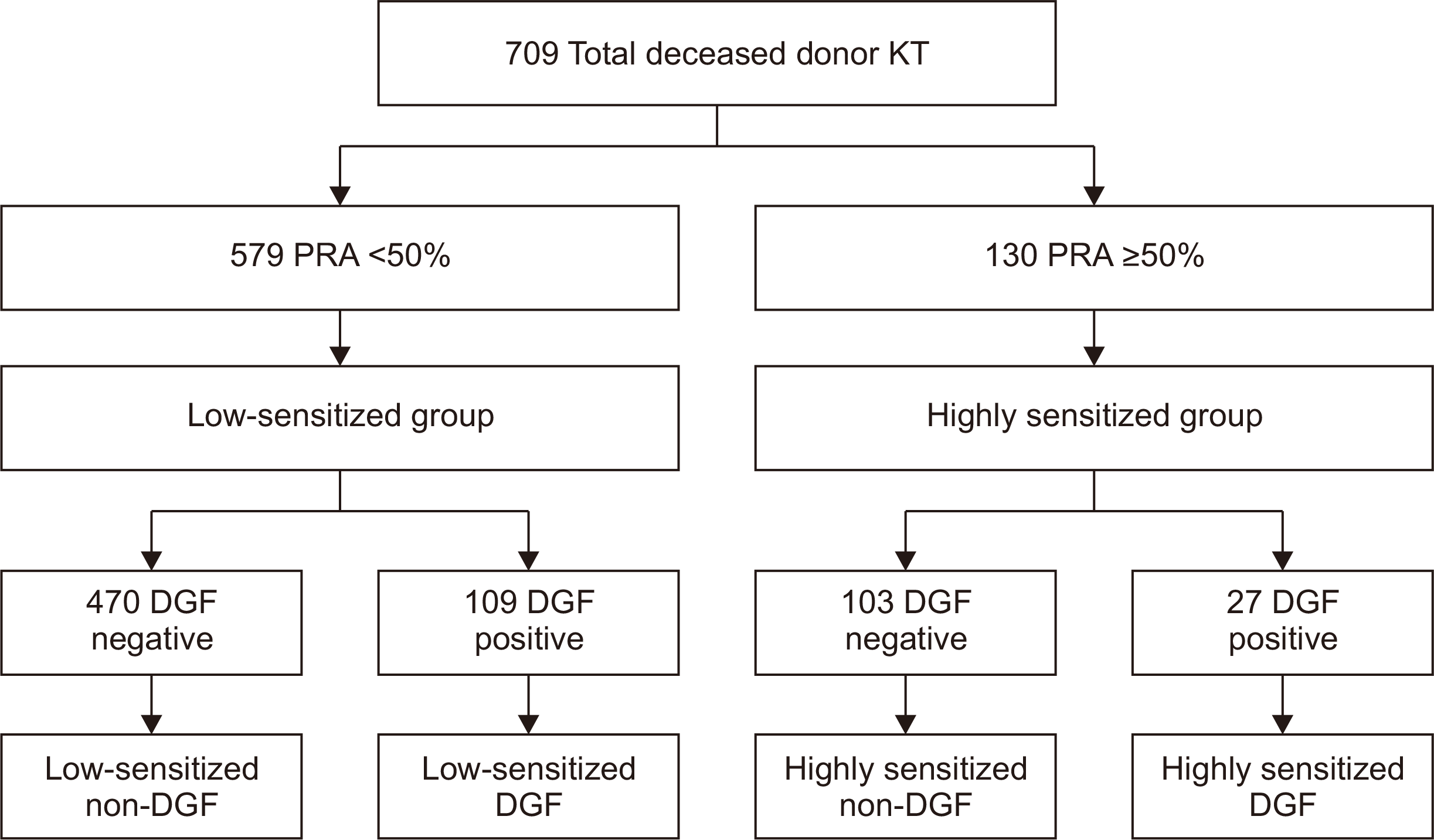
The distribution of patients in various groups is presented in Fig. 1. Highly sensitized KTRs were defined by a PRA level that was equal to or greater than 50% [17]. DGF was defined by the need for dialysis in the first week after KT [1]. Therefore, a total of 709 KTRs were classified into low-sensitized and highly sensitized groups based on a PRA level of 50%. The low-sensitized group included 579 (81.7%) patients and 130 (18.3%) were categorized under the highly sensitized group. Depending on the development of DGF, each group was classified into non-DGF and DGF subgroups. The low-sensitized group included 470 (81.2%) patients as a low-sensitized non-DGF subgroup and another 109 (18.8%) patients classified as the low-sensitized DGF subgroup. The highly sensitized group included 103 (79.2%) patients without DGF (highly sensitized non-DGF subgroup) and 27 (20.8%) with DGF (highly sensitized DGF subgroup). The median follow-up period in this study was 55.7 months (interquartile range, 32.2–85.4 months) and there was no difference among the four subgroups.

Clinical Parameters and Outcomes

We retrospectively reviewed the medical records of KTRs and DDs. We collected data for KTRs, including age, sex, body mass index (BMI), history of diabetes mellitus (DM) and hypertension (HTN), number of previous KTs, cause of end-stage renal disease, number of HLA mismatches, percentage of PRAs, type of induction therapy, maintenance immunosuppressants, and cold ischemic time. Additionally, we collected DD data including age, sex, BMI, history of DM and HTN, cause of death, last-day urine volume, central venous pressure and mean arterial pressure from hospitalization date until KT.
The primary outcome of this study was the incidence of biopsy-proven acute rejection (BPAR). Secondary outcomes included the change of allograft function, death-censored graft survival, and patient mortality among the four subgroups. The change in allograft function was measured by the estimated glomerular filtration rate (eGFR) using the Chronic Kidney Disease Epidemiology Collaboration equation [18]. The eGFR data were collected up to 3 years (1 week, 2 weeks, 1 month, 3 months, 6 months, 12 months, 2 years, and 3 years) after KT. Interpretation of protocol or indicated biopsy findings was based on the Banff classification 2013 [19]. The allograft biopsy included the period immediately after KT and could also include the DGF period. BPAR was defined as acute T-cell-mediated rejection (TCMR) or acute antibody-mediated rejection (ABMR) confirmed by allograft biopsy according to Banff classification. Death-censored allograft survival was defined as the period until dialysis was restarted or preemptive KT was performed, except for death of patient with a functioning allograft after KT. Patient death was defined as the period from KT until death for any cause.
We compared the clinical outcomes among low-sensitized non-DGF, low-sensitized DGF, highly sensitized non-DGF, and highly sensitized DGF subgroups. We also investigated whether high sensitization and DGF had a synergistic effect on the clinical outcomes.

Statistical Analysis

Continuous variables with normal distribution were expressed as mean±standard deviation. Student t-test was used for the analysis. Continuous variables with non-normal distribution were expressed as medians and interquartile ranges and analyzed using the one-way analysis of variance (one-way ANOVA) test. Categorical variables were expressed as counts and percentages and analyzed using chi-square or Fisher's exact test. The risk of BPAR incidence among the four subgroups was analyzed by binary logistic regression analysis. Comparison of death-censored allograft survival, and patient mortality among the four subgroups was investigated via Cox regression analysis. As confounding variables for multivariate analysis of these clinical outcomes, sex, age, history of DM, previous KT history, and numbers of HLA mismatch were considered in KTRs. Sex, age, and DM history were considered in DDs. The P-values less than 0.05 were considered statistically significant. All statistical analyses were performed using IBM SPSS ver. 21.0 (IBM Corp., Armonk, NY, USA) and the MedCalc statistical software ver. 15.5 (MedCalc Software, Ostend, Belgium).

RESULTS

Comparison of Clinical and Laboratory Parameters According to Pre-sensitization and Development of DGF

We described baseline characteristics of the patients in the four subgroups as shown in Table 1 [20]. The development of DGF in the low-sensitized and highly sensitized groups was 109 (18.8%) and 27 (20.8%), respectively, and there was no significant difference. Age, height, weight, and BMI were not different among the four groups both in KTRs and DDs. The proportion of male KTRs was lower or tended to be lower in highly sensitized groups regardless of DGF than in low-sensitized groups. The proportion of re-transplant KT history was significantly higher in two highly sensitized subgroups than in low-sensitized group. Anti-thymocyte globulin (ATG) was more frequently used than anti-IL-2 receptor antibody as induction agent in two highly sensitized subgroups. The combined use of RTX was more frequent in the two highly sensitized subgroups. Almost all patients received a triple-drug maintenance immunosuppressive regimen with tacrolimus, mycophenolate mofetil (or mycophenolic acid), and steroids.

Comparison of BPAR According to Pre-sensitization and Development of DGF

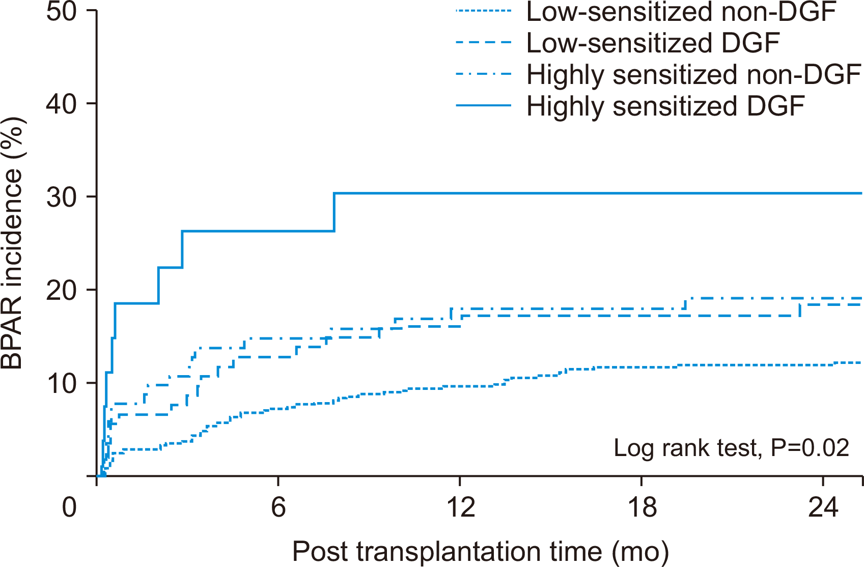
Three cases of allograft biopsy were performed during the DGF period. Two of the three allograft biopsies performed during the DGF period had BPAR. BPAR was detected in 57 cases (12.1%) in the low-sensitized non-DGF subgroup, 20 cases (18.3%) in the low-sensitized DGF subgroup, 21 cases (20.4%) in the highly sensitized non-DGF subgroup, and eight cases (29.6%) in the highly sensitized DGF subgroup. TCMR including mixed rejection occurred statistically significantly more frequently in the highly sensitized DGF subgroup than in the other two non-DGF subgroups (P=0.039, vs. low-sensitized non-DGF; P=0.018 vs. highly sensitized non-DGF). However, in ABMR including mixed rejection, none of the subgroups showed statistically significant differences (Table 2). In the crude model of BPAR incidence, the hazard ratio of two highly sensitized subgroups was significantly higher than in the low-sensitized non-DGF subgroup (reference subgroup) (P=0.029; hazard ratio [HR], 1.856; 95% confidence interval [CI], 1.067–3.228 vs. highly sensitized non-DGF and P=0.012; HR, 3.051; 95% CI, 1.277–7.291 vs. highly sensitized DGF). The low-sensitized DGF subgroup tended to manifest a higher BPAR incidence than the reference subgroup, but it was not significant (P=0.087; HR, 1.628; 95% CI, 0.931–2.846). Similarly, in the adjusted model, BPAR incidence of two highly sensitized subgroups compared with the reference subgroup was statistically significantly higher (P=0.029; HR, 1.866; 95% CI, 1.065–3.269 vs. highly sensitized-non-DGF and P=0.005; HR, 3.631; 95% CI, 1.481–8.904 vs. highly sensitized-DGF). Within two highly sensitized subgroups, the BPAR risk tended to be higher in the highly sensitized DGF subgroup than in the highly sensitized non-DGF group (P=0.235; HR, 1.638; 95% CI, 0.725–3.700). In addition, in the adjusted model, BPAR incidence was independently influenced by elderly recipients above 65 years of age and with greater than three HLA mismatches (P=0.019; HR, 0.451; 95% CI, 0.232–0.877 and P=0.006; HR, 2.521; 95% CI, 1.295–4.904) (Table 3, Fig. 2).

Comparison of Allograft Function According to Pre-sensitization and Development of DGF

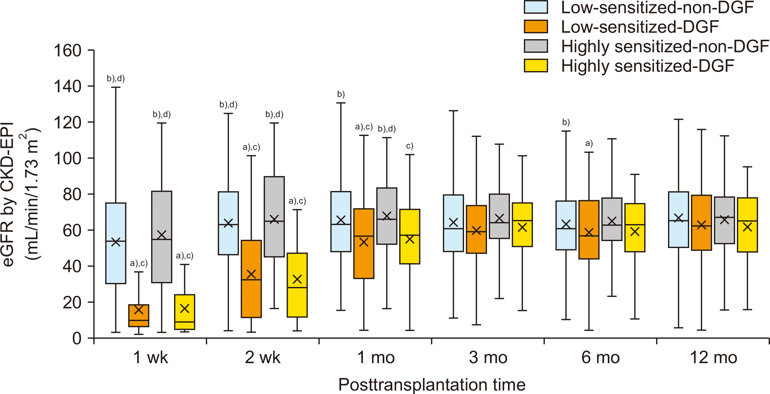
The allograft function was significantly lower by one month after KT in two DGF subgroups compared with the other two non-DGF subgroups. However, in two DGF subgroups, allograft function was improved gradually after KT, and there was no statistically significant difference among the four subgroups from 3 months to 1 year after KT (Fig. 3). No statistically significant differences were found in allograft function after KT between two non-DGF subgroups or between two DGF subgroups.

Comparison of Death-Censored Allograft Survival According to Pre-sensitization and Development of DGF

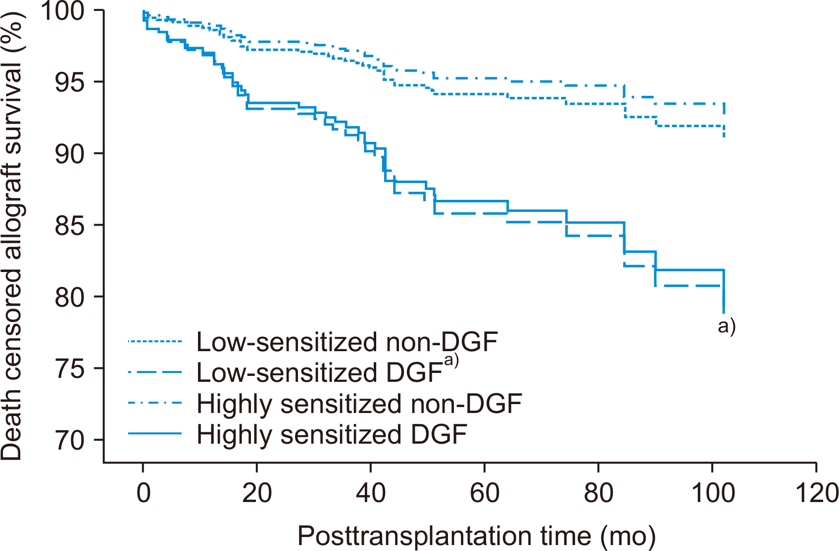
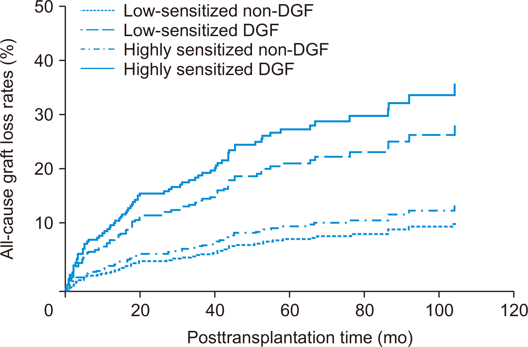
Death-censored allograft loss up to 10 years after KT occurred in 26 cases (5.5%) in the low-sensitized non-DGF subgroup, 14 cases (12.8%) in the low-sensitized DGF subgroup, five cases (4.9%) in the highly sensitized non-DGF subgroup, and four cases (14.8%) in the highly sensitized DGF subgroup. Acute or chronic rejection was the most common cause of death-censored allograft loss in all four subgroups (Supplementary Table 1). In the crude model, death-censored allograft survival rate was significantly lower only in the low-sensitized DGF subgroup than in the reference subgroup (P=0.010; HR, 2.352; 95% CI, 1.226–4.510). In the adjusted model, death-censored allograft survival rate was significantly lower only in the low-sensitized-DGF subgroup than in the reference subgroup (P=0.006; HR, 2.523; 95% CI, 1.307–4.872) (Table 4, Fig. 4).

Comparison of Patient Mortality According to Pre-sensitization and Development of DGF

Patient death up to 10 years after KT occurred in 18 cases (3.8%) in the low-sensitized non-DGF subgroup, nine cases (8.3%) in the low-sensitized DGF subgroup, five cases (4.9%) in the highly sensitized non-DGF subgroup, and three cases (11.1%) in the highly sensitized DGF subgroup. Among the causes of patient death, infection was the most common in all four subgroups during the observation period. We observed infection-related death in 10 cases (55.6%) in the low-sensitized non-DGF subgroup, five cases (55.6%) in the low-sensitized DGF subgroup, three cases (60.0%) in the highly sensitized non-DGF subgroup, and three cases (100.0%) in the highly sensitized DGF subgroup (Supplementary Table 1). In the univariate Cox regression model, patient mortality tended to be higher in the two DGF groups, but was not statistically significant (P=0.058 vs. low-sensitized-DGF; P=0.541 vs. highly sensitized non-DGF; P=0.080 vs. highly sensitized DGF) (Table 4, Fig. 5).

DISCUSSION

In this study, we found that DGF in highly sensitized patients after DDKT increased BPAR compared with pre-sensitization or DGF alone. The results suggest that in pre-sensitized patients, the development of DGF can result in adverse synergistic effect for allograft outcomes, especially higher incidence of allograft rejection.
The proportion of study population using ATG as induction therapy in the pre-sensitized group was 63.8%, which was higher than that of the low-sensitized group (30.4%). In addition, in the pre-sensitized group, the proportion of patients exposed to combined RTX as induction therapy was 36.9%. The risk factors for development of DGF have been associated with various immunological and non-immunological factors in KT donors and recipients [3]. In our study, the DGF group manifested significantly higher acute kidney injury in DDs than the non-DGF group, but there was no significant difference in other risk factors for DGF, such as recipients who were obese or diabetic, with prolonged cold ischemic time, and expanded criteria donor. A higher proportion of patients using ATG or combined RTX in the pre-sensitized group might have reduced the risk of developing DGF associated with pre-sensitization. Until recently, the effect of ATG compared with other induction therapies or the effect of combined RTX as an induction therapy in the development of DGF has been debated; however, some studies have reported promising results [21,22].
In this study, our main objective was to investigate whether development of DGF can increase the risk for allograft rejection in pre-sensitized patients. Previous reports showed that the development of DGF increased the expression of HLA antigens associated with ischemia-reperfusion injury [23]. In addition, ischemia-reperfusion injury not only triggers innate immunity including complement system via toll-like receptor signaling, but also activates dendritic cells [24,25]. These dendritic cells mature in the lymphatic system and activate T cells and B cells by upregulation of co-receptors such as CD80, CD86, and CD40 to the cell surface [26]. Therefore, we hypothesized that the combination of DGF and pre-sensitization may augment the activation of allo-immune responses after DDKT. As a result, in this study, the highly sensitized DGF subgroup reported the highest incidence of BPAR.
In addition, in the adjusted model for BPAR incidence in this study, we found that elderly recipients and HLA mismatch were independent factors affecting BPAR incidence. In this study, when the age of KTR was below 65 years, the incidence of BPAR was twice higher than that of elderly KTRs (16.4% vs 8.4%). Previous studies by Meier-Kriesche et al. [27] and Tullius and Milford [28] reported that fewer acute rejections occurred with increasing KTR age. Our findings were consistent with the previous studies. Although the lower incidence of acute rejection in elderly recipients is unclear, it might be primarily associated with impaired cellular immunity, and alterations in B cells might be secondary to T cell dysfunction [29,30].
In this study, BPAR incidence was 2.5-fold higher in patients with more than three HLA mismatches than in those with fewer than three HLA mismatches (17.3% vs 7.7%). A few previous studies have reported that higher HLA mismatches increase acute rejection, which was consistent with our findings [31,32]. HLA mismatch is known to be associated with immunogenicity, which has been attributed to amino acid polymorphism in donor HLA [33]. In addition, Kosmoliaptsis et al. [33] showed that the differences in epitopes formed by polymorphic amino acids can be used as a predictive model compared with traditional methods, such as the difference in HLA mismatches in the prognosis after KT. In our study, no HLA mismatch method was used, suggesting the need for further studies.
HLA donor-specific antibodies (HLA-DSAs) measured by single antigen bead (SAB) method had superior impact for the clinical outcomes of KTRs than that of previous tests for sensitization [34,35]. Recently, Wehmeier et al. [35] also reported that the presence of HLA-DSA using SAB method reflected ABMR and allograft loss better than broadness of sensitization using PRA. However, the cut-off value for the titer of HLA-DSA varied from mean fluorescence intensity (MFI) 1,000 to 10,000 for each center or study [10,36]. We also performed post-hoc subgroup analysis of KTRs with HLA-DSA measured via SAB assay. Positive HLA-DSA was defined as MFI 1,000 or higher. However, only nine KTRs carried HLA-DSA in the DGF subgroup. In the univariate binary regression analysis performed via post-hoc subgroup analysis, the BPAR incidence did not show significant difference. Furthermore, no significant difference in death-censored allograft survival and patient mortality in post hoc subgroup analysis was detected in univariate Cox regression analysis. Differences among participating centers may be due to the recent introduction of the HLA-DSA test in DDKT recipients in our study. In addition, HLA-DQ was included in the SAB method only recently.
We also investigated the impact of RTX on acute rejection in patients combined with DGF and pre-sensitization. In our previous study, it was found that the use of RTX in highly sensitized patients with PRA of 50% or more significantly reduced the incidence of rejection [15]. In contrast, in this study, the BPAR incidence of the highly sensitized group was 18/82 (22.0%) without RTX and 11/48 (22.9%) with RTX, and there was no significant difference (P=0.898). However, in our previous study, only 18.8% (6/32) of the patients in the RTX group were included as DDKT patients. In this study, the number included in the RTX group was small, which may be insufficient to determine a significant effect. Therefore, further large-scale investigations are needed.
In contrast, death-censored allograft survival was only associated with the development of DGF; however, it was not affected by pre-sensitization. Previous meta-analyses reported that DGF increased allograft loss, and recent studies also showed lower death-censored allograft survival with DGF development [5,37]. Several studies reported that the development of DGF was reduced by identifying and modifying the risk factors for DGF [2-4]. However, until now, no specific treatment for the development of DGF is available. Therefore, long-term outcomes involving DGF may not be modified. In this study, the incidence of death-censored allograft loss in two highly sensitized subgroups was 2/83 (2.4%) with ATG use and 5/47 (10.6%) with basiliximab use, 1/48 (2.1%) with combined RTX use and 6/82 (7.3%) without combined RTX use (P=0.098, P=0.259). Although no significant differences were found among the highly sensitized subgroups in our study, death-censored allograft loss tended to be lower with ATG use or combined RTX use. A previous randomized trial and a recent study reported no difference in allograft loss between those who used ATG and basiliximab [38,39]. In the previous randomized controlled trial, death-censored allograft loss was reported in 11/138 (8.0%) with combined RTX use, which was lower than 18/142 (12.7%) in the group without RTX use; however, there was no statistically significant difference [22]. However, neither of these studies had a high level of sensitization. In addition, there were limitations associated with short observation period or single-center study. Therefore, the use of enhanced induction therapy or combined RTX in a large study population involving highly sensitized patients may be associated with a reduction in death-censored allograft loss.
Meanwhile, the two DGF subgroups tended to show increased patient mortality, but it was not statistically significant and had little effect on patient mortality by pre-sensitization. The relationship between development of DGF and patient mortality is still disputed. Yarlagadda et al. [5] reported that the development of DGF did not increase patient mortality via meta-analysis of six previous studies. Studies included in this meta-analysis used a 5-year mortality rate and if the observation period exceeded 5 years, there may be a difference in the mortality rate depending on the DGF development, especially in those returning to dialysis after allograft loss. In contrast, in another recent study, Tapiawala et al. [40] reported that the development of DGF increased patient mortality. Based on an analysis of 50,000 subjects included in the US Renal Data System, the investigators reported that DGF increased the mortality rate 6 and 12 months later. Therefore, DGF development may be associated with an increase in patient mortality. In addition, we used censoring to analyze when there was follow-up loss or no death during the observation period; therefore, the number of patient deaths might be higher.
The study has limitations related to the retrospective design, which increases the possibility of selection bias. Further, a total of 709 patients were analyzed, but the number of patients in the highly sensitized DGF subgroup was only 27. In addition, few cases of clinical outcomes were detected in this subgroup, including eight cases of BPAR, three cases of death-censored graft loss, and three cases of patient death. In the absence of serial biopsies, the impact of the rejection reaction may have been underestimated, and we might not understand the impact of chronic allograft tissue injury. Finally, the standard of pre-sensitization was set to the level of PRA. Therefore, large-scale investigations using SAB for HLA-DSA are needed in the future.
In conclusion, the combination of high sensitization and DGF development could increase the risk of BPAR, as the most important factor in allograft outcome, comparing with high sensitization or DGF alone. Therefore, enhanced desensitization and strict monitoring after KT may be necessary to prevent DGF development in highly sensitized patients before KT.

Supplementary Materials

kjt-35-3-149-supple.pdf
Supplementary materials can be found via https://doi.org/10.4285/kjt.21.0014.

ACKNOWLEDGMENTS

Conflict of Interest
No potential conflict of interest relevant to this article was reported.
Funding/Support
This study was supported by a research grant from the Korean Society for Transplantation and by Research Fund of Seoul St. Mary’s Hospital, The Catholic University of Korea.
This study was supported by research grant from the Korean Society for Transplantation (2021-00-01007-015).
Author Contributions
Conceptualization: BHC. Data curation: SGK, SH, HL, YSK, KJ, SH, CWY, WYP, BHC. Formal analysis: SGK, SHE, WYP, BHC. Funding acquisition: BHC. Methodology: BHC. Project administration: BHC. Visualization: SGK. Writing–original draft: SGK. Writing–review & editing: SGK, WYP, BHC.

REFERENCES

1. Yarlagadda SG, Coca SG, Garg AX, Doshi M, Poggio E, Marcus RJ, et al. 2008; Marked variation in the definition and diagnosis of delayed graft function: a systematic review. Nephrol Dial Transplant. 23:2995–3003. DOI: 10.1093/ndt/gfn158. PMID: 18408075. PMCID: PMC2727302.
crossref
2. Irish WD, Ilsley JN, Schnitzler MA, Feng S, Brennan DC. 2010; A risk prediction model for delayed graft function in the current era of deceased donor renal transplantation. Am J Transplant. 10:2279–86. DOI: 10.1111/j.1600-6143.2010.03179.x. PMID: 20883559.
crossref
3. Siedlecki A, Irish W, Brennan DC. 2011; Delayed graft function in the kidney transplant. Am J Transplant. 11:2279–96. DOI: 10.1111/j.1600-6143.2011.03754.x. PMID: 21929642. PMCID: PMC3280444.
crossref
4. Ponticelli C. 2014; Ischaemia-reperfusion injury: a major protagonist in kidney transplantation. Nephrol Dial Transplant. 29:1134–40. DOI: 10.1093/ndt/gft488. PMID: 24335382.
crossref
5. Yarlagadda SG, Coca SG, Formica RN Jr, Poggio ED, Parikh CR. 2009; Association between delayed graft function and allograft and patient survival: a systematic review and meta-analysis. Nephrol Dial Transplant. 24:1039–47. DOI: 10.1093/ndt/gfn667. PMID: 19103734.
crossref
6. Wu WK, Famure O, Li Y, Kim SJ. 2015; Delayed graft function and the risk of acute rejection in the modern era of kidney transplantation. Kidney Int. 88:851–8. DOI: 10.1038/ki.2015.190. PMID: 26108067.
crossref
7. Dikow R, Becker LE, Schaier M, Waldherr R, Gross ML, Zeier M. 2010; In renal transplants with delayed graft function chemokines and chemokine receptor expression predict long-term allograft function. Transplantation. 90:771–6. DOI: 10.1097/TP.0b013e3181f009ef. PMID: 20697328.
crossref
8. Viglietti D, Abboud I, Hill G, Vernerey D, Nochy D, Antoine C, et al. 2015; Kidney allograft fibrosis after transplantation from uncontrolled circulatory death donors. Transplantation. 99:409–15. DOI: 10.1097/TP.0000000000000228. PMID: 25222117.
crossref
9. Jordan SC, Pescovitz MD. 2006; Presensitization: the problem and its management. Clin J Am Soc Nephrol. 1:421–32. DOI: 10.2215/CJN.01651105. PMID: 17699241.
crossref
10. Filippone EJ, Farber JL. 2015; Humoral immune response and allograft function in kidney transplantation. Am J Kidney Dis. 66:337–47. DOI: 10.1053/j.ajkd.2015.03.033. PMID: 25987262.
crossref
11. Loupy A, Hill GS, Jordan SC. 2012; The impact of donor-specific anti-HLA antibodies on late kidney allograft failure. Nat Rev Nephrol. 8:348–57. DOI: 10.1038/nrneph.2012.81. PMID: 22508180.
crossref
12. Sethi S, Najjar R, Peng A, Mirocha J, Vo A, Bunnapradist S, et al. 2019; Allocation of the highest quality kidneys and transplant outcomes under the new kidney allocation system. Am J Kidney Dis. 73:605–14. DOI: 10.1053/j.ajkd.2018.12.036. PMID: 30929853.
crossref
13. Montgomery RA, Lonze BE, King KE, Kraus ES, Kucirka LM, Locke JE, et al. 2011; Desensitization in HLA-incompatible kidney recipients and survival. N Engl J Med. 365:318–26. DOI: 10.1056/NEJMoa1012376. PMID: 21793744.
crossref
14. Marfo K, Lu A, Ling M, Akalin E. 2011; Desensitization protocols and their outcome. Clin J Am Soc Nephrol. 6:922–36. DOI: 10.2215/CJN.08140910. PMID: 21441131.
crossref
15. Hwang SD, Chung BH, Oh EJ, Choi BS, Park CW, Kim YS, et al. 2015; Effect of pretransplant rituximab use on posttransplant clinical outcomes in patients with high panel reactive antibody scores. Nephron. 130:239–44. DOI: 10.1159/000435924. PMID: 26182858.
crossref
16. Vo AA, Petrozzino J, Yeung K, Sinha A, Kahwaji J, Peng A, et al. 2013; Efficacy, outcomes, and cost-effectiveness of desensitization using IVIG and rituximab. Transplantation. 95:852–8. DOI: 10.1097/TP.0b013e3182802f88. PMID: 23511212.
crossref
17. Lieber SR, Perez FV, Tabossi MR, Persoli LB, Marques SB, Mazzali M, et al. 2007; Effect of panel-reactive antibody in predicting crossmatch selection of cadaveric kidney recipients. Transplant Proc. 39:429–31. DOI: 10.1016/j.transproceed.2007.01.045. PMID: 17362748.
crossref
18. Levey AS, Stevens LA, Schmid CH, Zhang YL, Castro AF 3rd, Feldman HI, et al. 2009; A new equation to estimate glomerular filtration rate. Ann Intern Med. 150:604–12. DOI: 10.7326/0003-4819-150-9-200905050-00006. PMID: 19414839. PMCID: PMC2763564.
crossref
19. Sis B, Mengel M, Haas M, Colvin RB, Halloran PF, Racusen LC, et al. 2010; Banff '09 meeting report: antibody mediated graft deterioration and implementation of Banff working groups. Am J Transplant. 10:464–71. DOI: 10.1111/j.1600-6143.2009.02987.x. PMID: 20121738.
crossref
20. Metzger RA, Delmonico FL, Feng S, Port FK, Wynn JJ, Merion RM. 2003; Expanded criteria donors for kidney transplantation. Am J Transplant. 3 Suppl 4:114–25. DOI: 10.1034/j.1600-6143.3.s4.11.x. PMID: 12694055.
crossref
21. Noël C, Abramowicz D, Durand D, Mourad G, Lang P, Kessler M, et al. 2009; Daclizumab versus antithymocyte globulin in high-immunological-risk renal transplant recipients. J Am Soc Nephrol. 20:1385–92. DOI: 10.1681/ASN.2008101037. PMID: 19470677. PMCID: PMC2689909.
crossref
22. van den Hoogen MW, Kamburova EG, Baas MC, Steenbergen EJ, Florquin S, M Koenen HJ, et al. 2015; Rituximab as induction therapy after renal transplantation: a randomized, double-blind, placebo-controlled study of efficacy and safety. Am J Transplant. 15:407–16. DOI: 10.1111/ajt.13052. PMID: 25612493.
crossref
23. Shoskes DA, Parfrey NA, Halloran PF. 1990; Increased major histocompatibility complex antigen expression in unilateral ischemic acute tubular necrosis in the mouse. Transplantation. 49:201–7. DOI: 10.1097/00007890-199001000-00045. PMID: 2105546.
crossref
24. Li K, Fazekasova H, Wang N, Sagoo P, Peng Q, Khamri W, et al. 2011; Expression of complement components, receptors and regulators by human dendritic cells. Mol Immunol. 48:1121–7. DOI: 10.1016/j.molimm.2011.02.003. PMID: 21397947. PMCID: PMC3084445.
crossref
25. van Werkhoven MB, Damman J, van Dijk MC, Daha MR, de Jong IJ, Leliveld A, et al. 2013; Complement mediated renal inflammation induced by donor brain death: role of renal C5a-C5aR interaction. Am J Transplant. 13:875–82. DOI: 10.1111/ajt.12130. PMID: 23398742.
crossref
26. Crabtree GR. 1989; Contingent genetic regulatory events in T lymphocyte activation. Science. 243:355–61. DOI: 10.1126/science.2783497. PMID: 2783497.
crossref
27. Meier-Kriesche HU, Ojo A, Hanson J, Cibrik D, Lake K, Agodoa LY, et al. 2000; Increased immunosuppressive vulnerability in elderly renal transplant recipients. Transplantation. 69:885–9. DOI: 10.1097/00007890-200003150-00037. PMID: 10755545.
28. Tullius SG, Milford E. 2011; Kidney allocation and the aging immune response. N Engl J Med. 364:1369–70. DOI: 10.1056/NEJMc1103007. PMID: 21410395.
crossref
29. Martins PN, Pratschke J, Pascher A, Fritsche L, Frei U, Neuhaus P, et al. 2005; Age and immune response in organ transplantation. Transplantation. 79:127–32. DOI: 10.1097/01.TP.0000146258.79425.04. PMID: 15665758.
crossref
30. Weiskopf D, Weinberger B, Grubeck-Loebenstein B. 2009; The aging of the immune system. Transpl Int. 22:1041–50. DOI: 10.1111/j.1432-2277.2009.00927.x. PMID: 19624493.
crossref
31. Wissing KM, Fomegné G, Broeders N, Ghisdal L, Hoang AD, Mikhalski D, et al. 2008; HLA mismatches remain risk factors for acute kidney allograft rejection in patients receiving quadruple immunosuppression with anti-interleukin-2 receptor antibodies. Transplantation. 85:411–6. DOI: 10.1097/TP.0b013e31816349b5. PMID: 18322434.
crossref
32. Foster BJ, Dahhou M, Zhang X, Platt RW, Smith JM, Hanley JA. 2014; Impact of HLA mismatch at first kidney transplant on lifetime with graft function in young recipients. Am J Transplant. 14:876–85. DOI: 10.1111/ajt.12643. PMID: 24612783.
crossref
33. Kosmoliaptsis V, Mallon DH, Chen Y, Bolton EM, Bradley JA, Taylor CJ. 2016; Alloantibody responses after renal transplant failure can be better predicted by donor-recipient HLA amino acid sequence and physicochemical disparities than conventional HLA matching. Am J Transplant. 16:2139–47. DOI: 10.1111/ajt.13707. PMID: 26755448. PMCID: PMC5021128.
crossref
34. Vlad G, Ho EK, Vasilescu ER, Colovai AI, Stokes MB, Markowitz GS, et al. 2009; Relevance of different antibody detection methods for the prediction of antibody-mediated rejection and deceased-donor kidney allograft survival. Hum Immunol. 70:589–94. DOI: 10.1016/j.humimm.2009.04.018. PMID: 19375470.
crossref
35. Wehmeier C, Hönger G, Cun H, Amico P, Hirt-Minkowski P, Georgalis A, et al. 2017; Donor specificity but not broadness of sensitization is associated with antibody-mediated rejection and graft loss in renal allograft recipients. Am J Transplant. 17:2092–102. DOI: 10.1111/ajt.14247. PMID: 28245084.
crossref
36. Tambur AR, Herrera ND, Haarberg KM, Cusick MF, Gordon RA, Leventhal JR, et al. 2015; Assessing antibody strength: comparison of MFI, C1q, and titer information. Am J Transplant. 15:2421–30. DOI: 10.1111/ajt.13295. PMID: 25930984.
crossref
37. Shamali A, Kassimatis T, Phillips BL, Burton H, Kessaris N, Callaghan C. 2019; Duration of delayed graft function and outcomes after kidney transplantation from controlled donation after circulatory death donors: a retrospective study. Transpl Int. 32:635–45. DOI: 10.1111/tri.13403. PMID: 30685880.
38. Brennan DC, Daller JA, Lake KD, Cibrik D, Del Castillo D. Thymoglobulin Induction Study Group. 2006; Rabbit antithymocyte globulin versus basiliximab in renal transplantation. N Engl J Med. 355:1967–77. DOI: 10.1056/NEJMoa060068. PMID: 17093248.
crossref
39. Jeong ES, Lee KW, Kim SJ, Yoo HJ, Kim KA, Park JB. 2019; Comparison of clinical outcomes of deceased donor kidney transplantations, with a focus on three induction therapies. Korean J Transplant. 33:118–27. DOI: 10.4285/jkstn.2019.33.4.118.
crossref
40. Tapiawala SN, Tinckam KJ, Cardella CJ, Schiff J, Cattran DC, Cole EH, et al. 2010; Delayed graft function and the risk for death with a functioning graft. J Am Soc Nephrol. 21:153–61. DOI: 10.1681/ASN.2009040412. PMID: 19875806. PMCID: PMC2799285.
crossref

Fig. 1
Patient distribution. Of the 709 deceased donor kidney transplantation (KT) recipients included in this study, 579 had panel-reactive antibody (PRA) less than 50%, and 130 had PRA 50% or more. Of the deceased-donor kidney transplantation (DDKT) recipients with a PRA of less than 50%, 470 did not develop delayed graft function (DGF), and 109 developed DGF. Among DDKT recipients with a PRA of 50% or more, 103 did not develop DGF and 27 developed DGF.
kjt-35-3-149-f1.tif
Fig. 2
Comparison of biopsy-proven acute rejection (BPAR) incidence in the four subgroups by Kaplan-Meier analysis. DGF, delayed graft function.
kjt-35-3-149-f2.tif
Fig. 3
Comparison of changes in allograft function after kidney transplantation among patients in the four subgroups classified according to human leukocyte antigen pre-sensitization and delayed graft function (DGF) development. eGFR, estimated glomerular filtration rate; CKD, chronic kidney disease; EPI, epidemiology collaboration. a)P<0.05 vs. low-sensitized non-DGF; b)P<0.05 vs. low-sensitized DGF; c)P<0.05 vs. highly sensitized-non-DGF; d)P<0.05 vs. highly sensitized DGF.
kjt-35-3-149-f3.tif
Fig. 4
Comparison of death-censored allograft survival in the four subgroups. Compared with low-sensitized non-delayed graft function (non-DGF) subgroup, death-censored allograft survival was decreased in low-sensitized DGF subgroup (P=0.006). a)P<0.05 vs. low-sensitized non-DGF.
kjt-35-3-149-f4.tif
Fig. 5
Comparison of patient mortality in the four subgroups. Compared with low-sensitized non-delayed graft function (non-DGF) subgroup, patient mortality tended to be higher in the two DGF subgroups, but was not statistically significant (P=0.058 vs. low-sensitized DGF, P=0.080 vs. highly sensitized DGF). P>0.05 for each comparison.
kjt-35-3-149-f5.tif
Table 1
Baseline demographic and immunologic characteristics of study population
Variable Low-sensitized
non-DGF (n=470)
Low-sensitized
DGF (n=109)
Highly sensitized
non-DGF (n=103)
Highly sensitized
DGF (n=27)
Recipient
Age (yr) 49.8±9.8 50.5±10.0 50.1±9.8 52.6±9.1
Elderly (≥65 yr) 88 (18.7) 15 (13.8) 22 (21.4) 6 (22.2)
Male sex 297 (63.2)c) 65 (59.6)c) 45 (43.7)a),b) 12 (44.4)
BMI (kg/m2) 23.1±4.8 22.6±4.6 23.0±3.5 23.0±3.1
Dialysis vintagee) 8.1±9.5 9.2±11.6 10.0±12.2 36.8±34.9
Primary renal disease
DM 90 (19.1) 18 (16.5) 12 (11.7) 4 (14.8)
HTN 88 (18.7) 18 (16.5) 13 (12.6) 2 (7.4)
CGN 198 (42.1) 52 (47.7) 61 (59.2) 19 (70.4)
Others 94 (20.0) 21 (19.3) 17 (16.5) 2 (7.4)
History of HTN 374 (79.6)d) 90 (82.6)d) 79 (76.7) 17 (63.0)a),b)
History of DM 101 (21.5) 25 (22.9) 14 (13.6) 7 (25.9)
Previous KT 28 (6.0)c),d) 10 (9.2)c),d) 33 (32.0)a),b) 12 (44.4)a),b)
Donor
Age (yr) 46.0±14.4 45.1±14.3 45.9±14.3 45.9±13.0
Elderly (age≥ 65 yr) 88 (18.7)b) 10 (9.2)a) 13 (12.6) 3 (11.1)
Male sex 309 (65.7)c) 75 (68.8) 78 (75.7)a) 21 (77.8)
BMI (kg/m2) 23.4±3.8 23.6±3.5 23.5±3.5 23.0±3.0
HTN 99 (21.1) 21 (19.3) 15 (14.6) 3 (11.1)
DM 49 (10.4) 7 (6.4) 7 (6.8) 3 (11.1)
Cold ischemic time (hr) 4.2±2.1 4.8±2.2c) 3.9±2.1b) 4.5±2.4
SCD vs. ECDf)
SCD 318 (67.7) 80 (73.4) 78 (75.7) 21 (77.8)
ECD 152 (32.3) 29 (26.6) 25 (24.3) 6 (22.2)
AKI by KDIGO 240 (51.1)b),d) 76 (69.7)a),c) 48 (46.6)b),d) 20 (74.1)a),c)
KDPI (%) 62.8±25.9 62.9±22.2 59.0±24.5 63.5±23.1
Immunologic parameter
Mismatch number
<3 99 (21.1) 17 (15.6)d) 18 (17.5) 9 (33.3)b)
≥3 371 (78.9) 92 (84.4)d) 85 (82.5) 18 (66.7)b)
HLA-DSA 7/434 (1.6)c),d) 4/88 (4.5)c),d) 24/100 (24.0)a),b) 5/24 (20.8)a),b)
class I 2/434 (0.5)c),d) 0/88 (0.0)c),d) 9/100 (9.0)a),b) 3/24 (12.5)a),b)
class II 5/434 (1.2)b),c),d) 4/88 (4.5)a),c) 18/100 (18.0)a),b) 4/24 (16.7)a)
HLA-DSA, MFI
1,000 to <3,000 2/434 (0.5) 4/88 (4.5) 5/100 (5.0) 2/24 (8.3)
3,000 to <5,000 1/434 (0.2) 0/88 (0.0) 10/100 (10.0) 0/24 (0.0)
5,000 to <10,000 3/434 (0.7) 0/88 (0.0) 4/100 (4.0) 1/24 (4.2)
≥10,000 1/434 (0.2) 0/88 (0.0) 5/100 (5.0) 2/24 (8.3)
Mean MFI 3244.5c),d) 2401.7c),d) 7199.7a),b) 7391.4a),b)
Induction therapy
Basiliximab 336 (71.5)b),c),d) 67 (61.5)a),c) 35 (34.0)a),b) 12 (44.4)a)
ATG 134 (28.5)b),c),d) 42 (38.5)a),c) 68 (66.0)a),b) 15 (55.6)a)
Combined rituximab 2 (0.4)c),d) 1 (0.9)c),d) 41 (39.8)a),b) 7 (25.9)a),b)
Maintenance immunosuppression
Tac-MMF/Myf-steroid 468 (99.6) 107 (98.2) 103 (100.0) 27 (100.0)
CSA-MMF/Myf-steroid 2 (0.4) 1 (0.9) 0 0
SRL-MMF/Myf-steroid 0 1 (0.9) 0 0

Values are presented as mean±standard deviation or number (%). The values ​​for HLA-DSA were only for patients who had undergone HLA-DSA testing. In addition, the number of patients undergoing HLA-DSA test in each group was expressed in denominator.

DGF, delayed graft function; BMI, body mass index; DM, diabetes mellitus; HTN, hypertension; CGN, chronic glomerulonephritis; KT, kidney transplantation; SCD, standard criteria donors; ECD, expanded criteria donors; AKI, acute kidney injury; KDIGO, kidney disease improving global outcomes; KDPI, kidney donor profile index; HLA, human leukocyte antigen; DSA, donor-specific antibody; MFI, mean fluorescence intensity; ATG, antithymocyte globulin; Tac, tacrolimus; MMF, mycophenolate mofetil; Myf, mycophenolic acid; CSA, cyclosporine A; SRL, sirolimus.

a)P<0.05 vs. low-sensitized non-DGF; b)P<0.05 vs. low-sensitized DGF; c)P<0.05 vs. highly sensitized non-DGF; d)P<0.05 vs. highly sensitized DGF; e)For patients with a previous kidney transplant history, the years from the restart of dialysis to the date of kidney transplant was defined as the dialysis vintage; f)ECD was defined as all donors older than 60 years and donors older than 50 years with any two of the following criteria: (1) hypertension, (2) cerebrovascular cause of brain death, or (3) pre-retrieval serum creatinine level >1.5 mg/dL [20].

Table 2
Comparison of BPAR across the four subgroups
Variable Low-sensitized
non-DGF
Low-sensitized
DGF
Highly sensitized
non-DGF
Highly sensitized
DGF
BPAR 57 (12.1)b),c) 20 (18.3) 21 (20.4)a) 8 (29.6)a)
BPAR type
TCMR 38 (66.7) 14 (70.0) 6 (28.6) 4 (50.0)
ABMR 9 (15.8) 2 (10.0) 6 (28.6) 1 (12.5)
Mixed 6 (10.5) 1 (5.0) 0 2 (25.0)
Others 4 (7.0) 3 (15.0) 9 (42.9) 1 (12.5)
TCMR including mixed rejection 44 (9.4)c) 15 (13.8) 6 (5.8)c) 6 (22.2)a),b)
ABMR including mixed rejection 15 (3.2) 3 (2.8) 6 (5.8) 3 (11.1)

Values are presented as number (%); the percentage in the BPAR type is for the total number of BPARs. A total of 303 KTRs underwent allograft biopsy, of which BPAR was identified in 106 KTRs (35.0%). This is the result of BPAR which was confirmed at least once in KTRs who underwent repeated biopsy.

BPAR, biopsy proven acute rejection; DGF, delayed graft function; TCMR, T-cell-mediated rejection; ABMR, antibody-mediated rejection; KTR, kidney transplantation recipient.

a)P<0.05 vs. low-sensitized non-DGF; b)P<0.05 vs. highly sensitized non-DGF, c)P<0.05 vs. highly sensitized DGF.

Table 3
Univariate and multivariate binary logistic regression analysis for BPAR incidence
Variable Crude model Adjusted modela)


HR 95% CI P-value HR 95% CI P-value
Low-sensitized non-DGF Reference Reference
Low-sensitized DGF 1.628 0.931–2.846 0.087 1.532 0.871–2.692 0.138
Highly sensitized non-DGF 1.856 1.067–3.228 0.029 1.866 1.065–3.269 0.029
Highly sensitized DGF 3.051 1.277–7.291 0.012 3.631 1.481–8.904 0.005
Elderly recipient - - - 0.451 0.232–0.877 0.019
MN (≥3) - - - 2.521 1.295–4.904 0.006

BPAR, biopsy proven acute rejection; HR, hazard ratio; CI, confidence interval; DGF, delayed graft function; MN, mismatch number.

a)Adjusted by several recipient and donor factors. Recipient factors included sex, age, history of diabetes mellitus, previous kidney transplant history, and human leukocyte antigen mismatch number. And donor factors included sex, age, and history of diabetes mellitus. Elderly was defined as 65 years or older.

Table 4
Univariate and multivariate Cox proportional hazards analysis for death-censored allograft survival and patient mortality
Crude model Adjusted modela)


HR 95% CI P-value HR 95% CI P-value
Death-censored allograft survival - - - - - -
Low-sensitized non-DGF Reference Reference
Low-sensitized DGF 2.352 1.226–4.510 0.010 2.523 1.307–4.872 0.006
Highly sensitized non-DGF 0.776 0.270–2.227 0.637 0.805 0.280–2.312 0.686
Highly sensitized DGF 2.281 0.689–7.545 0.177 2.366 0.714–7.835 0.159
Patient mortality - - - - - -
Low-sensitized non-DGF - Reference - - - -
Low-sensitized DGF 2.173 0.974–4.848 0.058 - - -
Highly sensitized non-DGF 1.363 0.505–3.676 0.541 - - -
Highly sensitized DGF 2.977 0.877–10.107 0.080 - - -

HR, hazard ratio; CI, confidence interval; DGF, delayed graft function.

a)Adjusted by several recipient and donor factors. Recipient factors included sex, age, history of diabetes mellitus, previous kidney transplant history, and human leukocyte antigen mismatch number. And donor factors included sex, age, and history of diabetes mellitus.

TOOLS
Similar articles