

 PDF
PDF Citation
 Print
Print


Introduction
Unique anatomy and differences in venous drainage of the cranium are important considerations for the clinician and observation of these aberrations may be important in diagnosis and treatment. Clinical cases involving the spread of infection from the middle ear to the intracranial venous system, as well as cases involving chronic otitis media, have all presented with an abnormal presence of a petrosquamous sinuses [1, 2]. Here the authors report an unusual finding of a bilateral presentation of a rare venous sinus, the venous sinus of Kelch.
Go to : 
Case Report
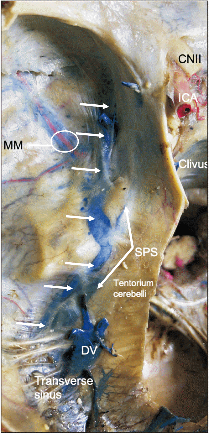
During the routine dissection of a male cadaver aged 75-year-old at death, a venous sinus (Fig. 1) was identified traveling from the superior orbital fissure anteriorly, over the floor of the middle cranial fossa, over the petrous part of the temporal bone, to drain into the transverse sinus. The variant was found on left and right sides (Fig. 2) but the right sided sinus was damaged before it could be photographed. Anteriorly, the sinus communicated with the superior ophthalmic vein, which drained primarily into the cavernous sinus. Along the floor of the middle cranial fossa, the venous sinuses communicated with the middle meningeal veins (Fig. 1). The superior petrosal sinus traveled medial to the sinus and did not communicate with it. The variant venous sinus was approximately 8 mm in width at its anterior and posterior extremes but was dilated to 1.2 cm as it crossed over the petrous part of the temporal bone. The sinuses were roughly 7.2 cm long. No additional anatomical variations or intracranial pathology were noted in this specimen.
Go to :
Discussion
The dural venous sinus of Kelch is a rare venous sinus of the skull base and one which has been infrequently reported. It effectively unites the veins of the superior orbital fissure to the transverse sinus. To our knowledge, cadaveric findings of this sinus have only been reported once before and these were of a variant of the sinus. Moreover, a bilaterally present sinus has not been reported.
Embryology
In the early embryo at the 5 to 8 mm stage, the brain is covered by a continuous plexus which drains both laterally and ventrally through three stems, the anterior, the middle, and the posterior. By the 11 to 14 mm embryonic stage, each subdivision of the brain is drained laterally into the dural plexuses by primitive pia-arachnoidal vessels. During the 17 to 20 mm stage of development, middle and posterior plexuses have joined to form the sigmoid sinus while the two parts of the anterior dural plexus; the tentorial sinus, and the marginal sinus border the expanding hemisphere caudally. At the 22 to 24 mm embryonic stage, the supraorbital vein no longer drains into the stem of the anterior dural sinus, which has diminished and is continuous with the prootic sinus. The prootic sinus is a stem of the middle dural plexus which incorporates into the stem of the posterior dural plexus and empties into the sigmoid sinus. Growth of the brain has elongated the tentorial sinus, and the marginal sinus now constitutes the medial end of the primitive transverse sinus.
By the 40 mm stage, the cavernous sinuses are developing, and are medial extensions of the prootic sinus which is now continuous with the newly developed inferior petrosal sinus. A new lateral tributary of the prootic sinus now anastomoses with a primitive temporal emissary vein derived from the sigmoid sinus to form the petrosquamous sinus. At the 60 to 80 mm stage, cerebral growth and the development of the otic capsule has led to the formation of the superior petrosal sinus. The primitive transverse sinus has moved backward on the sigmoid sinus and has anastomosed with the tentorial sinus. The prootic sinus is now continuous with the petrosquamous sinus. In the adult, remnants of the tentorial sinus may persist and anastomose with the cavernous sinus which is now joined with the superior petrosal sinus. It would be at this stage that one would expect to find the sinus of Kelch being formed (Fig. 3). The petrosquamous sinus and the remnants of the prootic sinus become diploic veins and drain the dura and bone when the middle meningeal sinus drain through the foramen ovale and into the sagittal sinus [3].
 | Fig. 3Fetal development of the intracranial venous sinuses. In this schematic drawing, note that the superior ophthalmic vein are developing and are near the more posteriorly located transverse sinus. This position would be the most likely stage where communication (dotted line) could occur between these two systems e.g., the venous sinus of Kelch.
|
Comparative anatomy
In primates, there are large evolutionary differences in the orbitotemporal venous sinuses. In primitive primates, these sinuses are large and drain the brain. The cranio-orbital sinus empties into the postglenoid emissary vein which also receives drainage from the petrosquamous sinus. In humans, this network of orbitotemporal sinuses is modified and persists as part of the middle meningeal vein system which no longer drains the brain, the orbit, or the temporal fossa, and the petrosquamous sinus is attenuated or non-existent [4]. Due to these evolutionary differences, the presence of a venous sinus of Kelch may indicate the remnant of the cranio-orbital sinus in humans [5].
Sinus of Kelch
Reports of a venous sinus of Kelch are rare in the literature. Knott [6] briefly described such a sinus as an accessory sinus which passes backward from the superior orbital fissure over the upper border of the petrous part of the temporal bone to terminate into the transverse sinus. Hollinshead [7] stated similarities between the sinus of Hyrtl and the sinus of Kelch and indicated that they may actually describe the same structure. More recently, unilaterally present sinus of Hyrtl have been described in the literature and bilateral occurrences appear to have not been previously reported [8-10]. We previously reported a unilateral variant of the sinus of Kelch found in an 88-year-old at death female cadaver [11]. In this specimen, the sinus of Kelch, which was approximately 3 mm in diameter posteriorly and 6 mm in diameter anteriorly, drained posteriorly into the superior petrosal sinus rather than into the transverse sinus. On the ipsilateral side of this same specimen, an enlarged arachnoid granulation was identified protruding through the foramen rotundum. However, in the case described herein, the only intracranial anatomical variations identified were the two venous sinuses of Kelch.
In conclusion, knowledge of the variant anatomy of the intradural venous sinuses is important to anatomists and clinicians alike. Clinically, such knowledge can minimize misdiagnoses such as when anatomical variations are noted on imaging. Surgically, such an understanding can avoid intraoperative complications such as iatrogenic hemorrhage.
Go to :
 XML Download