Journal List > Anat Cell Biol > v.54(3) > 1147572

Naderi, Ahangar, Badakhshan, Ghasemi, and Shaki: Zinc and selenium supplement mitigated valproic acid-induced testis toxicity by modulating the oxidative redox balance in male rats

Abstract

Valproic acid (VPA) is widely used antiepileptic agent which is associated with reproductive toxicity via impairment in oxidative redox. Zinc (Zn) and selenium (Se) are trace element with antioxidant effect that known to be essential for spermatogenesis. In the current study, the protective effect of co-administration of Zn and Se on VPA-induced reproductive toxicity in male rats was evaluated. Forty-eight male rats were divided into 8 groups of six (n=6): Control group (treated with normal saline); VPA only (250, 500, 1,000 mg/kg) group; VPA (500 mg/kg) plus Zn (2 mg/kg) group; VPA (500 mg/kg) plus Se (1.5 mg/kg) group; VPA (500 mg/kg) plus a combination of Zn and Se group; and VPA+vitamin E (20 mg/kg) group. The Animals were sacrificed after 28 days of treatment and sperm analysis was taken. Also, evaluation of oxidative stress markers including malondialdehyde (MDA), protein carbonyl (PC), glutathione (GSH) and histopathological changes were done on testis tissue. Morphological changes and a significant decrease in motility and sperm count in rats treated with VPA were observed. Also, an increase in oxidative stress marker, including MDA and PC and a decrease in GSH level was evident in VPA group. Zn and Se administration was able to protect against sperm abnormality, ameliorate the histological change in testis tissue, and suppressed the increase in oxidative stress markers induced by VPA. These results indicated that combination therapy with Zn and Se showed better an ameliorative effect than each one alone. Therefore, it can be suggested as an effective supplement for reproductive impairment in VPA-treated patient.

Introduction

Infertility is one of the main health problems in human life, and nearly about 30% of this problem is related to dysfunction in the male reproductive system [1]. Spermatogenesis is a sensitive and complex process that various factors can disturb this process and lead to decline sperm quantity and quality and reduce male fertility [2]. One of the most common reasons of male infertility is long term use of some drugs [3].
Valproic acid (VPA) is a commonly prescribed antiepileptic agent which is used for widespread of neurologic disorder such as bipolar affective disorders and migraine [4-6].
Its pharmacologic action is mediated via an increase in the neurotransmitter level, such as γ-amino butyric acid and glutamate [7]. In spite of the high efficacy of VPA in the treatment of many neurological diseases, but VPA induced toxicity such as hepatotoxicity [8] and also male reproductive toxicity is a main concern for patients [9]. Previous studies showed the VPA can interfere with the male reproductive system function and lead to decrease of sperm count, motility and morphological abnormality [10]. However, limited studies investigated the mechanism VPA induced the male reproductive toxicity.
One of the most importantly suggested mechanisms is oxidative stress [10]. Oxidative stress occurred when there is an imbalance between the production and removal of reactive oxygen species (ROS) [11, 12]. Previous studies showed that over production of ROS or significant depletion of antioxidant system (e.g., glutathione [GSH]) lead to oxidative damage in different tissue after VPA treatment [12, 13].
In addition, both animals [14] and human [15] studies revealed that VPA mediated male infertility is accompanied by increased oxidative stress in testis.
Zinc (Zn) is known as the second most important trace metals in the body after iron. It has critical roles in human body as a structural component of proteins and as a cofactor in various enzymes especially antioxidant enzymes [16].
On the other hand, severe Zn depletion could increase oxidative stress biomarkers in rat plasma [17]. Interestingly, previous studies showed that the plasma level of Zn in VPA-treated rats was significantly depressed compared to control groups [17]. Several evidences supported the protective effect of Zn against oxidative damage to molecules, organelles, and cells in vitro and in vivo [18].
Selenium (Se) is a cofactor of glutathione peroxidase, which is a cyto-antioxidant enzyme. Se enhances the availability of GSH, which is one of the most abundant intrinsic antioxidants that help in preventing lipid peroxidation and the resulting cell damage [19].
Therefore, in this study, we investigated the protective effects of Zn and Se against VPA induce reproductive toxicity in male rats via assessment of oxidative stress parameter, semen analysis and pathology evaluation of testicular tissue.

Material and Methods

Chemical and reagents

Valproic acid sodium salt, sodium selenite (Na2SeO3), and zinc sulfate were purchased from Sigma Aldrich (St. Louis, MO, USA). All other chemicals were of analytical grade and were purchased from standard commercial suppliers.

Animals and treatments

Male Wistar rats from our own breeding colony were used in the study (ethical code. IR.MAZUMS.REC.94-1764). The animals were caged individually and kept under standard laboratory conditions (temperature25°C±1°C, natural light–dark cycle). The rats had free access to drinking water and commercial standard laboratory diet. Rats (n=48) weighting approximately 220–250 g were randomly divided into 8 experimental groups composed of six animals each, and treated as follows: Group 1 (control group) received normal saline; Group 2–4 received different dose of VPA (250, 500 and 1,000 mg/kg); Group 5 received (VPA 500 mg/kg+Zn 2 mg/kg); Group 6 received (VPA 500 mg/kg+Se 1.5 mg/kg); Group 7 received (VPA 500 mg/kg+combination of Zn+Se); Group 8 received (VPA+vitamin E 20 mg/kg as a positive control) by gavage for 28 consecutive days.

Measurement of lipid peroxidation

Lipid peroxidation (LPO) was measured in the testes tissue using thiobarbituric acid reacting substance and was expressed in terms of malondialdehyde (MDA) content. Sample aliquots were incubated with 10% trichloroacetic acid and 0.67% thiobarbituric acid. The mixture was heated in a boiling water bath for 30 minutes, an equal volume of n-butanol was added, and the final mixture was centrifuged. The absorbance of the samples was determined at 532 nm. The results were expressed as μM MDA [12].

Measurement protein carbonyl in testis and epididymides

Protein carbonyl content was measured by a method based on the reaction of carbonyls with 2,4-dinitrophenylhydrazine forming dinitro phenyl hydrazone, a yellow compound. The standard curve was prepared by using different bovine serum albumin concentrations and the slope was used to express the levels of carbonyl protein as µM protein [20].

Measurement of glutathione

Content GSH content was determined using 5,5-dithiobis (2-nitrobenzoic acid) as the indicator and the developed yellow color was read at 412 nm on a spectrophotometer [21].

Sperm count and motility assay

The epididymal sperm suspension was prepared in 1 ml of phosphate buffered saline (PBS) at pH 7.2 and incubated at 32°C for 10 minutes. An aliquot from the suspension (1 ml) was diluted in a ratio of 1:40 with PBS. A sample of the diluted suspension was charged into a hemocytometer. The total sperm count in eight squares (except the central erythrocyte area) of one mm2 each was determined and multiplied by 5×10⁴ to get the total count [22]. Then the same eight squares were also examined for motile sperms and the percentage of motile sperm were recorded [23].

Sperm morphology assay

A fine epididymal sperm suspension was made and stained with 0.2 ml of 1% aqueous eosin. About one drop of stained suspension was placed on a clean slide and was dried. Slides were examined for abnormalities in five hundred sperms per animal and were classified into normal and abnormal sperms. Further, the abnormal sperms were designated under head abnormalities and tail abnormalities. The head abnormalities were further sub-grouped as amorphous, hookless and banana shaped, and the tail abnormalities as coiled/folded and broken [22, 23].

Histopathological assay

The left testis were removed and fixed at 4% buffered formaldehyde solution for 24 hours for histological examination. The samples were embedded in paraplast, cut into 5 mm thick sections and stained with hematoxylin and eosin (H&E) for light microscopic evaluation. Six aleatory fields of each section of the testis were qualitatively analyzed [9]. Spermatogenesis was assessed using the Johnsen scoring system [24].

Statistical analysis

Results are presented as mean±standard deviation from six times assay. All statistical analyses were performed using the SPSS software (version 13; SPSS Inc., Chicago, IL, USA) and one-way ANOVA test, followed by the post hoc Tukey test was used. Statistical significance was set at P<0.05.

Results

Effects of zinc and selenium on lipid peroxidation in rat testis
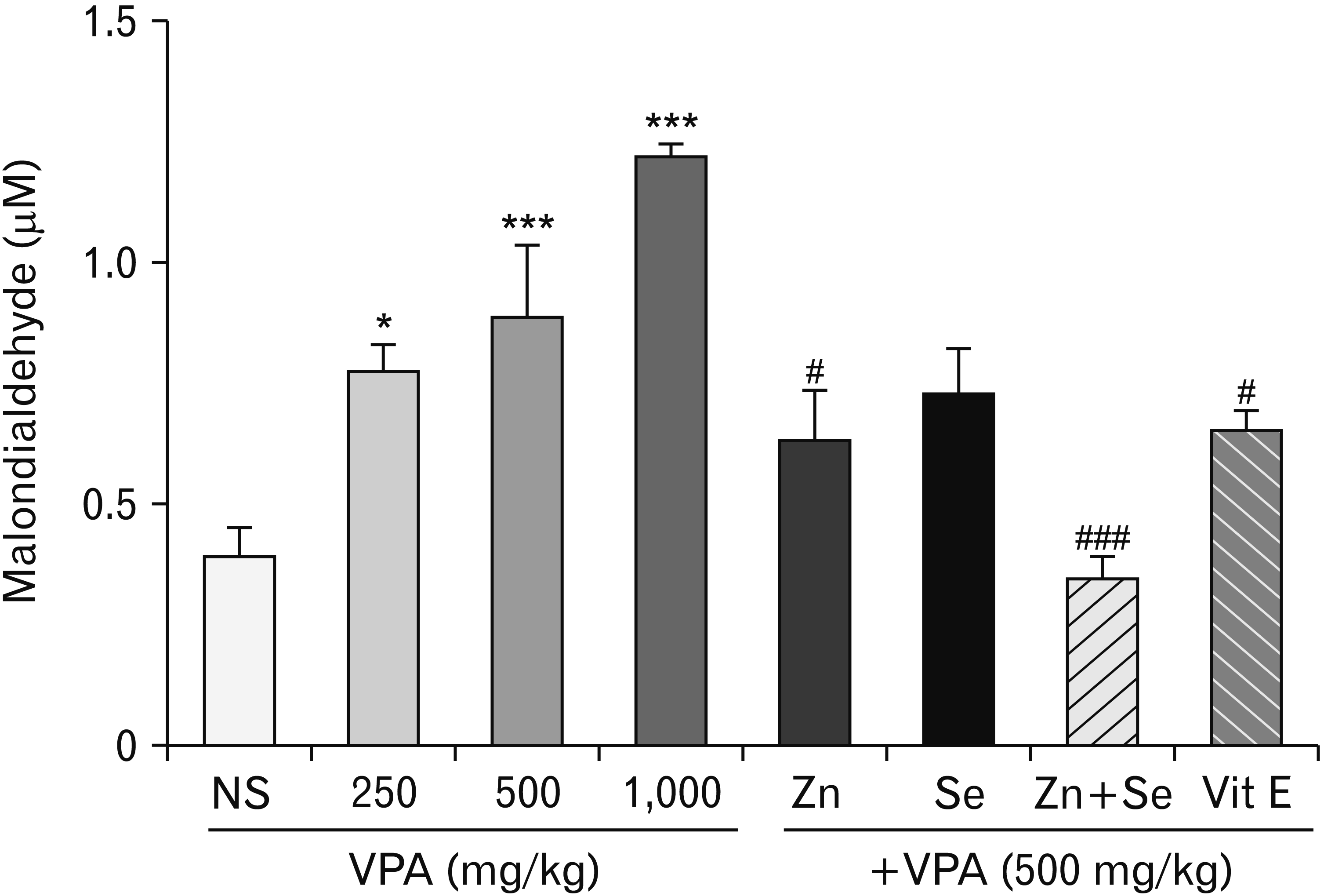
MDA levels were significantly increased in VPA-treated rats compared to control group. We observed significant differences in MDA concentration between VPA+Zn and VPA+combination of Zn and Se compared to VPA (500 mg)-treated rats (Fig. 1).

Effects of zinc and selenium on protein carbonyl in rat testis
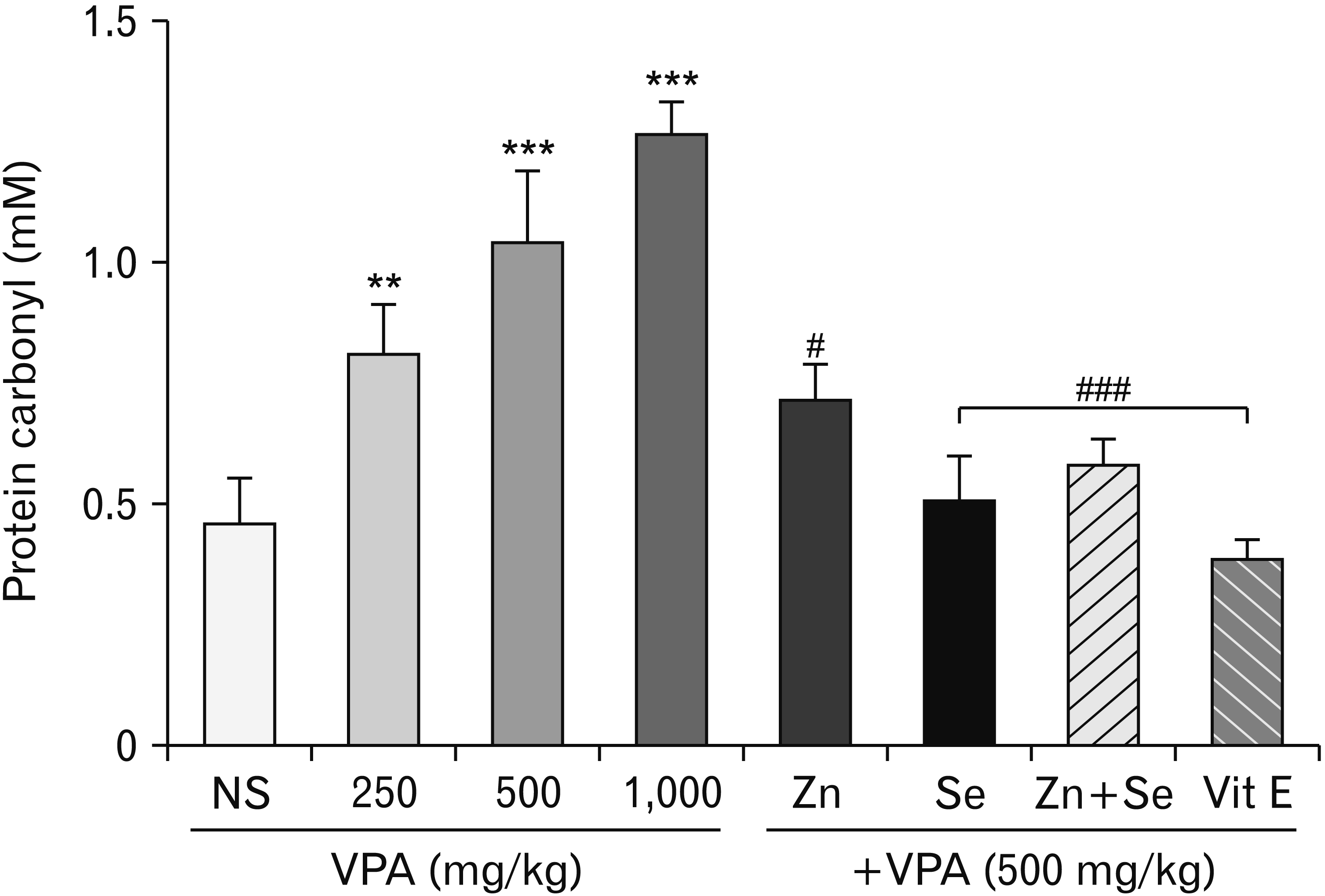
The protein concentration was found to be increased as a consequence of oxidative stress in VPA (500 mg)-treated rats compared with the control group. Significant difference in protein carbonyl concentration between VPA+Zn, VPA+Se, and VPA+combination of Zn and Se groups compared to VPA-treated rats were observed. Also, the Administration of Se and combinations of Zn+Se showed significant difference in protein carbonyl concentration from the Zn alone (Fig. 2).

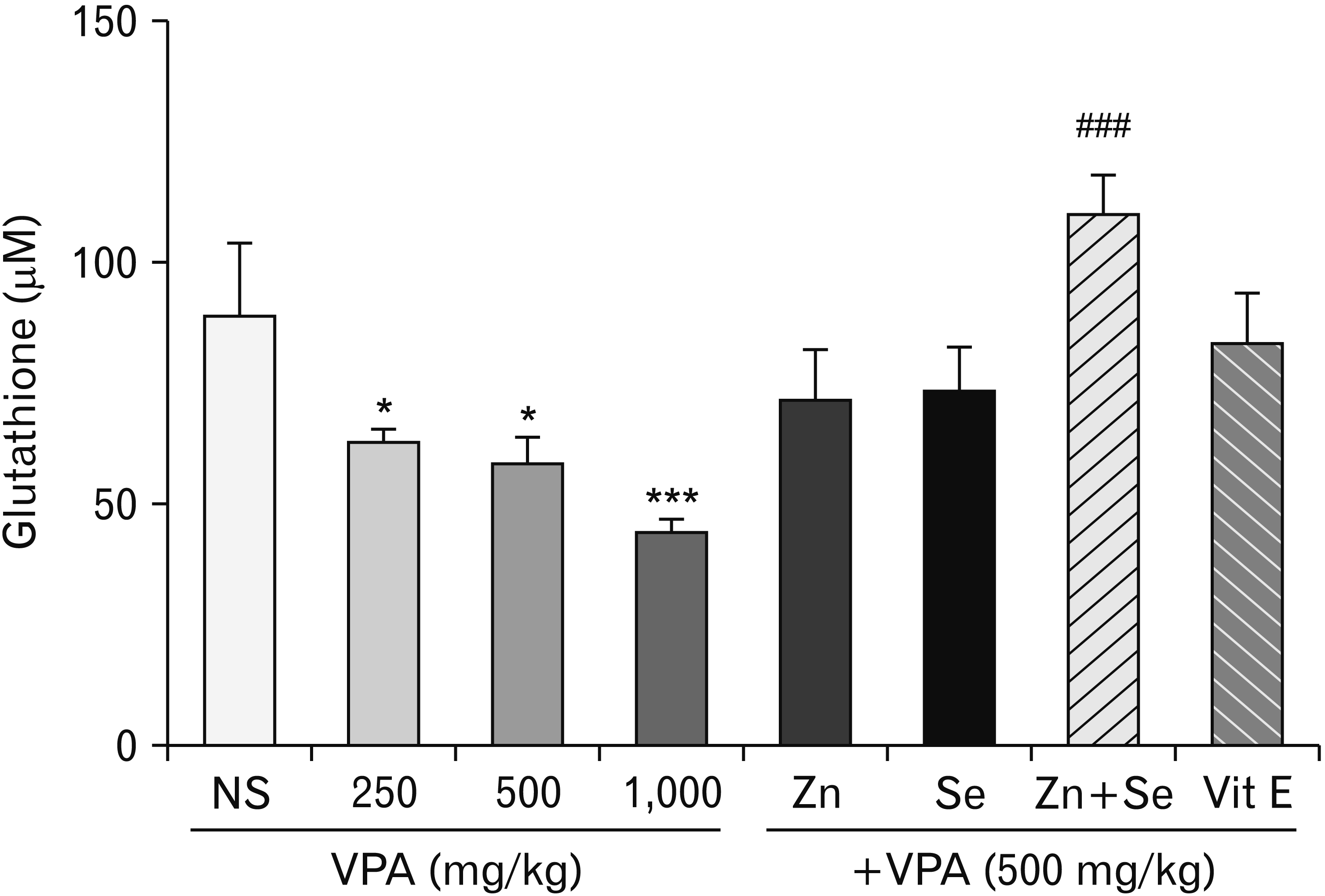
Effects of zinc and selenium on glutathione in rat testis

We yielded a significant decrease in GSH level after VPA administration. Also, co-administration of Zn and Se showed significant difference in GSH concentration of Se or Zn alone (Fig. 3).

Effects of zinc and selenium on sperm motility

Spermatozoa motility decreased in rats treated with VPA compared to control groups, while co-administration of Zn+Se, Zn and Se alone showed significant difference in sperm motility compared to VPA-treated groups (Table 1).

Effects of zinc and selenium on sperm morphology

Light microscopical analysis testicular sections assessed qualitative morphology sperm cells. Sperm morphology affected in rats treated with VPA compared to control groups, while co- administration of Zn+Se, Se and Zn alone significantly restore the altered (Table 1).

Effects of zinc and selenium on sperm count

Sperm concentration was affected by treatment with VPA, the combination of Zn+Se, Se and Zn alone increased sperm count, but showed no significant difference in sperm count compared to VPA-treated groups (Table 1).

Histopathological examination
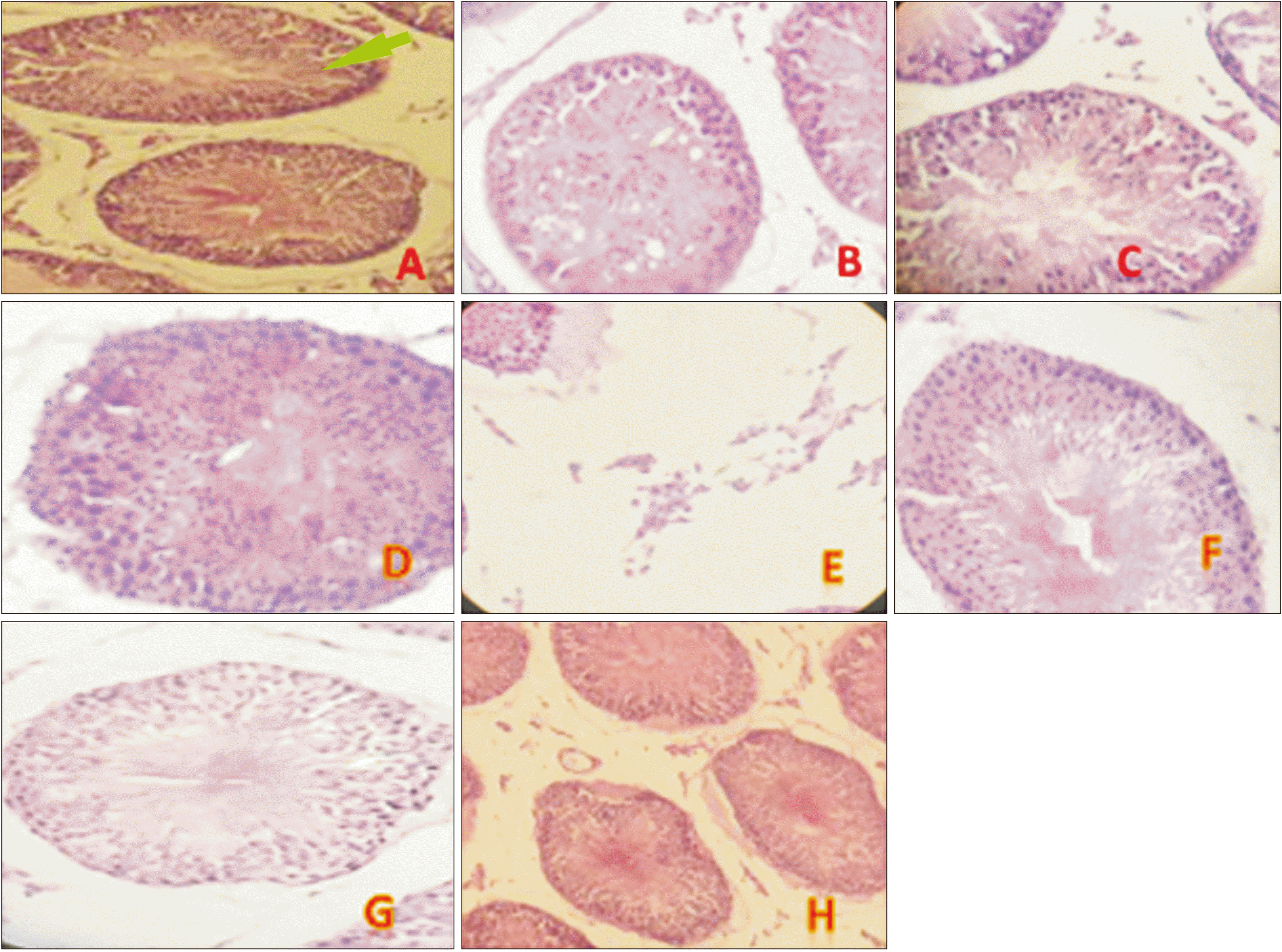
The histological appearance of the testicular tissues of the control group was normal in appearance (Fig. 4A). VPA induced histopathological variations in the testis such as decrease in germinal cell layer thickness (Fig. 4B), vacuolization and degenerative changes in germinal cells (Fig. 4C), impaired spermatogenesis, stop maturation in some parts (Fig. 4D), increase in leydig cells (Fig. 4E) An improvement was showed in the groups which received combination of Zn and Se, thus showing its protective action (Fig. 4F–H).
Spermatogenesis was evaluated using the Johnsen scoring system (Table 2).

Discussion

VPA is frequently used for the treatment of various epileptic states, but its side effect such as reproductive toxicity limited its use [25]. It has been well shown that abnormalities in the sperm analysis were seen in patients treated with VPA [26].
On the other hand, in animal studies, oral administration of VPA for 60 days in rats significantly decreased testicular weight, sperm cell concentration, live sperms, and percentage of progressively motile spermatozoa and increased percentage of morphologically abnormal spermatozoa [27].
The mechanism by which VPA treatment leads to impaired sperm quality has not been completely known. An increasing of evidence suggested that oxidative stress may be involved in VPA testis toxicity [28].
Some organs such as testis known to be particularly sensitive to oxidative stress and can lead to significant damage in this tissue [29]. In fact, both the testis and the sperm cells are rich in polyunsaturated fatty acids, which make them susceptible to ROS attack and consequently result in oxidative damage [30-32]. We observed an increase of LPO, protein carbonyl level and significant decrease of GSH content as indicators of oxidative damage to cellular macromolecules after VPA treatment in rat testis.
Therefore, this study confirmed the role of oxidative damage in VPA induced reproductive toxicity.
In addition, VPA induced histopathological changes such as a decrease in germinal cell layer thickness, degenerative changes in germinal cells, vacuolization in Sertoli cells, impaired spermatogenesis, stop maturation in some parts, increase in Leydig cells and impaired spermatogenesis in testis tissue of rats (Fig. 4).
This study has demonstrated significant effects of Zn and Se on rat spermatogenesis. As shown by the Johnsen scores, VPA toxicity is related with impaired spermatogenesis, On the other hand, Zn and Se supplementation is related with amelioration of the impairment induced by VPA toxicity, thus resulting in a higher Johnsen score and obviously, normal spermatogenesis (Table 1).
Also, morphological changes and decrease in motility and sperm count in rats treated with VPA were observed. Previous studies showed that Zn concentration of plasma was significantly decreased after chronic VPA treatment. So, Zn deficiency may be considered as a mechanism involved in oxidative damage due to VPA therapy [33]. Several evidences demonstrated a protective role of Zn against oxidative damage to molecules, organelles, and cells in vitro [18]. Interestingly, Bedwal et al. [34] reported that prostate has a higher Zn content than any other organ of the human body.
Indeed, it was shown that Zn deficiency leads to gonadal dysfunction, decreased weight of testes and caused shrinkage of seminiferous tubules [34]. A recent study has shown that Zn-deficient in the diet paves promote the cell damage in the rat testis. Moreover, Zn-deficiency increases LPO in various rat tissues, while the Zn supplementation restored this impairment [35]. On the other hand, the administration of high dose of Zn prevented tissue damage due to various toxic agents such as acetaminophen, bromobenzene, carbon tetrachloride, pesticide, nickel, endotoxin and cadmium [35-39].
In our study, Zn administration restore the impairment of sperm motility and morphological changes and improve pathological changes in testis tissue. Also, oxidative stress markers in testis tissue of VPA-treated rats significantly reduced by Zn administration. Therefore, the antioxidant effect of Zn can be considered as a possible protective mechanism against VPA induced testis toxicity.
Moreover, Se is another trace elements in all biological systems such as reproduction and its antioxidant effects have well known [40]. Testis and epididymis contain excessive concentrations of Se which demonstrating its important role during spermatogenesis for improvement of semen quality. Also, this study showed Se ameliorated VPA-induced oxidative stress in the testis tissue. On the other hand, VPA induced pathological changes in testis tissue as well as abnormalities in the sperm analysis restored after administration of Se.
In this study, we observed Zn and Se administration considerably inhibited oxidative stress in VPA-treated rats. Interestingly, determination of sperm count, motility and morphological changes which are sensitive biomarkers of testicular damage showed that co-administration of Zn and Se has a better effect than each one alone against VPA-induced sperm abnormality. This study demonstrated that VPA treatment caused oxidative damage and spermatogenesis abnormalities in rat testis tissue. Also, Zn and Se administration ameliorated these disturbances caused by VPA, whereas Se and Zn together exert a better effect against the observed oxidative stress and reproductive toxicity induced by VPA. Therefore, use of Zn and Se supplement can be suggested for attenuation of the side effects of VPA therapy.

Acknowledgements

The article was extracted from Pharm.D. Thesis of Faezeh Badakhshan and supported by a grant (no. 1764) from the research council of Mazandaran University of Medical Sciences, Sari, Iran (ethical code: IR.MAZUMS.REC.94-1764).

Notes

Author Contributions

Conceptualization: NA, FS. Data acquisition: MN, FB. Data analysis or interpretation: MG, FS. Drafting of the manuscript: MN, FB. Critical revision of the manuscript: NA, FS. Approval of the final version of the manuscript: NA, FS.

Conflict of Interest

No potential conflict of interest relevant to this article was reported.

References

1. Isidori AM, Pozza C, Gianfrilli D, Isidori A. 2006; Medical treatment to improve sperm quality. Reprod Biomed Online. 12:704–14. DOI: 10.1016/S1472-6483(10)61082-6. PMID: 16792845.
crossref
2. Bener A, Al-Ansari AA, Zirie M, Al-Hamaq AO. 2009; Is male fertility associated with type 2 diabetes mellitus? Int Urol Nephrol. 41:777–84. DOI: 10.1007/s11255-009-9565-6. PMID: 19381857.
crossref
3. Jungwirth A, Giwercman A, Tournaye H, Diemer T, Kopa Z, Dohle G, Krausz C. 2012; European Association of Urology guidelines on male infertility: the 2012 update. Eur Urol. 62:324–32. DOI: 10.1016/j.eururo.2012.04.048. PMID: 22591628.
crossref
4. Emekli-Alturfan E, Alev B, Tunali S, Oktay S, Tunali-Akbay T, Ozturk LK, Yanardag R, Yarat A. 2015; Effects of edaravone on cardiac damage in valproic acid induced toxicity. Ann Clin Lab Sci. 45:166–72. PMID: 25887870.
5. Blumenfeld A, Gennings C, Cady R. 2012; Pharmacological synergy: the next frontier on therapeutic advancement for migraine. Headache. 52:636–47. DOI: 10.1111/j.1526-4610.2011.02058.x. PMID: 22221151.
crossref
6. Soares-Weiser K, Bravo Vergel Y, Beynon S, Dunn G, Barbieri M, Duffy S, Geddes J, Gilbody S, Palmer S, Woolacott N. 2007; A systematic review and economic model of the clinical effectiveness and cost-effectiveness of interventions for preventing relapse in people with bipolar disorder. Health Technol Assess. 11:iii–iv. ix-206DOI: 10.3310/hta11390. PMID: 17903393.
crossref
7. Bialer M. 2012; Why are antiepileptic drugs used for nonepileptic conditions? Epilepsia. 53 Suppl 7:26–33. DOI: 10.1111/j.1528-1167.2012.03712.x. PMID: 23153207.
crossref
8. Lewis JH, Zimmerman HJ, Garrett CT, Rosenberg E. 1982; Valproate-induced hepatic steatogenesis in rats. Hepatology. 2:870–3. DOI: 10.1002/hep.1840020622. PMID: 6815046.
crossref
9. Ourique GM, Saccol EM, Pês TS, Glanzner WG, Schiefelbein SH, Woehl VM, Baldisserotto B, Pavanato MA, Gonçalves PB, Barreto KP. 2016; Protective effect of vitamin E on sperm motility and oxidative stress in valproic acid treated rats. Food Chem Toxicol. 95:159–67. DOI: 10.1016/j.fct.2016.07.011. PMID: 27424124.
crossref
10. Kose-Ozlece H, Ilık F, Cecen K, Huseyınoglu N, Serım A. 2015; Alterations in semen parameters in men wıth epilepsy treated with valproate. Iran J Neurol. 14:164–7. PMID: 26622982. PMCID: PMC4662690.
11. Valko M, Leibfritz D, Moncol J, Cronin MT, Mazur M, Telser J. 2007; Free radicals and antioxidants in normal physiological functions and human disease. Int J Biochem Cell Biol. 39:44–84. DOI: 10.1016/j.biocel.2006.07.001. PMID: 16978905.
crossref
12. Shokrzadeh M, Zamani E, Mehrzad M, Norian Y, Shaki F. 2015; Protective effects of propofol against Methamphetamine-induced neurotoxicity. Toxicol Int. 22:92–9. DOI: 10.4103/0971-6580.172250. PMID: 26862267. PMCID: PMC4721183.
crossref
13. Shaki F, Pourahmad J. 2013; Mitochondrial toxicity of depleted uranium: protection by Beta-glucan. Iran J Pharm Res. 12:131–40. PMID: 24250581. PMCID: PMC3813223.
14. Girish C, Shweta O, Raj V, Balakrishnan S, Varghese RG. 2014; Ellagic acid modulates sodium valproate induced reproductive toxicity in male Wistar rats. Indian J Physiol Pharmacol. 58:416–22. PMID: 26215011.
15. Chen SS, Shen MR, Chen TJ, Lai SL. 1992; Effects of antiepileptic drugs on sperm motility of normal controls and epileptic patients with long-term therapy. Epilepsia. 33:149–53. DOI: 10.1111/j.1528-1157.1992.tb02298.x. PMID: 1733750.
crossref
16. Ackland ML, Michalczyk A. 2006; Zinc deficiency and its inherited disorders- a review. Genes Nutr. 1:41–9. DOI: 10.1007/BF02829935. PMID: 18850219. PMCID: PMC3454812.
crossref
17. Daffron JC, Kasarskis EJ. 1984; Effect of valproic acid on zinc metabolism in the rat. Toxicol Lett. 23:321–5. DOI: 10.1016/0378-4274(84)90028-6. PMID: 6441315.
crossref
18. Bray TM, Bettger WJ. 1990; The physiological role of zinc as an antioxidant. Free Radic Biol Med. 8:281–91. DOI: 10.1016/0891-5849(90)90076-U. PMID: 2187766.
crossref
19. Hsu PC, Guo YL. 2002; Antioxidant nutrients and lead toxicity. Toxicology. 180:33–44. DOI: 10.1016/S0300-483X(02)00380-3. PMID: 12324198.
crossref
20. Dalle-Donne I, Rossi R, Giustarini D, Milzani A, Colombo R. 2003; Protein carbonyl groups as biomarkers of oxidative stress. Clin Chim Acta. 329:23–38. DOI: 10.1016/S0009-8981(03)00003-2. PMID: 12589963.
crossref
21. Naderi M, Ahangar N, Shaki F. 2016; Zinc and selenium protects against sodium valproate induced nephrotoxicity through modulation of oxidative stress. J Mazandaran Univ Med Sci. 26:111–22.
22. Bairy L, Paul V, Rao Y. 2010; Reproductive toxicity of sodium valproate in male rats. Indian J Pharmacol. 42:90–4. DOI: 10.4103/0253-7613.64503. PMID: 20711373. PMCID: PMC2907022.
crossref
23. Narayana K. 2008; An aminoglycoside antibiotic gentamycin induces oxidative stress, reduces antioxidant reserve and impairs spermatogenesis in rats. J Toxicol Sci. 33:85–96. DOI: 10.2131/jts.33.85. PMID: 18303187.
crossref
24. Omu AE, Al-Azemi MK, Al-Maghrebi M, Mathew CT, Omu FE, Kehinde EO, Anim JT, Oriowo MA, Memon A. 2015; Molecular basis for the effects of zinc deficiency on spermatogenesis: an experimental study in the Sprague-dawley rat model. Indian J Urol. 31:57–64. DOI: 10.4103/0970-1591.139570. PMID: 25624578. PMCID: PMC4300574.
crossref
25. Khan S, Ahmad T, Parekh CV, Trivedi PP, Kushwaha S, Jena G. 2011; Investigation on sodium valproate induced germ cell damage, oxidative stress and genotoxicity in male Swiss mice. Reprod Toxicol. 32:385–94. DOI: 10.1016/j.reprotox.2011.09.007. PMID: 22001255.
crossref
26. Isojärvi JI, Löfgren E, Juntunen KS, Pakarinen AJ, Päivänsalo M, Rautakorpi I, Tuomivaara L. 2004; Effect of epilepsy and antiepileptic drugs on male reproductive health. Neurology. 62:247–53. DOI: 10.1212/01.WNL.0000098936.46730.64. PMID: 14745062.
crossref
27. Soliman GA, Abla A. 1999; Effects of antiepileptic drugs carbamazepine and sodium valproate on fertility of male rats. Dtsch Tierarztl Wochenschr. 106:110–3. PMID: 10220947.
28. Hamza AA, Amin A. 2007; Apium graveolens modulates sodium valproate-induced reproductive toxicity in rats. J Exp Zool A Ecol Genet Physiol. 307:199–206. DOI: 10.1002/jez.357. PMID: 17351917.
crossref
29. Tomascik-Cheeseman LM, Coleman MA, Marchetti F, Nelson DO, Kegelmeyer LM, Nath J, Wyrobek AJ. 2004; Differential basal expression of genes associated with stress response, damage control, and DNA repair among mouse tissues. Mutat Res. 561:1–14. DOI: 10.1016/j.mrgentox.2004.02.011. PMID: 15238225.
crossref
30. Agarwal A, Gupta S, Sikka S. 2006; The role of free radicals and antioxidants in reproduction. Curr Opin Obstet Gynecol. 18:325–32. DOI: 10.1097/01.gco.0000193003.58158.4e. PMID: 16735834.
crossref
31. Aitken RJ, Baker MA. 2006; Oxidative stress, sperm survival and fertility control. Mol Cell Endocrinol. 250:66–9. DOI: 10.1016/j.mce.2005.12.026. PMID: 16412557.
crossref
32. Aitken RJ, Roman SD. 2008; Antioxidant systems and oxidative stress in the testes. Oxid Med Cell Longev. 1:15–24. DOI: 10.4161/oxim.1.1.6843. PMID: 19794904. PMCID: PMC2715191.
crossref
33. Ahangar N, Naderi M, Noroozi A, Ghasemi M, Zamani E, Shaki F. 2017; Zinc deficiency and oxidative stress involved in valproic acid induced hepatotoxicity: protection by zinc and selenium supplementation. Biol Trace Elem Res. 179:102–9. DOI: 10.1007/s12011-017-0944-z. PMID: 28124216.
crossref
34. Bedwal RS, Bahuguna A. 1994; Zinc, copper and selenium in reproduction. Experientia. 50:626–40. DOI: 10.1007/BF01952862. PMID: 8033970.
crossref
35. Amara S, Abdelmelek H, Garrel C, Guiraud P, Douki T, Ravanat JL, Favier A, Sakly M, Ben Rhouma K. 2008; Preventive effect of zinc against cadmium-induced oxidative stress in the rat testis. J Reprod Dev. 54:129–34. DOI: 10.1262/jrd.18110. PMID: 17420618.
crossref
36. Cagen SZ, Klaassen CD. 1979; Protection of carbon tetrachloride-induced hepatotoxicity by zinc: role of metallothionein. Toxicol Appl Pharmacol. 51:107–16. DOI: 10.1016/0041-008X(79)90013-9. PMID: 524364.
crossref
37. Sidhu P, Garg ML, Dhawan DK. 2004; Protective role of zinc in nickel induced hepatotoxicity in rats. Chem Biol Interact. 150:199–209. DOI: 10.1016/j.cbi.2004.09.012. PMID: 15535990.
crossref
38. Malhotra A, Dhawan DK. 2014; Current view of zinc as a hepatoprotective agent in conditions of chlorpyrifos induced toxicity. Pestic Biochem Physiol. 112:1–6. DOI: 10.1016/j.pestbp.2014.04.007. PMID: 24974110.
crossref
39. Alcorn JM, Fierer J, Chojkier M. 1992; The acute-phase response protects mice from D-galactosamine sensitization to endotoxin and tumor necrosis factor-alpha. Hepatology. 15:122–9. DOI: 10.1002/hep.1840150121. PMID: 1727788.
40. Ahsan U, Kamran Z, Raza I, Ahmad S, Babar W, Riaz MH, Iqbal Z. 2014; Role of selenium in male reproduction - a review. Anim Reprod Sci. 146:55–62. DOI: 10.1016/j.anireprosci.2014.01.009. PMID: 24613013.

Fig. 1
Effects of Selenium (Se) and Zinc (Zn) on valproic acid (VPA)-induced lipid peroxidation in testis tissue. Data were expressed as mean. NS, normal salin (control group); Vit E, vitamin E. *P<0.05 compared to control. ***P<0.001 compared to control. #P<0.05 compared to VPA (500 mg/kg). ###P<0.001 compared to VPA (500 mg/kg).
acb-54-3-387-f1.tif
Fig. 2
Effects of Selenium (Se) and Zinc (Zn) on valproic acid (VPA)-induced protein carbonyl in testis tissue. Data were expressed as mean. NS, normal salin (control group); Vit E, vitamin E. **P<0.01 compared to control. ***P<0.001 compared to control. #P<0.05 compared to VPA (500 mg/kg). ###P<0.001 compared to VPA (500 mg/kg).
acb-54-3-387-f2.tif
Fig. 3
Effects of Selenium (Se) and Zinc (Zn) on valproic acid (VPA)-induced GSH oxidation in testis tissue. Data were expressed as mean. NS, normal salin; Vit E, vitamin E. *P<0.05 compared to control. ***P<0.001 compared to control. ###P<0.001 compared to VPA (500 mg/kg).
acb-54-3-387-f3.tif
Fig. 4
H&E-stained testis histology pictures (with ×40 objective lens) showing normal testis. Morphology (control) (A), VPA-treated groups showed a decrease in germinal cell layer thickness (B), degenerative. changes and vacuolization in germinal cells (C), impaired spermatogenesis, stop maturation in some parts (D), increase in leydig cells (E). VPA+Zn-treated group (F), and VPA+Se-treated group (G) showed a normal germinal cell layer thickness and improve maturation. (H) VPA+Zn+Se-treated groups (combination therapy) showed better effect than Zn and Se alone. VPA, valproic acid; Zn, zinc; Se, selenium.
acb-54-3-387-f4.tif
Table 1
Effects of VPA on sperm parameters
Group Sperm abnormality (%) Sperm motility (%) Sperm count (×106/ml)
NS 13.6±3 94.1±1 39±4
VPA 47±4a) 71±1a) 24.3±2.5a)
VPA+Se 35±3c) 85.3±1.5d) 26±3
VPA+Zn 34.2±3c) 84±1d) 26.6 ±4
VPA+(Zn+Se) 30.8±3d) 81.1±1d) 26.6±2.5
VPA+Vit E 10±3d) 81.7±0.7d) 34±2b)

Values are presented as mean±SD. VPA, valproic acid; Se, selenium; Zn, zinc; Vit E, vitamin E. Sperm morphology, motility and count were evaluated in control (normal saline), VPA, VPA+Se, VPA+Zn, VPA+Zn+Se and VPA+Vit E groups. a)Significantly different from control group (P<0.001). b)Significantly different from VPA-treated group (P<0.05). c)Significantly different from VPA-treated group (P<0.01). d)Significantly different from VPA-treated group (P<0.001).

Table 2
Effects of Zn and Se on testicular weight and histology
Parameter NS VPA VPA+Se VPA+Zn VPA+Sn+Zn VPA+Vit E
Testicular weight (g) 1.65±0.09 1.36±0.11a) 1.56±0.06c) 1.59±0.08d) 1.63±0.05e) 1.54±0.07b)
Johnson score 9.3±0.74 4±0.57a) 5.8±0.68c) 7±0.81e) 8.3±0.74e) 6.3±0.74d)

Values are presented as mean±SD. NS, normal salin (control group); PA, valproic acid; Se, selenium; Zn, zinc; Sn, selenium; Vit E, vitamin E. Johnsen score - scale of 10 to 1: 10, normal spermatogenesis with open lumen; 9, many spermatozoa with obliteration of lumen; 8, only a few sprmtozoa; 7, no spermatozoa but many spermatids present; 6, no spermatozoa and only a few spermatids; 5, no sperms/spermatids but several sprmatocytes; 4, only a few spermatocytes present; 3, spermatogonia only germ cells present; 2, no germ cells but sertoli cells only present; 1, no cells in tubular section. a)Significantly different from control group (P<0.0001). b)Significantly different from VPA-treated group (P<0.05). c)Significantly different from VPA-treated group (P<0.01). d)Significantly different from VPA-treated group (P<0.001). e)Significantly different from VPA-treated group (P<0.0001).

TOOLS
Similar articles